An Introduction to the Human Body
Before we can study any single organ, we need two things: a map and a compass. This chapter gives you both. You will learn how the body is built up in nested levels from atoms to a whole person, meet the eleven organ systems that share the work of staying alive, and discover the one idea that runs underneath all of physiology — homeostasis, the body's relentless drive to hold its internal world steady. Then we will learn the precise language anatomists use to describe where things are, so that for the rest of the book, "left," "above," and "in front" never mean the wrong thing.
By the end of this chapter you can
- Distinguish anatomy from physiology and explain why structure and function are always studied together.
- Order the six levels of structural organization from chemical to organism, and give an example at each level.
- Name the eleven organ systems, state the main job of each, and predict which system a given organ belongs to.
- Define homeostasis and diagram a negative feedback loop using its three parts — receptor, control center, and effector.
- Contrast negative and positive feedback and give a real example of each.
- Describe the anatomical position and use directional terms in correct pairs (superior/inferior, proximal/distal, and the rest).
- Identify the three body planes and the body cavities, and locate a structure by region or quadrant.
Section 1The levels of organization
Two questions, always asked together
Everything in this book grows out of two words. Anatomy asks what is it and where is it — the shape, the parts, the position of a structure. Physiology asks how does it work — the function, the process, the job that structure performs. The two are inseparable, because in the body structure always fits function. The wall of an air sac in the lung is thin as tissue paper precisely so that oxygen can cross it in a fraction of a second; the wall of the heart's main pumping chamber is thick and muscular precisely because it must launch blood to the whole body. Change the structure and you change what the tissue can do — a rule you will see over and over.
Small parts build bigger parts
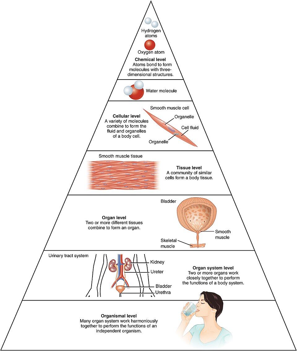
The body is organized like a set of nesting dolls, each level built from the one below it. There are six levels, and it is worth being able to recite them in order:
| Level | What it is | Example |
|---|---|---|
| 1. Chemical | Atoms bond into molecules | Hydrogen + oxygen → a water molecule; amino acids → a protein |
| 2. Cellular | Molecules assemble into the smallest living unit | A muscle cell, a neuron, a red blood cell |
| 3. Tissue | Similar cells working together on one task | Cardiac muscle tissue; nervous tissue |
| 4. Organ | Two or more tissue types forming a structure with a job | The heart, the stomach, the skin |
| 5. Organ system | Organs cooperating on a large function | The cardiovascular system (heart + vessels + blood) |
| 6. Organism | All systems together = one living person | You |
Read the ladder in either direction and it tells a story. Going up, tiny parts combine into ever-larger, more capable structures until they add up to a living human. Going down, any disease can be traced to a lower level: a heart attack is an organ problem that begins with cells starved of oxygen because a molecule (cholesterol) built up in a vessel wall. Learning to move up and down this ladder is one of the most useful habits in all of A&P.
Zoom in and out through all six levels in the interactive below.
- Anatomy = structure and position; physiology = function and process; structure always fits function.
- The six levels, low to high: chemical → cellular → tissue → organ → organ system → organism.
- The cell is the smallest living unit and the dividing line between chemistry and life.
- Disease can be traced down the ladder to the tissue, cell, or molecule where it began.
Section 2The organ systems
Eleven teams, one goal
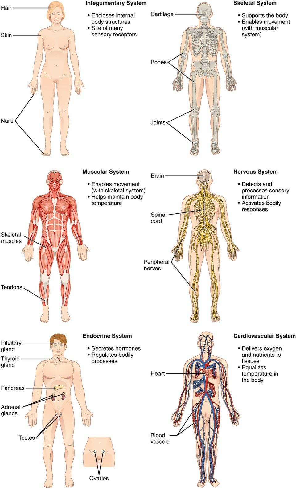
Keeping a human alive is too big a job for any one organ, so the work is divided among eleven organ systems. They are not really separate — they overlap and depend on one another constantly — but grouping them by their main function makes the body learnable. A helpful way to hold them is to sort them by the kind of job they do: covering and support, control and communication, moving substances, and continuing the species.
| System | Main job | Signature organs |
|---|---|---|
| Integumentary | Covers and protects; temperature, sensation, vitamin D | Skin, hair, nails |
| Skeletal | Support, protection, movement levers, blood-cell formation, mineral storage | Bones, cartilage, joints |
| Muscular | Movement, posture, heat | Skeletal muscles |
| Nervous | Fast control and communication | Brain, spinal cord, nerves |
| Endocrine | Slow, hormonal control | Thyroid, pancreas, adrenal, pituitary |
| Cardiovascular | Transports blood, O₂, nutrients, wastes | Heart, blood vessels, blood |
| Lymphatic / Immune | Fluid return and defense against pathogens | Lymph nodes, spleen, thymus, vessels |
| Respiratory | Gas exchange — O₂ in, CO₂ out | Lungs, airways |
| Digestive | Breaks down food, absorbs nutrients, removes waste | Stomach, intestines, liver, pancreas |
| Urinary | Filters blood, balances water/salts/pH, makes urine | Kidneys, ureters, bladder |
| Reproductive | Produces offspring and sex hormones | Ovaries/uterus; testes |
No system works alone
The magic is in the cooperation. Take one breath and follow it: the respiratory system pulls in oxygen, but it is the cardiovascular system that carries that oxygen to the tissues, the muscular system (the diaphragm) that did the pulling, the nervous system that set the breathing rate, and the skeletal system (the rib cage) that gave the lungs a frame to expand inside. When you meet a disease later in the course, ask which systems it touches — the answer is almost never just one.
Tap each system in the interactive to watch its organs light up on the body.
- There are eleven organ systems; know each system's main job and a signature organ.
- Systems constantly cooperate — a single act like breathing draws on five or more at once.
- Some organs serve two systems (the pancreas is digestive and endocrine).
Section 3Homeostasis & feedback
The single big idea of physiology
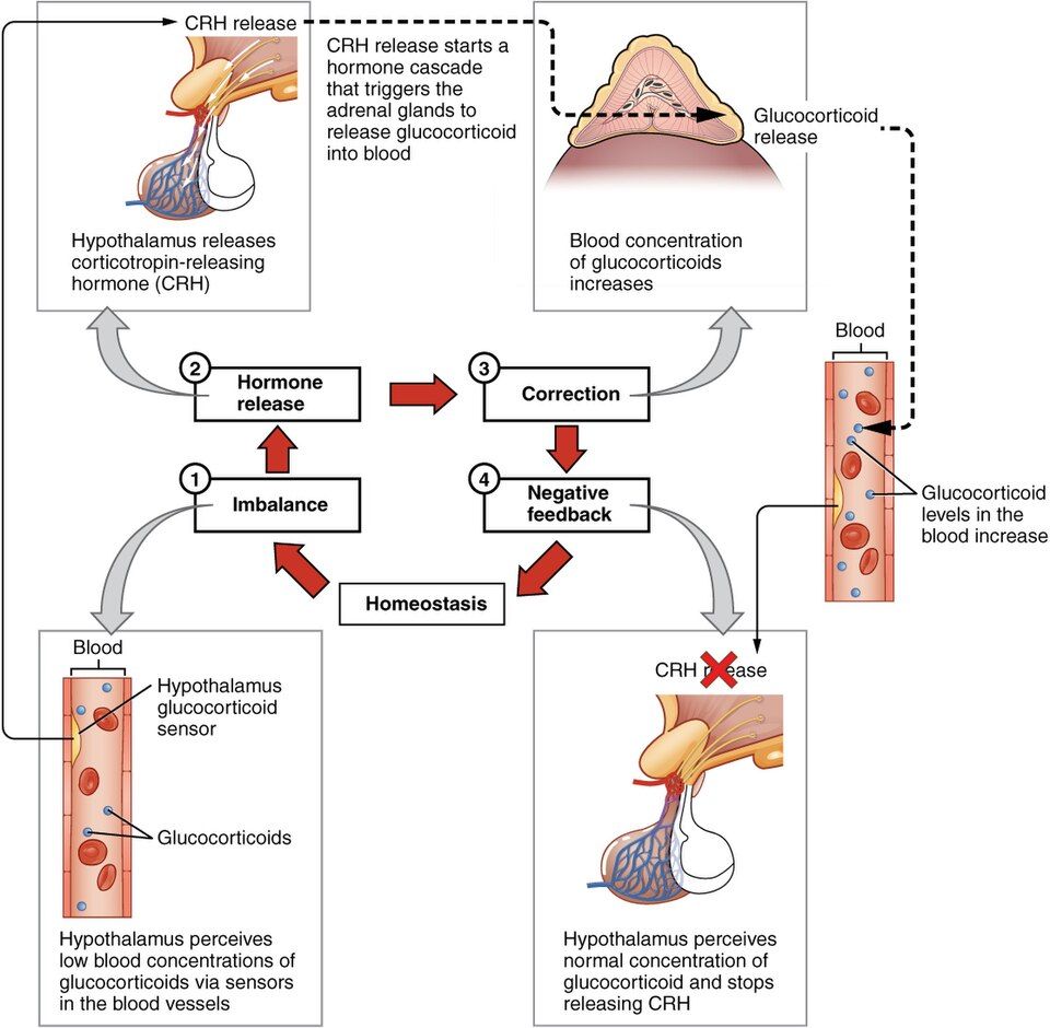
If you remember one word from this whole course, make it homeostasis — the maintenance of a stable internal environment despite a changing outside world. Your body temperature hovers near 37 °C whether you are in a snowstorm or a sauna; your blood glucose, blood pH, and fluid volume all stay penned inside narrow ranges. They do not stay perfectly still — they wobble gently around a set point, like a tightrope walker making constant tiny corrections. That dynamic balance is health. Push a value too far outside its range and cells stop working; push it further and the person dies. Nearly every disease in the rest of this book is, at heart, a failure of homeostasis.
The negative feedback loop: three parts
The body keeps values steady mainly through negative feedback, and every negative feedback loop has the same three components. Learn them once and you can read any loop in the body:
- A receptor (a sensor) detects the change and reports it.
- A control center (usually the brain or an endocrine gland) compares the reading to the set point and decides on a response.
- An effector (a muscle or gland) carries out the response, which pushes the value back toward normal.
It is called negative feedback because the response opposes (negates) the original change — if the value went up, the loop brings it down, and vice versa. The classic example is body temperature. Step into the cold: temperature receptors in your skin fire; the control center (the hypothalamus) reads "too cold"; and it drives effectors — blood vessels constrict to keep heat in, and muscles shiver to make heat. Temperature climbs back to the set point, and the loop quiets down. A home thermostat works exactly the same way: sense, compare, correct.
Positive feedback: the rare amplifier
Occasionally the body wants the opposite — a signal that amplifies itself until an event is finished. This is positive feedback, and it is rare precisely because it is explosive. In childbirth, the baby's head stretches the cervix, which triggers oxytocin, which strengthens contractions, which stretches the cervix more — a snowballing cycle that builds until delivery ends it. Blood clotting works the same way: the first platelets to stick release chemicals that recruit more platelets, which recruit more, sealing the wound fast. Positive feedback always needs a built-in stop (birth, or a finished clot); without one it would run away.
| Feature | Negative feedback | Positive feedback |
|---|---|---|
| Effect on the change | Opposes / reverses it | Amplifies / continues it |
| Result | Returns to set point (stability) | Drives toward a completion event |
| Frequency | The rule — most loops | The exception — a few |
| Examples | Temperature, glucose, blood pressure, pH | Childbirth, blood clotting, lactation |
Knock a value off its set point in the interactive and watch a negative feedback loop pull it home.
- Homeostasis is a stable internal environment held near a set point; losing it is the root of most disease.
- A negative feedback loop has three parts — receptor → control center → effector — and its response opposes the change to restore normal.
- Positive feedback amplifies a change until a completion event stops it: childbirth, clotting, milk let-down.
Section 4The language of position
Start from the anatomical position
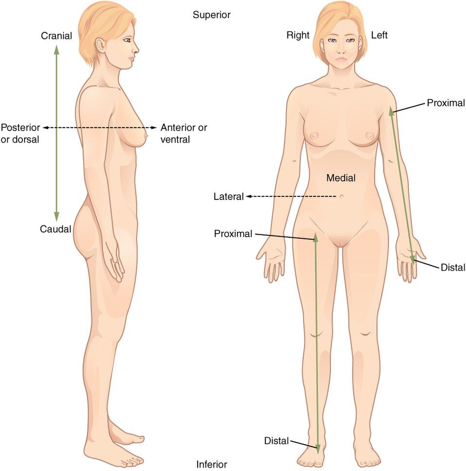
To describe where things are without confusion, everyone agrees to start from the same pose: the anatomical position. The body stands upright, facing forward, feet slightly apart, arms at the sides with the palms turned forward. Every directional term is defined from this pose, so a structure's description never changes even if the real patient is lying down or upside down. One more rule that trips up students: "right" and "left" always mean the patient's own right and left, not yours as you look at them.
The directional pairs
Directional terms come in opposing pairs, which makes them easy to learn two at a time:
| Term | Meaning | Example |
|---|---|---|
| Superior / Inferior | Above / below (toward head / toward feet) | The heart is superior to the stomach |
| Anterior / Posterior | Front / back (ventral / dorsal) | The sternum is anterior to the heart |
| Medial / Lateral | Toward the midline / away from it | The nose is medial to the eyes |
| Proximal / Distal | Nearer to / farther from the trunk (limbs) | The elbow is proximal to the wrist |
| Superficial / Deep | Toward the surface / away from it | The skin is superficial to the muscle |
Two of these pairs deserve extra care. Proximal and distal are used mainly for the limbs and always measured from where the limb attaches to the trunk — so the shoulder is proximal, the fingertips distal. Medial and lateral are measured from the body's imaginary midline, so structures near the center are medial and structures out toward the sides are lateral.
See each pair drawn on a body in anatomical position below.
- All terms are defined from the anatomical position (upright, facing forward, palms forward).
- Right and left always mean the patient's right and left.
- Learn the pairs: superior/inferior, anterior/posterior, medial/lateral, proximal/distal, superficial/deep — and use them relatively ("X is [term] to Y").
Section 5Planes & sections
Three ways to slice
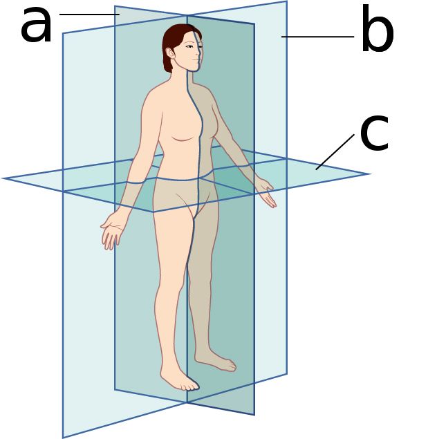
To look inside the body — on a cadaver table or on a CT scan — we imagine cutting it along flat surfaces called planes. There are three, and they are perpendicular to one another:
- The sagittal plane divides the body into left and right. A cut exactly down the midline is a midsagittal section; an off-center one is parasagittal.
- The frontal (coronal) plane divides the body into front and back (anterior and posterior).
- The transverse (horizontal) plane divides the body into top and bottom (superior and inferior) — the "slice" view you see on a CT.
Why does this matter to a nurse or clinician? Because medical images are always taken in one of these planes, and reading a scan means knowing which slice you are looking at. A transverse CT of the abdomen shows organs in cross-section; a sagittal MRI of the spine, viewed from the side, shows the vertebrae stacked top-to-bottom (superior to inferior). Naming the plane is the first step in orienting yourself to any image.
Watch each plane sweep through the body in the interactive below.
- Sagittal = left/right; frontal (coronal) = front/back; transverse = top/bottom.
- A midsagittal cut makes equal mirror halves; other sagittal cuts (parasagittal) do not.
- Medical images (CT, MRI) are read by first naming their plane.
Section 6Cavities & regions
Where the organs live
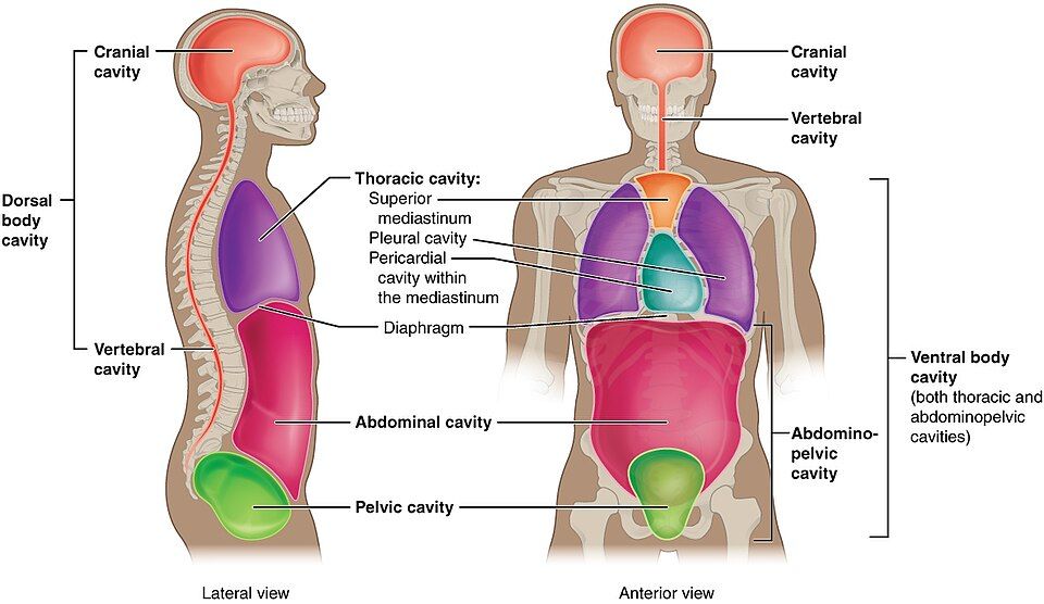
The organs are not loose inside you — they are housed in enclosed spaces called body cavities that protect them and let them move. There are two main groups. The dorsal (posterior) cavity runs along the back and holds the nervous system: the cranial cavity (the brain) and the vertebral cavity (the spinal cord). The larger ventral (anterior) cavity holds most other organs and is split by the diaphragm into the thoracic cavity above (heart and lungs) and the abdominopelvic cavity below (digestive organs, then the pelvic organs).
Serous membranes: the slippery linings
Organs in the ventral cavity are wrapped in thin, double-layered serous membranes that secrete a slick fluid so organs can glide without friction. Each membrane has two layers with paired names — a parietal layer lining the cavity wall and a visceral layer hugging the organ itself: parietal and visceral pleura around the lungs, parietal and visceral pericardium around the heart, and parietal and visceral peritoneum around the abdominal organs. When one of these membranes becomes inflamed, you get the "-itis" diseases — pleuritis, pericarditis, peritonitis — and the friction or fluid buildup causes real trouble.
Mapping the abdomen: regions and quadrants
Because so much can go wrong in the belly, clinicians divide the abdominopelvic surface into a grid to describe exactly where a pain or a mass is. The simple version uses four quadrants (right and left, upper and lower — RUQ, LUQ, RLQ, LLQ); the detailed version uses nine regions (a tic-tac-toe grid: the central umbilical region, the epigastric above it, the hypogastric below, and flanking regions on each side). Saying "RLQ pain" instantly narrows the suspects — the appendix lives there.
Explore the cavities, membranes, and the region/quadrant grids in the interactive.
- The dorsal cavity (cranial + vertebral) holds the CNS; the ventral cavity splits at the diaphragm into thoracic and abdominopelvic.
- Serous membranes — pleura, pericardium, peritoneum — reduce friction; their inflammation gives the "-itis" diseases.
- The abdomen maps into 4 quadrants or 9 regions; location narrows the diagnosis.
The chapter in one breath
- Anatomy is structure, physiology is function, and structure always fits function.
- The body is built in six nested levels: chemical → cellular → tissue → organ → organ system → organism, with the cell as the smallest living unit.
- Eleven organ systems share the work of survival and constantly cooperate; know each one's main job.
- Homeostasis is the stable internal environment that defines health; most disease is a failure of it.
- Negative feedback (receptor → control center → effector) opposes change to restore a set point; positive feedback amplifies change to complete an event (childbirth, clotting).
- Anatomical language starts from the anatomical position; directional terms are used in relative pairs, and right/left mean the patient's.
- Three planes (sagittal, frontal, transverse) slice the body; organs sit in dorsal and ventral cavities lined by serous membranes; the abdomen maps into quadrants and regions.
ReferenceWords worth knowing
A quick reference for the terms in this chapter — skim it now, or circle back whenever a word trips you up.
- Anatomical position
- The standard reference pose — standing upright, facing forward, arms at the sides with palms forward — from which all directional terms are defined.
- Anatomy
- The study of the body's structures and their physical relationships (what a part is and where it lies).
- Anterior (ventral)
- Toward the front of the body.
- Control center
- The part of a feedback loop (often the brain or an endocrine gland) that compares a sensed value to the set point and directs a response.
- Distal
- Farther from the point where a limb attaches to the trunk (the fingers are distal to the elbow).
- Effector
- The muscle or gland in a feedback loop that carries out the corrective response.
- Frontal (coronal) plane
- A vertical plane dividing the body into anterior (front) and posterior (back) portions.
- Homeostasis
- The maintenance of a relatively stable internal environment despite external change, kept near a set point by feedback.
- Medial
- Toward the midline of the body.
- Negative feedback
- A control loop whose response opposes and reverses the original change, restoring the value to its set point; the body's main homeostatic mechanism.
- Organ
- A structure made of two or more tissue types that performs a specific function (for example, the heart or stomach).
- Physiology
- The study of how the body's structures function and the processes that keep it alive.
- Positive feedback
- A control loop whose response amplifies the original change until a completion event stops it (childbirth, blood clotting).
- Proximal
- Nearer to the point where a limb attaches to the trunk (the elbow is proximal to the wrist).
- Sagittal plane
- A vertical plane dividing the body into left and right portions; a midline cut is midsagittal.
- Serous membrane
- A thin, double-layered membrane (pleura, pericardium, peritoneum) that lines a ventral cavity and secretes fluid to reduce friction around organs.
- Set point
- The target value a homeostatic variable is regulated around (for example, ~37 °C for body temperature).
- Superior
- Toward the head or the upper part of the body.
- Transverse (horizontal) plane
- A plane dividing the body into superior and inferior portions; produces a cross-sectional view.