The Tissue Level of Organization
In the last chapter we zoomed in on the single cell; now we take one step back up the ladder. Cells rarely work alone — they gather into tissues, teams of similar cells that share a job and, together, do something no lone cell could. The remarkable part is how few kinds there are: your entire body, every organ in it, is stitched together from just four primary tissues. This chapter introduces all four — how each looks under the microscope, where it lives, and what it does — and ends with the story every tissue eventually has to tell: how it heals when it is hurt.
By the end of this chapter you can
- Define tissue and name the four primary tissue types, giving the general job of each.
- List the shared features of epithelial tissue and classify any epithelium by its layering and cell shape.
- Describe the common blueprint of all connective tissue — cells scattered in an extracellular matrix of fibers and ground substance — and identify its major subtypes.
- Distinguish the three muscle tissues by striations, voluntary vs. involuntary control, and cell shape.
- Diagram a neuron, label its parts, and trace how nervous tissue carries a signal in one direction.
- Outline the four stages of tissue repair and contrast regeneration with fibrosis (scarring).
- Explain how the four tissues combine to build organs and body membranes.
Section 1The four tissue types
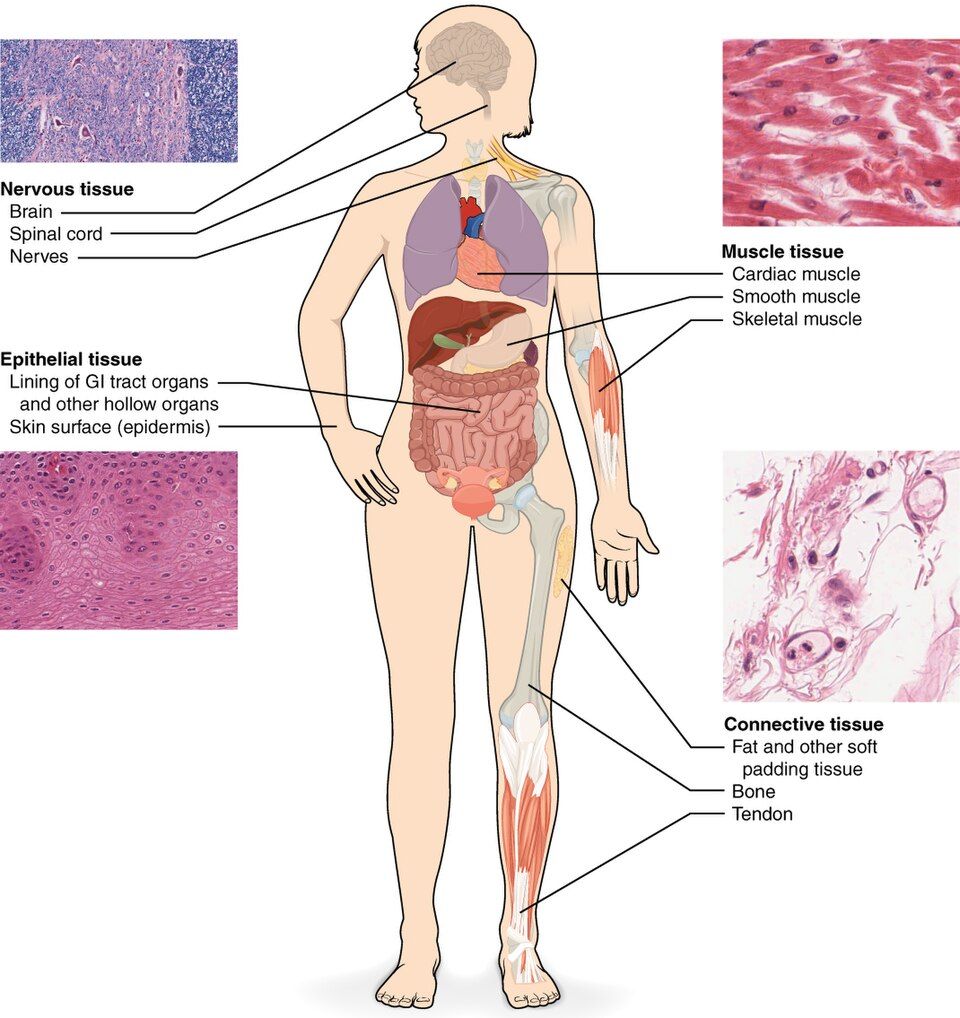
A society of cells
A tissue is a group of similar cells that work together to carry out a shared function. That is the whole definition, but the word "together" is doing a lot of work. The cells of a tissue are not just neighbors who happen to look alike — they are coordinated, held in place by junctions or by the material they secrete around themselves, and specialized for one kind of job. The branch of biology that studies tissues under the microscope is histology, and it is essentially anatomy at very high magnification: instead of asking what an organ looks like to the naked eye, we ask what it is made of.
Four families do all the work
What makes the whole subject learnable is that no matter how complicated an organ appears, it is assembled from only four primary tissue types. Learn these four and you have the vocabulary to describe any structure in the body. Each family answers a different fundamental need — a body must cover and line its surfaces, bind and support its parts, move, and communicate.
| Tissue | What it does | Where you find it |
|---|---|---|
| Epithelial | Covers surfaces, lines cavities, forms glands; protects, absorbs, secretes | Skin surface, gut lining, kidney tubules, gland cells |
| Connective | Binds, supports, protects, stores, and transports; the body's packing material | Bone, fat, tendon, cartilage, blood |
| Muscle | Contracts to generate movement and force; also makes heat | Skeletal muscles, heart wall, walls of hollow organs |
| Nervous | Senses, processes, and rapidly transmits electrical signals | Brain, spinal cord, peripheral nerves |
Notice how different the four are in how much room the cells give one another. Epithelial cells are packed together so tightly there is almost no space between them — that is what makes a good barrier. Connective tissue does the opposite: its cells are few and far apart, floating in a sea of non-living material they secrete. That single contrast — crowded cells versus scattered cells in a matrix — is one of the fastest ways to tell the first two tissues apart on a slide.
From tissues to organs and membranes
An organ is simply two or more tissue types layered together so that the whole does something none of them could do alone. The stomach is the clearest example: an epithelial lining that secretes acid and mucus, a connective tissue layer carrying its vessels and nerves, smooth muscle that churns, and nervous tissue that times the churning — four tissues, one organ. The simplest of these combinations is a body membrane, a thin sheet that covers or lines a surface, and there are only four kinds. The cutaneous membrane is the skin itself: stratified squamous epithelium over dense irregular connective tissue, and the only membrane that is dry. A mucous membrane lines every cavity that opens to the outside — airway, gut, urinary and reproductive tracts — and pairs an epithelium with an underlying loose connective layer called the lamina propria. A serous membrane lines the closed ventral cavities and wraps the organs inside them (pleura, pericardium, peritoneum), joining a simple squamous epithelium called mesothelium to a thin areolar layer, and it secretes a watery fluid so organs glide. The synovial membrane is the odd one out: it lines joint cavities and is made of connective tissue only, with no epithelium at all.
Meet all four tissues side by side in the interactive below.
- A tissue is a group of similar cells performing a shared function; studying them is histology.
- The body has four primary tissues: epithelial (cover/line), connective (bind/support), muscle (move), and nervous (communicate).
- Epithelial cells are tightly packed; connective cells are scattered in a matrix — a quick way to tell them apart.
- Tissues layer together to build organs and the four body membranes: cutaneous (skin, dry), mucous (lines cavities open to the outside), serous (lines closed ventral cavities), and synovial (joint cavities, connective tissue only).
Section 2Naming an epithelium
What every epithelium has in common
Epithelial tissue covers every free surface of the body and lines every cavity, tube, and duct — the outer skin, the inside of your gut, the airways, the blood vessels. Because it stands at every boundary, it shares a set of features built for that role. Its cells are tightly packed with very little material between them, so almost nothing crosses without going through a cell. Every epithelium has polarity: an apical surface that faces the open space (often decorated with microvilli or cilia) and a basal surface anchored to a thin basement membrane that glues it to the connective tissue below. Epithelium is avascular — it has no blood vessels of its own and must be fed by diffusion from the tissue underneath — yet it is richly supplied with nerves, and it regenerates quickly, replacing cells lost to the constant wear of being a surface.
The two-word naming rule
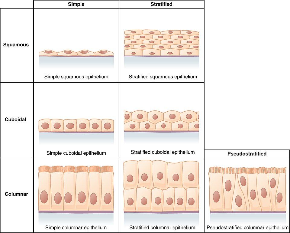
Here is the payoff of this section: every epithelium is named by the same simple formula, so once you learn the rule you can name a tissue you have never seen. The name has two parts — first the number of layers, then the shape of the cells at the surface.
- Layers. One layer of cells is simple; two or more stacked layers is stratified.
- Shape. Flat, scale-like cells are squamous; cube-shaped cells are cuboidal; tall, column-shaped cells are columnar.
Snap the two halves together and you have the name: a single flat layer is simple squamous; many stacked flat layers make stratified squamous; one row of tall cells is simple columnar. The structure even predicts the job. Thin simple epithelia are ideal where things must cross quickly — simple squamous lines the air sacs of the lung and the walls of capillaries because oxygen and nutrients slip across it in an instant. Thick stratified epithelia are built for protection — stratified squamous forms the surface of your skin and the lining of the mouth and esophagus, layers you can afford to lose and rebuild.
| Epithelium | Structure | Best-known job and location |
|---|---|---|
| Simple squamous | One layer of flat cells | Rapid diffusion — lung air sacs, capillary and vessel lining (endothelium) |
| Simple cuboidal | One layer of cube cells | Secretion and absorption — kidney tubules, gland ducts |
| Simple columnar | One layer of tall cells | Absorption and secretion — lining of the stomach and intestines |
| Stratified squamous | Many stacked flat layers | Protection against abrasion — skin surface, mouth, esophagus, vagina |
| Pseudostratified columnar | One layer that looks layered | Ciliated lining of the airways that sweeps mucus upward |
| Transitional | Stretchy, shape-changing layers | Bladder and ureters — stretches as they fill |
Two entries in that table are the famous exceptions. Pseudostratified columnar epithelium is really a single layer — every cell touches the basement membrane — but because the nuclei sit at different heights it looks stacked, hence "pseudo" (false) stratified. Transitional epithelium is the shape-shifter of the urinary tract: its cells slide over one another and flatten as the bladder fills, then bunch back up as it empties.
Build an epithelium's name box by box and watch it appear in the interactive.
- Epithelia are tightly packed, polar (apical vs. basal), sit on a basement membrane, are avascular, and regenerate fast.
- Name any epithelium as layers (simple = one, stratified = many) + cell shape (squamous, cuboidal, columnar).
- Simple epithelia favor absorption/secretion/diffusion; stratified epithelia favor protection; pseudostratified and transitional are the special cases.
Section 3Connective tissue
One recipe, endless variety
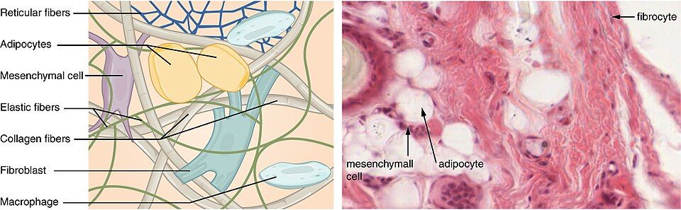
Connective tissue is the most abundant and the most diverse tissue in the body, and at first it can seem like a grab-bag: bone, fat, tendon, cartilage, and even blood all belong to it. What ties them together is a single shared blueprint. Unlike epithelium, where the cells are everything, connective tissue is mostly extracellular matrix — a non-living material the cells secrete and then sit inside. Its cells are relatively few and widely scattered; the character of the tissue comes almost entirely from the matrix around them. This is the key insight of the whole section: change the matrix and you change the tissue. Make the matrix a firm rubbery gel and you get cartilage; harden it with calcium salts and you get bone; make it liquid and you get blood. Adipose (fat) is the instructive exception: its cells store the lipid inside themselves, each swollen with a single droplet, so the matrix is squeezed down to almost nothing — fill the cells rather than the matrix and you get fat.
The matrix: fibers and ground substance
The extracellular matrix always has two ingredients. First are the protein fibers, of which there are three kinds: strong, rope-like collagen fibers that resist pulling; stretchy elastic fibers that snap back into shape; and fine, mesh-like reticular fibers that form supporting scaffolds. Second is the ground substance, the material that fills the space between the fibers and cells — anywhere from watery to gel-like to rock-solid, depending on the tissue. The cells themselves usually come in two flavors named by a simple convention: a cell whose name ends in -blast is the young, active builder that secretes matrix (a fibroblast, osteoblast, or chondroblast), while one ending in -cyte is the mature cell that maintains it (a fibrocyte, osteocyte, or chondrocyte).
| Connective tissue | Matrix | Job and location |
|---|---|---|
| Loose (areolar) | Soft, gel-like, loosely woven fibers | All-purpose packing and cushioning; underlies epithelia everywhere |
| Adipose (fat) | Cells so full of fat that matrix is minimal | Energy storage, insulation, padding around organs |
| Dense regular | Tightly packed parallel collagen | Strong pull in one direction — tendons and ligaments |
| Cartilage | Firm, rubbery gel | Flexible support — nose, ears, joint surfaces, discs |
| Bone (osseous) | Collagen hardened with calcium salts | Rigid support and protection; mineral storage |
| Blood | Liquid matrix (plasma) | Transport of cells, gases, nutrients, and wastes |
Reading that table top to bottom, you can watch a single idea flex through every possible stiffness — from a soft cushion, to a padded fat pad, to a tough cable, to a rubbery shock absorber, to solid mineral, all the way to a flowing liquid. Same recipe, radically different results, tuned entirely by what the cells put into the matrix.
Rebuild the matrix in the interactive and watch one tissue turn into the next.
- All connective tissue follows one recipe: scattered cells in an extracellular matrix of fibers + ground substance.
- Three fiber types — collagen (strength), elastic (stretch), reticular (scaffold); -blast cells build the matrix and -cyte cells maintain it.
- Changing the matrix yields the whole family: loose, adipose, dense, cartilage, bone, and blood.
Section 4Muscle tissue
Cells built to shorten
Muscle tissue has one talent that no other tissue has: it can contract, actively shortening to pull on something. Its cells are packed with two protein filaments — actin and myosin — that slide past one another to shorten the cell, and everything about a muscle cell's shape serves that pull. When these filaments line up in orderly, repeating rows, they catch the light in alternating bands and the tissue looks striated (striped); when they are arranged less regularly, the tissue looks smooth. Contraction not only moves the body but also generates much of your body heat — which is why shivering warms you up.
The three muscle tissues
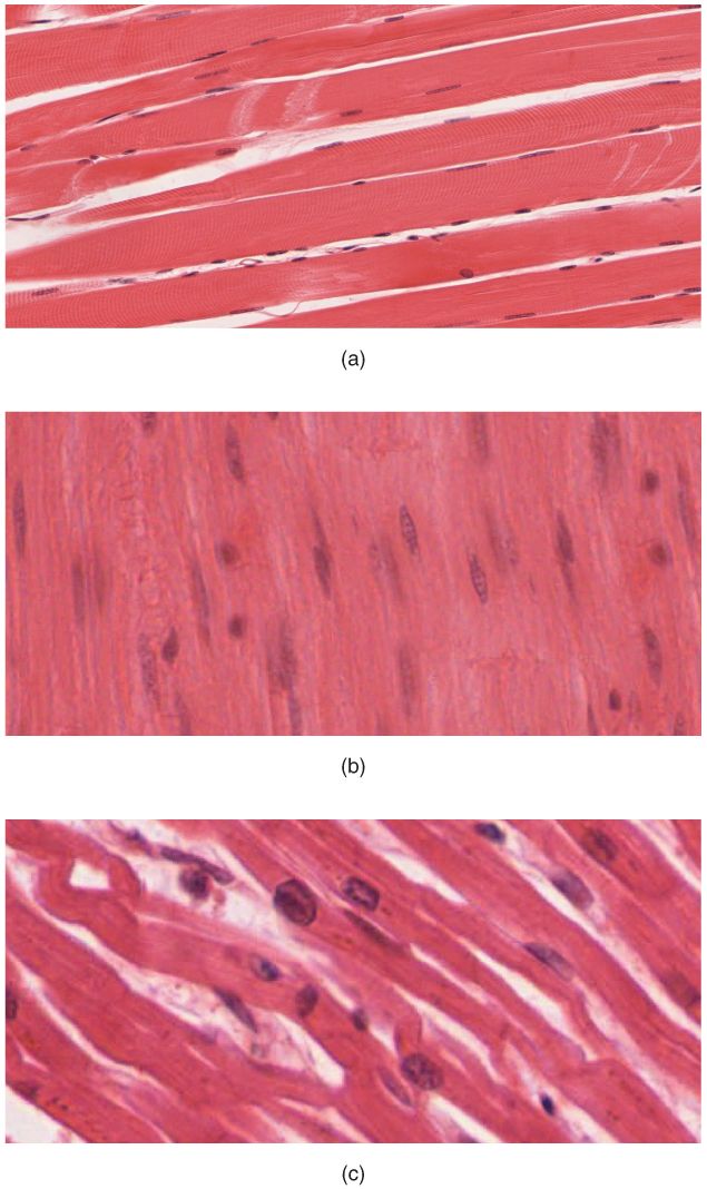
There are exactly three muscle tissues, and you can tell them apart with three questions: Is it striped? Can you control it consciously? What shape are its cells? Skeletal muscle is the meat that moves your bones — it is striated, voluntary (under conscious control), and made of long, cylindrical, multinucleated fibers. Cardiac muscle is found only in the wall of the heart — it is striated but involuntary, and its short, branching cells are joined end-to-end by intercalated discs that let the whole heart contract as one coordinated unit. Smooth muscle lines the walls of hollow organs like the gut, blood vessels, and bladder — it is non-striated, involuntary, and built of small, tapered, spindle-shaped cells that squeeze those organs slowly and tirelessly.
| Feature | Skeletal | Cardiac | Smooth |
|---|---|---|---|
| Striations | Striated | Striated | Non-striated |
| Control | Voluntary | Involuntary | Involuntary |
| Cell shape | Long cylinders | Short, branched | Tapered spindles |
| Nuclei per cell | Many | One (or two) | One |
| Location | Attached to bones | Heart wall only | Walls of hollow organs |
A useful mental shortcut: the two striated muscles (skeletal and cardiac) are the fast, powerful ones, while the one smooth muscle is the slow, steady one. And only one of the three — skeletal — answers to your will. The heart and the gut run themselves.
Watch each of the three muscles contract in the interactive below.
- Muscle tissue contracts using sliding actin and myosin filaments; orderly filaments make it look striated.
- Skeletal = striated, voluntary, long multinucleated fibers; cardiac = striated, involuntary, branched with intercalated discs; smooth = non-striated, involuntary, spindle cells.
- Tell them apart with three questions: striped? voluntary? cell shape?
Section 5Nervous tissue
The neuron: a cell shaped like a message
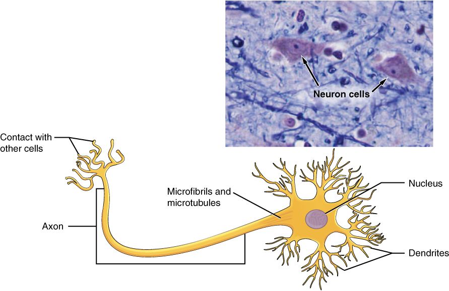
Nervous tissue senses changes, makes decisions, and sends electrical signals faster than any other tissue in the body. Its star cell is the neuron, and the neuron's strange shape is really a map of how a signal flows through it — in one end, out the other. Signals arrive at the bushy dendrites, short branching extensions that receive input from other cells. They funnel into the cell body (or soma), which holds the nucleus and does the cell's housekeeping. If the incoming signal is strong enough, the neuron fires an electrical pulse that races away down the axon, a single long fiber that carries the message toward the next cell. Many axons are wrapped in a fatty myelin sheath that acts like insulation on a wire, letting the signal jump along much faster. The traffic is strictly one-way: dendrite → cell body → axon, every time.
| Part | What it is | Its job in the signal |
|---|---|---|
| Dendrites | Short, branching extensions | Receive incoming signals from other cells |
| Cell body (soma) | Contains nucleus and organelles | Integrates the input; keeps the cell alive |
| Axon | Single long fiber | Sends the electrical pulse toward the next cell |
| Myelin sheath | Fatty insulating wrap on the axon | Speeds the signal along the axon |
| Axon terminals | Branched tips of the axon | Pass the signal to the next cell at a synapse |
Neuroglia: the support crew
Neurons get the glory, but they are at least matched in number by neuroglia (or glial cells), the support cells of nervous tissue. Careful counts put the two roughly one-to-one across the whole brain, with glia outnumbering neurons in the cerebral cortex and neurons far outnumbering glia in the cerebellum. Glia do not carry the signal themselves; instead they nourish neurons, insulate their axons with myelin, hold the tissue together, and defend it against invaders. Two properties define what nervous tissue as a whole can do: excitability, the ability to respond to a stimulus by generating an electrical signal, and conductivity, the ability to carry that signal from one place to another. Together they let this tissue act as the body's high-speed communication network.
Tap a part of the neuron and watch a signal fire in the interactive.
- The neuron carries a one-way signal: dendrites receive → cell body integrates → axon transmits.
- The fatty myelin sheath insulates the axon and speeds the signal; neuroglia support and protect neurons.
- Nervous tissue is defined by excitability and conductivity; mature neurons rarely divide, so CNS damage often heals poorly.
Section 6Tissue repair
Two ways to fix a wound
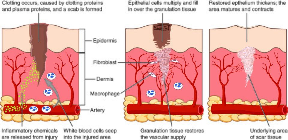
When a tissue is injured, the body can mend it in one of two ways. The better outcome is regeneration — the damaged cells are replaced with new cells of the very same type, and the tissue is restored to normal, as if the injury never happened. The fallback is fibrosis, in which the gap is filled with dense fibrous scar tissue instead of the original cell type. Scar tissue plugs the hole and restores strength, but it does not do the original job — scar in the heart cannot beat, and scar in the skin has no hair or sweat glands. Which path a tissue takes depends largely on how well its cells can divide: fast-dividing tissues like skin and gut regenerate beautifully, while tissues whose cells barely divide — cardiac muscle, neurons — tend to scar.
The healing playbook
Whatever the tissue, the body runs the same four-step repair program in order. Think of it as a construction project on an emergency timeline: first stop the flooding, then clear the site, then rebuild the frame, then finish the surfaces.
| Stage | What happens | Purpose |
|---|---|---|
| 1. Hemostasis | Blood clots seal the broken vessels; a scab forms | Stop the bleeding |
| 2. Inflammation | Vessels widen; immune cells arrive to clear debris and germs | Clean up the wound |
| 3. Proliferation | New capillaries and collagen build pink granulation tissue; epithelium migrates across | Rebuild the tissue |
| 4. Remodeling | Collagen is reorganized and strengthened; the scar matures and shrinks | Restore strength over weeks to months |
The visible signs of the second stage — redness, heat, swelling, and pain — are the classic marks of inflammation, and although they feel like the problem, they are actually the body doing its job. The widened, leaky vessels that cause the redness and swelling are what deliver the clotting factors, infection-fighters, and clean-up crews the wound needs. Inflammation is not the injury; it is the repair beginning.
Step through a healing wound from clot to scar in the interactive below.
- Healing happens by regeneration (same cells restored) or fibrosis (scar tissue fills the gap); which one depends on how well the cells divide.
- The repair sequence is always hemostasis → inflammation → proliferation → remodeling.
- Inflammation (redness, heat, swelling, pain) is the repair process at work, not the injury itself.
The chapter in one breath
- A tissue is a group of similar cells sharing a job; the whole body is built from just four of them.
- Epithelial tissue covers and lines; it is tightly packed, polar, avascular, and named by layers (simple/stratified) + cell shape (squamous/cuboidal/columnar).
- Connective tissue binds and supports; its cells sit scattered in an extracellular matrix of fibers and ground substance — change the matrix and you get cartilage, bone, or blood, while adipose is the exception that fills its cells with lipid instead.
- Muscle tissue contracts: skeletal (striated, voluntary), cardiac (striated, involuntary, intercalated discs), and smooth (non-striated, involuntary).
- Nervous tissue communicates; the neuron carries a one-way signal from dendrites → cell body → axon, supported by neuroglia.
- Injured tissue heals by regeneration or fibrosis, always through hemostasis → inflammation → proliferation → remodeling.
- How well a tissue repairs comes down mostly to its blood supply and how readily its cells divide.
ReferenceWords worth knowing
A quick reference for the terms in this chapter — skim it now, or circle back whenever a word trips you up.
- Apical surface
- The free, exposed surface of an epithelial cell that faces the open space, cavity, or lumen; often bears microvilli or cilia.
- Areolar tissue
- The most common loose connective tissue — a soft, all-purpose packing material that underlies epithelia and cushions organs.
- Axon
- The single long fiber of a neuron that carries the electrical signal away from the cell body toward the next cell.
- Basement membrane
- A thin layer that anchors the basal surface of an epithelium to the connective tissue beneath it.
- Cardiac muscle
- Striated, involuntary muscle found only in the heart wall; its branched cells are joined by intercalated discs so the heart contracts as one.
- Collagen fiber
- The strong, rope-like protein fiber of connective tissue that resists pulling forces.
- Connective tissue
- The diverse tissue that binds, supports, protects, and transports; made of scattered cells in an extracellular matrix.
- Dendrite
- A short, branching extension of a neuron that receives incoming signals from other cells.
- Epithelial tissue
- Tightly packed tissue that covers body surfaces, lines cavities and tubes, and forms glands.
- Extracellular matrix
- The non-living material — fibers plus ground substance — that connective tissue cells secrete and sit within.
- Fibrosis
- Repair by the deposition of fibrous scar tissue in place of the original cell type; restores strength but not full function.
- Ground substance
- The material of the extracellular matrix that fills the space between cells and fibers; ranges from watery to gel-like to solid.
- Histology
- The microscopic study of tissues — anatomy at high magnification.
- Muscle tissue
- Tissue specialized to contract using actin and myosin filaments, generating movement, force, and heat.
- Nervous tissue
- Tissue specialized to sense, process, and rapidly transmit electrical signals; composed of neurons and neuroglia.
- Neuroglia
- The support cells of nervous tissue that nourish, insulate, hold together, and protect neurons.
- Neuron
- The signal-carrying cell of nervous tissue, with dendrites, a cell body, and an axon.
- Regeneration
- Repair in which damaged cells are replaced by new cells of the same type, restoring normal structure and function.
- Simple epithelium
- An epithelium one cell layer thick, suited to diffusion, absorption, and secretion.
- Squamous
- Describing flat, scale-like epithelial cells, ideal where substances must cross quickly.
- Stratified epithelium
- An epithelium of two or more stacked cell layers, built to protect against abrasion.
- Tissue
- A group of similar cells that work together to perform a shared function.