The Cardiovascular System: The Heart
Somewhere behind your breastbone, a muscle the size of your fist is squeezing about once a second, and it has never once taken a break. Over an average lifetime it will beat more than two and a half billion times and move enough blood to fill a small lake. This chapter opens that pump up. You will learn how the heart is built as two pumps in one, trace a single drop of blood on its full loop through the lungs and the body, watch the four valves keep everything flowing forward, and follow the tiny electrical spark the heart makes for itself — the spark that a machine at the bedside can read straight off the skin as an ECG.
By the end of this chapter you can
- Locate the heart in the mediastinum, name the layers of its wall and the sac around it, and explain why the left ventricle is the thickest chamber.
- Identify the four chambers and the great vessels, and state which blood (oxygen-rich or oxygen-poor) each one carries.
- Trace blood flow through the heart in order and distinguish the pulmonary from the systemic circuit.
- Name the four valves, place each one, and explain how chordae tendineae and pressure keep flow one-way.
- Describe the conduction system from SA node to Purkinje fibers and explain the purpose of the AV node delay.
- Break one heartbeat into the phases of the cardiac cycle and say what makes the “lub-dub” heart sounds.
- Match each wave of the ECG — P, QRS, and T — to the electrical event it records.
- Calculate cardiac output from heart rate and stroke volume, and predict how it changes.
Section 1The heart as a double pump
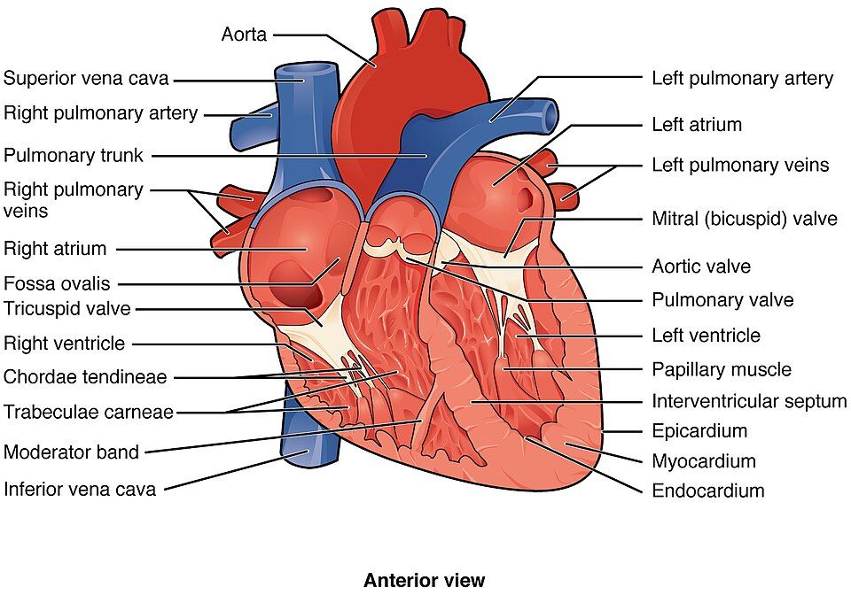
A fist-sized pump in the middle of the chest
The heart sits in the mediastinum, the space between the two lungs, resting on the diaphragm and tilted so that about two-thirds of it lies to the left of the midline. Its broad top, where the great vessels attach, is the base; its blunt, downward-and-leftward tip is the apex — the spot where you feel the beat against the chest wall. Around it is a tough double-walled sac, the pericardium. The outer fibrous pericardium anchors the heart and keeps it from overfilling; inside that, a two-layered serous pericardium secretes a slippery fluid into the pericardial cavity so the beating heart glides without friction. When that space fills with fluid or blood and squeezes the heart, the result — cardiac tamponade — is a true emergency.
Two pumps, four chambers
The single most useful idea in this whole chapter is that the heart is really two pumps bolted together, side by side, working in perfect step. The right heart receives oxygen-poor blood returning from the body and pushes it a short distance to the lungs. The left heart receives the freshly oxygenated blood coming back from the lungs and drives it out to the entire rest of the body. A solid muscular wall, the septum, keeps the two sides from mixing. Each pump has two chambers: a thin-walled atrium on top that receives blood, and a thick-walled ventricle below that ejects it. Blood always travels atrium → ventricle → out an artery.
| Chamber | Receives blood from | Sends blood to |
|---|---|---|
| Right atrium | Body, via the superior & inferior venae cavae | Right ventricle |
| Right ventricle | Right atrium | Lungs, via the pulmonary trunk & arteries |
| Left atrium | Lungs, via the four pulmonary veins | Left ventricle |
| Left ventricle | Left atrium | Whole body, via the aorta |
Three layers, and a heart that feeds itself
The wall itself has three layers. The inner endocardium is a smooth lining continuous with the vessels, so blood never touches rough tissue. The bulky middle myocardium is the cardiac muscle that actually does the pumping — and it is far thicker on the left. The outer epicardium is the visceral layer of the serous pericardium. Notice on the reference figure how the left ventricle wall dwarfs the right: it must generate roughly five times the pressure to push blood through the long systemic circuit, while the right ventricle only has to reach the nearby lungs. Finally, all that hard-working muscle needs its own blood supply, delivered by the coronary arteries that branch off the aorta. Block one and the muscle downstream starves — that is a heart attack, a myocardial infarction.
Tap through the chambers, vessels, and wall layers in the interactive below.
- The heart lies in the mediastinum, wrapped in a pericardial sac whose fluid lets it beat friction-free.
- It is two pumps in one: the right heart serves the lungs, the left heart serves the body, separated by the septum.
- Atria receive, ventricles eject; the wall is endocardium, myocardium, epicardium; the left ventricle is thickest because it faces the highest pressure.
- The coronary arteries feed the myocardium; blocking one causes a myocardial infarction.
Section 2Blood flow through the heart
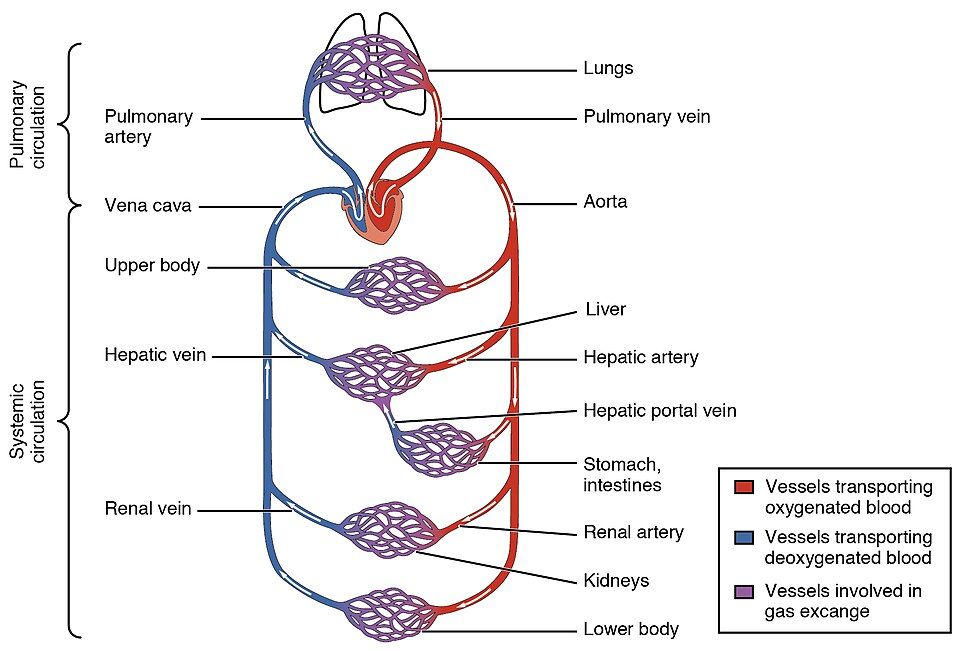
One drop, one full loop
Let us follow a single drop of blood all the way around. Returning from the body tired and oxygen-poor, it empties into the right atrium through the superior and inferior venae cavae. It drops through the tricuspid valve into the right ventricle, which squeezes it through the pulmonary valve into the pulmonary trunk and on to the lungs. In the lungs it unloads carbon dioxide and picks up a fresh load of oxygen. Now bright red, it flows back through the pulmonary veins into the left atrium, passes the mitral valve into the powerful left ventricle, and is launched through the aortic valve into the aorta to begin its journey around the body again.
| Step | Structure | Blood here is |
|---|---|---|
| 1 | Venae cavae → right atrium | Oxygen-poor |
| 2 | Tricuspid valve → right ventricle | Oxygen-poor |
| 3 | Pulmonary valve → pulmonary arteries → lungs | Oxygen-poor (arriving) |
| 4 | Pulmonary veins → left atrium | Oxygen-rich |
| 5 | Mitral valve → left ventricle | Oxygen-rich |
| 6 | Aortic valve → aorta → body | Oxygen-rich |
Two circuits, run in series
That single loop is really two loops chained together. The pulmonary circuit runs from the right heart to the lungs and back; its whole purpose is gas exchange. The systemic circuit runs from the left heart out to every other tissue — brain, gut, kidneys, muscles — and back. Because they are wired in series, the same drop must pass through both, and the two ventricles must pump the exact same volume over time. If the right side pumped even a little more than the left for long, blood would pile up in the lungs; if the left outran the right, it would pile up in the body. That balance is why heart failure on one side eventually drags down the other.
The great exception
Here is a rule worth burning in, along with the one place it seems to break. Arteries carry blood away from the heart; veins carry blood back toward it. Notice the definition says nothing about oxygen. Almost everywhere, arteries happen to be oxygen-rich and veins oxygen-poor — but the pulmonary vessels flip that. The pulmonary arteries carry oxygen-poor blood (away from the heart, toward the lungs), and the pulmonary veins carry oxygen-rich blood (back toward the heart). They are the exception that proves the real rule is about direction, not color.
Play or step a single drop through the whole loop in the interactive.
- The forward path: venae cavae → RA → tricuspid → RV → pulmonary valve → lungs → pulmonary veins → LA → mitral → LV → aortic valve → aorta.
- The pulmonary circuit (right heart ↔ lungs) and systemic circuit (left heart ↔ body) run in series, so both ventricles move equal volumes.
- Arteries lead away, veins lead back — a rule about direction; the pulmonary vessels are the oxygen-content exception.
Section 3The four one-way valves
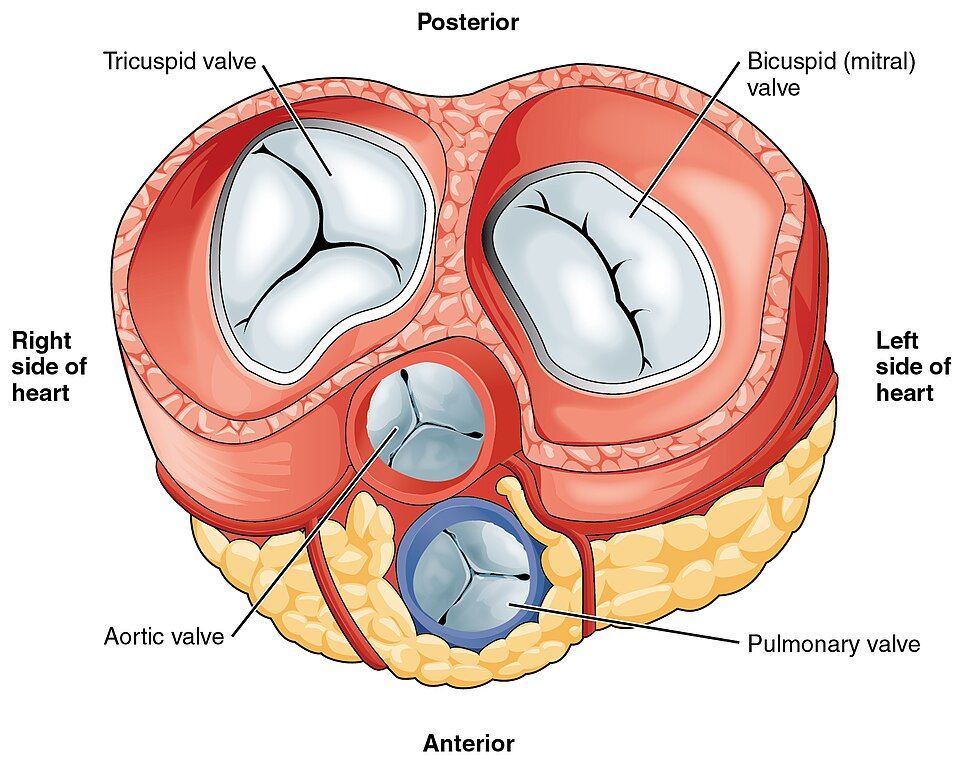
Four doors that only open one way
A pump is only as good as its valves, and the heart has four, each a flap-door that opens under forward pressure and slams shut against backward pressure. Two of them, the atrioventricular (AV) valves, sit between each atrium and its ventricle: the tricuspid valve on the right (three cusps) and the mitral or bicuspid valve on the left (two cusps). The other two, the semilunar valves, guard the exits where the great arteries leave: the pulmonary valve at the mouth of the pulmonary trunk and the aortic valve at the mouth of the aorta. Together they make sure that with every squeeze, blood can only go forward.
| Valve | Type | Location | Open when |
|---|---|---|---|
| Tricuspid | AV | Right atrium → right ventricle | Ventricles relaxed (filling) |
| Mitral (bicuspid) | AV | Left atrium → left ventricle | Ventricles relaxed (filling) |
| Pulmonary | Semilunar | Right ventricle → pulmonary trunk | Ventricles contracting (ejecting) |
| Aortic | Semilunar | Left ventricle → aorta | Ventricles contracting (ejecting) |
What holds them shut
The two valve types are built to resist backflow in different ways. When the ventricles contract hard, the pressure could blow the AV valve cusps up into the atria — so each cusp is tethered by tough strings, the chordae tendineae (“heart strings”), to little muscle bumps called papillary muscles on the ventricle floor. The papillary muscles tighten just before the squeeze, and the chords hold the cusps like the ropes on a parachute, letting them close flat but not turn inside out. The semilunar valves need no chords: their three cup-shaped pockets simply catch the blood that tries to fall back after ejection, and the weight snaps them shut.
When valves go wrong
Valves fail in exactly two ways, and the words are worth knowing. A stenotic valve is stiff and narrowed — it will not open all the way, so the heart must strain to push blood through a smaller hole. A regurgitant (or insufficient) valve is leaky — it will not close all the way, so blood sloshes backward with each beat. Either fault sets up turbulence, and turbulence you can hear through a stethoscope as a murmur. The normal, healthy sounds — the familiar “lub-dub” — are simply the valves snapping shut, which is the bridge into the cardiac cycle.
Tap each valve to watch it open and close in the interactive.
- Two AV valves (tricuspid right, mitral/bicuspid left) sit between atria and ventricles; two semilunar valves (pulmonary, aortic) guard the artery exits.
- Chordae tendineae and papillary muscles anchor the AV cusps so they cannot flip backward during the squeeze.
- Valves fail as stenosis (won’t open) or regurgitation (won’t close), producing murmurs; normal S1/S2 are valves snapping shut.
Section 4The conduction system
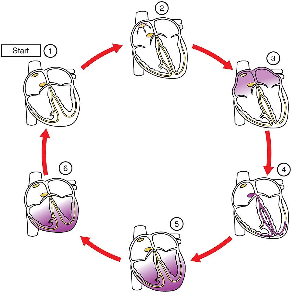
A heart that writes its own rhythm
Unlike the muscles in your arm, the heart does not wait for the brain to tell it to beat. A small population of specialized cells can depolarize all on their own, over and over, a property called autorhythmicity. The fastest of these cells — the sinoatrial (SA) node, tucked in the wall of the right atrium — fires about 60 to 100 times a minute and sets the pace for everyone else. That is why the SA node earns the nickname the heart’s pacemaker. Cut every nerve to the heart and it would keep right on beating; the nerves only speed it up or slow it down, as we will see.
The path of the spark
The impulse spreads in a precise, deliberate order so the chambers squeeze in the right sequence — atria first, then ventricles. From the SA node the signal sweeps across both atria, making them contract and top off the ventricles. It then reaches the atrioventricular (AV) node, the only electrical doorway between atria and ventricles, where it deliberately pauses for about a tenth of a second. From the AV node it races down the AV bundle (bundle of His), splits into the right and left bundle branches along the septum, and fans out through the Purkinje fibers into the ventricle walls. The ventricles then contract from the apex up, wringing blood toward the exits like squeezing a tube of toothpaste from the bottom.
| Component | Role | Intrinsic rate |
|---|---|---|
| SA node | Pacemaker; starts each beat, fires the atria | 60–100 / min |
| AV node | Gateway to the ventricles; delays the signal | 40–60 / min |
| AV bundle & branches | Carry the signal down the septum | — |
| Purkinje fibers | Spread the signal through ventricle walls | 20–40 / min |
Why the pause, and who controls the tempo
That AV-node delay is not a flaw — it is essential. It buys the atria just enough time to finish emptying into the ventricles before the ventricles slam shut and eject. Without it, all four chambers would contract at once and pump far less blood. Notice, too, the built-in backups: if the SA node quits, the AV node can take over at a slower 40 to 60 beats, and the Purkinje fibers slower still — a safety net that keeps a failing heart beating, just too slowly. Finally, the autonomic nervous system is the volume dial: sympathetic signals speed the SA node and strengthen contractions (fear, exercise), while the parasympathetic vagus nerve slows it back down at rest.
Follow the spark from the SA node all the way down in the interactive.
- The heart is autorhythmic; the SA node is the pacemaker, firing 60–100 times a minute.
- The path is SA node → AV node → AV bundle → bundle branches → Purkinje fibers, so atria contract before ventricles.
- The AV node delay lets the atria finish filling the ventricles; slower backup pacemakers protect against SA failure.
- Sympathetic nerves speed the heart, parasympathetic (vagus) nerves slow it.
Section 5The cardiac cycle
Squeeze and rest, over and over
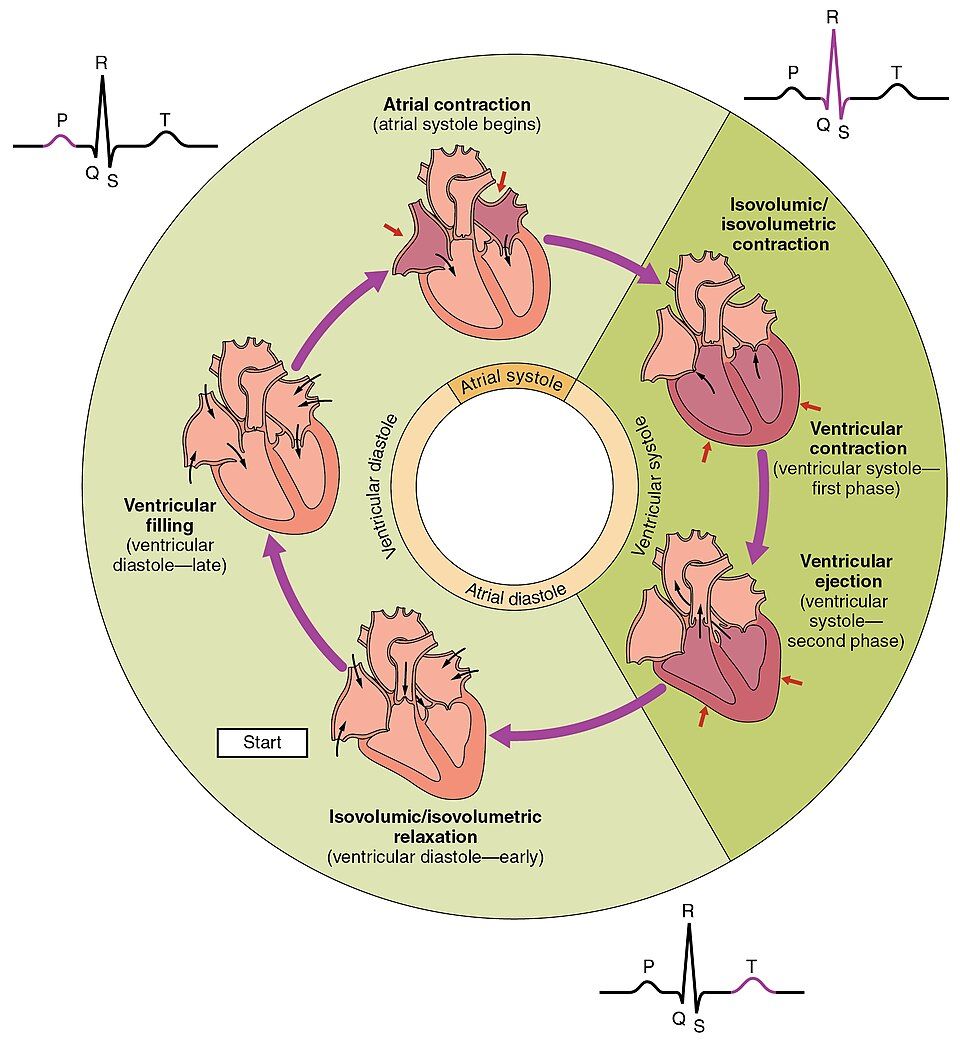
One heartbeat is a repeating rhythm of two states. Systole is contraction — a chamber squeezing to push blood out. Diastole is relaxation — a chamber loosening to fill back up. At a resting rate of about 75 beats a minute, the whole cycle takes roughly 0.8 seconds, and, importantly, the heart spends more of that time relaxing than contracting. That rest is not laziness: it is when the ventricles refill, and it is also the only time the coronary arteries can push blood into the thick heart muscle. Race the heart too fast and you steal from diastole — less filling, less coronary flow.
The phases, in order
Walk around the reference wheel and the sequence tells itself. During atrial systole the atria give a gentle squeeze that tops off the already-mostly-full ventricles. Then comes ventricular systole: first an isovolumetric contraction where all four valves are shut and pressure builds with no blood moving, then ventricular ejection as the semilunar valves pop open and blood surges into the arteries. Finally ventricular diastole: an isovolumetric relaxation with all valves shut again, then ventricular filling as the AV valves open and blood pours in — most of it passively, before the atria even contract.
| Phase | What happens | AV valves | Semilunar valves |
|---|---|---|---|
| Atrial systole | Atria squeeze; final 20–30% of filling | Open | Closed |
| Isovolumetric contraction | Ventricles tense; pressure builds, no ejection | Closed | Closed |
| Ventricular ejection | Ventricles push blood into the arteries | Closed | Open |
| Isovolumetric relaxation | Ventricles relax; pressure falls, no filling | Closed | Closed |
| Ventricular filling | Blood pours passively into ventricles | Open | Closed |
Lub-dub, and the numbers that matter
The two heart sounds are made by valves closing, not opening. S1, the low “lub,” is the AV valves snapping shut as ventricular systole begins; S2, the sharper “dub,” is the semilunar valves shutting as diastole begins. Between them the heart is ejecting; after S2 it is filling. A few numbers frame the whole thing. The volume in a ventricle at the end of filling is the end-diastolic volume (EDV); what remains after ejection is the end-systolic volume (ESV); the amount actually pumped per beat is the stroke volume (EDV − ESV). Multiply stroke volume by heart rate and you get cardiac output — the liters of blood the heart moves each minute, about 5 L at rest.
Step through each phase and hear the lub-dub in the interactive.
- Systole is contraction, diastole is relaxation; the resting cycle is about 0.8 s, mostly diastole.
- Order of phases: atrial systole → isovolumetric contraction → ejection → isovolumetric relaxation → filling.
- S1 (“lub”) = AV valves closing; S2 (“dub”) = semilunar valves closing.
- Stroke volume = EDV − ESV; cardiac output = heart rate × stroke volume (≈5 L/min at rest).
Section 6The ECG
Reading the heart’s electricity off the skin
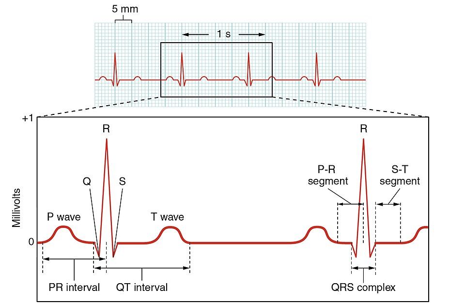
Every time the heart depolarizes, the wave of electrical change is large enough to reach the body surface, where electrodes on the skin can pick it up. Plotted over time, that signal is an electrocardiogram (ECG or EKG) — one of the most useful, painless, and inexpensive tests in all of medicine. It does not record the heartbeat itself but the electrical events that trigger it, so each bump on the tracing lines up with a step in the conduction system you just met. Learn to read three simple deflections and the whole picture opens up.
P, QRS, and T
The tracing repeats three main shapes. The small P wave is atrial depolarization — the SA node’s signal spreading across the atria just before they contract. The tall, spiky QRS complex is ventricular depolarization — big and sharp because the ventricles are so muscular. (Atrial repolarization happens here too, but it is buried inside the QRS and never gets its own wave.) The rounded T wave is ventricular repolarization, the ventricles electrically resetting so they can relax and fill again. Remember: depolarization triggers contraction, so on the tracing the electrical wave always comes a beat ahead of the mechanical squeeze.
| Feature | Electrical event | Mechanical partner |
|---|---|---|
| P wave | Atrial depolarization | Atria contract |
| PR interval | SA firing through the AV-node delay | Signal waits at the AV node |
| QRS complex | Ventricular depolarization | Ventricles contract |
| T wave | Ventricular repolarization | Ventricles relax |
Intervals, rate, and trouble
The flat stretches between the waves carry as much information as the waves themselves. The PR interval spans the start of the P wave to the start of the QRS — it is the atria firing plus that all-important AV-node pause; stretch it too long and you have a heart block. The QRS duration tells you how fast the ventricles are being activated, and the QT interval covers the whole span of ventricular electrical activity. You can even read the rate straight off the paper: the tracing runs at a fixed speed, so the spacing between two QRS peaks gives beats per minute. A tracing that is too fast is tachycardia, too slow is bradycardia, and a chaotic, quivering line with no real QRS at all — ventricular fibrillation — means the ventricles are twitching uselessly and no blood is moving. That is the rhythm a defibrillator is built to shock back into order.
Tap each wave to link it to its conduction event in the interactive.
- An ECG records the heart’s electrical events from the skin; each deflection maps to a conduction step.
- P = atrial depolarization, QRS = ventricular depolarization, T = ventricular repolarization; the electrical wave leads the squeeze.
- The PR interval holds the AV-node delay; a long one signals heart block.
- Rhythm names: tachycardia (fast), bradycardia (slow), ventricular fibrillation (chaotic, no output); ST elevation flags a heart attack.
The chapter in one breath
- The heart is a fist-sized double pump in the mediastinum, wrapped in a pericardial sac; the left ventricle is thickest because it faces the highest pressure.
- Blood flows atrium → ventricle → artery on each side: the right heart serves the pulmonary circuit (lungs), the left heart the systemic circuit (body), and the two run in series.
- Arteries lead away, veins lead back — a rule about direction; the pulmonary vessels flip the usual oxygen content.
- Four one-way valves — two AV (tricuspid, mitral), two semilunar (pulmonary, aortic) — keep flow forward; chordae tendineae hold the AV cusps shut.
- The heart is autorhythmic: the SA node paces it, the AV node delays the signal, and the Purkinje fibers deliver it to the ventricles.
- One beat = systole and diastole; S1/S2 are valves closing, and cardiac output = heart rate × stroke volume.
- The ECG reads the electrical events from the skin: P (atrial), QRS (ventricular depolarization), T (ventricular repolarization).
ReferenceWords worth knowing
A quick reference for the terms in this chapter — skim it now, or circle back whenever a word trips you up.
- Aorta
- The largest artery in the body; it carries oxygen-rich blood out of the left ventricle to begin the systemic circuit.
- Atrioventricular (AV) node
- The electrical gateway between the atria and ventricles; it delays the impulse about a tenth of a second so the atria can finish filling the ventricles.
- Atrioventricular (AV) valve
- A valve between an atrium and its ventricle — the tricuspid on the right, the mitral (bicuspid) on the left — held shut by chordae tendineae.
- Atrium
- An upper, thin-walled receiving chamber of the heart; the right atrium takes blood from the body, the left from the lungs.
- Cardiac cycle
- One complete heartbeat, made of contraction (systole) and relaxation (diastole) of the chambers.
- Cardiac output
- The volume of blood pumped by a ventricle each minute; equal to heart rate multiplied by stroke volume (about 5 L/min at rest).
- Chordae tendineae
- Tough fibrous cords that tie the AV valve cusps to the papillary muscles, keeping the cusps from flipping backward during contraction.
- Diastole
- The relaxation phase of a heart chamber, during which it fills with blood.
- Electrocardiogram (ECG)
- A recording of the heart’s electrical activity taken from the skin; its P, QRS, and T deflections map to conduction events.
- Endocardium
- The smooth inner lining of the heart, continuous with the lining of the blood vessels.
- Myocardium
- The thick middle layer of cardiac muscle that does the heart’s pumping; thickest in the left ventricle.
- Pericardium
- The double-walled sac around the heart; its inner serous layers secrete fluid so the beating heart moves without friction.
- Pulmonary circuit
- The loop of circulation from the right heart to the lungs and back, where blood picks up oxygen and drops off carbon dioxide.
- Purkinje fibers
- Fast-conducting fibers that spread the electrical impulse through the ventricle walls, triggering contraction from the apex up.
- QRS complex
- The tall, spiky deflection of the ECG marking ventricular depolarization, just before the ventricles contract.
- Semilunar valve
- A cup-shaped valve guarding a ventricle’s exit into a great artery — the pulmonary and aortic valves — that snaps shut against backflow.
- Sinoatrial (SA) node
- The heart’s natural pacemaker, in the wall of the right atrium; it fires 60–100 times a minute and sets the rhythm.
- Stroke volume
- The amount of blood a ventricle ejects in one beat; the end-diastolic volume minus the end-systolic volume.
- Systemic circuit
- The loop of circulation from the left heart out to all the body’s tissues and back to the right heart.
- Systole
- The contraction phase of a heart chamber, during which it ejects blood.
- Ventricle
- A lower, thick-walled pumping chamber of the heart; the right ejects blood to the lungs, the left to the whole body.