ASSESSING THE ABDOMEN, NUTRITION AND ELIMINATION
The abdomen is the one region where the four-step examination sequence you have used all semester changes: you inspect, then auscultate, then percuss, then palpate — because pressing on the belly first stirs the bowel and gives you sounds that were never really there. That single reordering is the signature of this chapter, and it is also a promise about how everything else here works: the abdomen does not announce itself the way a lung field or a pulse does. It hides a ruptured appendix behind a low-grade fever, a two-liter bladder behind restlessness, and a slow gastrointestinal bleed behind one dark stool nobody looked at. Nutrition and elimination belong in the same chapter because they are the abdomen’s inputs and outputs: what a patient can safely swallow, what their body did with it, and what came back out. You will learn the sequence, the numbers, the words to chart, and the findings that mean you pick up the phone now.
By the end of this chapter you can
- Map the four quadrants and nine regions of the abdomen and name the organs that lie beneath each.
- Perform the abdominal examination in the correct order — inspection, auscultation, percussion, palpation — and explain why auscultation moves ahead of percussion and palpation here and nowhere else.
- Distinguish expected from abnormal findings for contour, bowel sounds, bruits, percussion tones, liver span and palpation, and state what each abnormal finding suggests.
- Demonstrate focused maneuvers — Murphy sign, McBurney point, Rovsing sign, psoas, obturator, shifting dullness and fluid wave — and name the situations in which deep palpation is contraindicated.
- Screen nutritional status using BMI, waist circumference, percentage of unintentional weight loss, intake history and laboratory data, and explain why albumin is an inflammation marker rather than a nutrition marker.
- Carry out aspiration and enteral-feeding safety measures, including dysphagia screening, head-of-bed elevation and correct verification of feeding-tube placement.
- Assess bowel and urinary elimination using the Bristol Stool Form Scale, intake and output, daily weight, bladder scanning and post-void residual volumes, with age-appropriate values.
- Describe how abdominal, nutritional and elimination findings differ in infants, children, pregnancy and older adults, and how jaundice, pallor, cyanosis and erythema present across the full range of skin tones.
- Recognize the abdominal red flags that require immediate escalation, communicate them using SBAR, and distinguish tasks that may be delegated to UAP from those that remain with the registered nurse.
Section 1Mapping the Abdomen and Setting Up the Exam
Quadrants, regions and what lives beneath
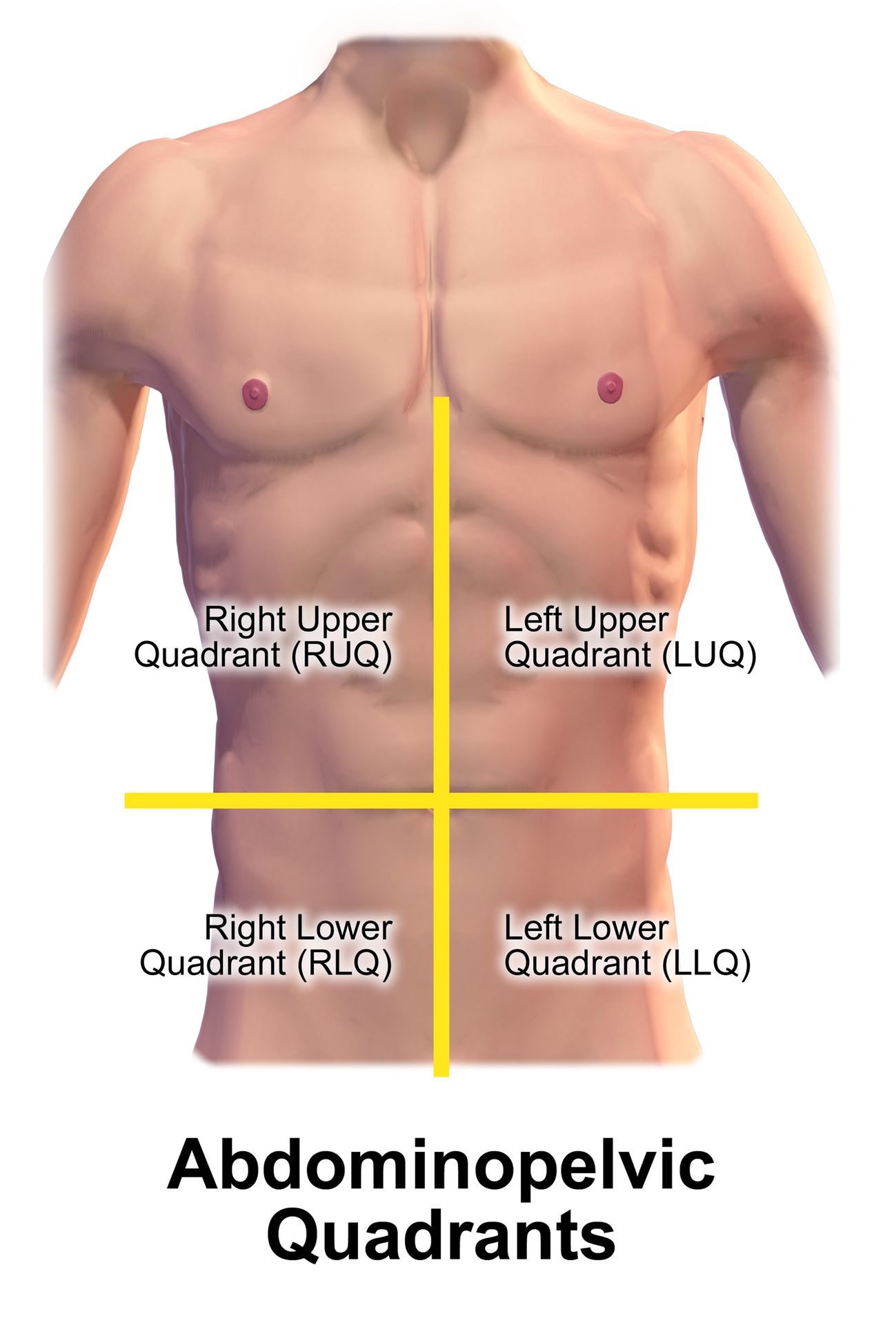
Draw two imaginary lines through the umbilicus — one vertical from the xiphoid process to the symphysis pubis, one horizontal — and you have the four quadrants that nurses use for charting and for handoff. This is the map you will speak in. When you tell a provider “tenderness in the right lower quadrant, guarding present,” you have said something anatomically specific in five words. When you say “the patient’s stomach hurts,” you have said nothing at all. The nine-region map (epigastric, umbilical, hypogastric or suprapubic down the midline; right and left hypochondriac, lumbar and inguinal on the sides) is more precise and appears in medical documentation and imaging reports, so you must be able to read it even if you chart in quadrants.
Underneath the surface, the organs are not evenly distributed, and knowing which structure sits where is what turns a location into a hypothesis. Right upper quadrant pain in a patient after a fatty meal points toward the gallbladder. Left upper quadrant pain after a bicycle-handlebar injury points toward the spleen. Midline suprapubic fullness in a patient who has not voided in eight hours points toward a distended bladder, not constipation. Pain also refers: gallbladder disease refers to the right scapula, splenic irritation to the left shoulder (Kehr sign), pancreatic pain straight through to the back, and ureteral colic from the flank down into the groin.
Two structures deserve a standing caution. The abdominal aorta runs just left of midline in the epigastric region; in an average-size adult it should measure less than 3 cm across and should pulsate forward, not sideways. A wide, expansile, laterally pulsating mass is a suspected abdominal aortic aneurysm, and you do not palpate deeply over it — you stop, take vital signs, and escalate. The other is any known or suspected abdominal mass in a child, particularly a Wilms tumor, which is never palpated repeatedly because of the theoretical risk of seeding tumor cells. Post a sign at the bedside if that diagnosis is under evaluation.
| Quadrant | Major structures beneath | Findings that commonly localize here |
|---|---|---|
| Right upper (RUQ) | Liver, gallbladder, duodenum, head of pancreas, right kidney and adrenal, hepatic flexure of colon | Cholecystitis (positive Murphy sign), hepatitis, hepatomegaly, duodenal ulcer, right lower lobe pneumonia referring down |
| Left upper (LUQ) | Stomach, spleen, left lobe of liver, body and tail of pancreas, left kidney and adrenal, splenic flexure | Splenomegaly or splenic rupture, gastritis, pancreatitis, gastric ulcer |
| Right lower (RLQ) | Cecum, appendix, right ovary and tube, right ureter, right spermatic cord | Appendicitis, ovarian cyst or torsion, ectopic pregnancy, ureteral stone, Crohn disease of the terminal ileum |
| Left lower (LLQ) | Sigmoid colon, descending colon, left ovary and tube, left ureter | Diverticulitis, constipation, ovarian pathology, ureteral stone |
| Midline | Abdominal aorta, bladder, uterus (when enlarged), umbilicus | Aortic aneurysm, urinary retention, pregnancy, umbilical hernia |
Positioning, exposure and the order that protects your findings
Set the room up before you touch anyone. Ask the patient to empty the bladder first — a full bladder is uncomfortable, distorts the suprapubic exam and makes a percussion finding you will then have to explain away. Position the patient supine with a small pillow under the head and another under slightly flexed knees; this relaxes the abdominal wall. Arms go at the sides or folded across the chest, never behind the head, which tightens the rectus muscles and manufactures rigidity you will misread as guarding. Drape so the abdomen is exposed from the xiphoid to the symphysis pubis while the chest and groin stay covered. Warm your hands and your stethoscope diaphragm, keep your fingernails short, and use a low light angled across the abdomen so contour and peristaltic waves cast shadows.
Now the sequence, and this is the paragraph to memorize. Everywhere else in the body you inspect, palpate, percuss and auscultate. In the abdomen you inspect, auscultate, percuss and palpate. The reason is mechanical: percussion and palpation stimulate the bowel and change its motility, so sounds heard afterward tell you about your own hands rather than about the patient. Auscultating before you press preserves the baseline. Two further ordering rules follow from patient comfort: examine the painful quadrant last, and if you perform a test for rebound tenderness, perform it at the very end, because it hurts and it will make everything after it uninterpretable.
Ask the patient to point with one finger to where it hurts before you begin. Watch the face, not the hand, during palpation — a patient who has been told to be brave will wince before they will complain. Talk the whole way through: “I am going to press lightly here, tell me the moment it is uncomfortable.” If the patient tenses, distract with conversation or ask them to breathe out slowly through the mouth; voluntary guarding relaxes with distraction, while involuntary guarding from peritoneal irritation does not. That distinction is one of the most clinically useful things in this chapter.
The focused history that steers the exam
The abdominal history is where most of the diagnostic yield lives, and it is nursing work, not something you wait for the provider to do. Use a structured symptom analysis — OLDCARTS (onset, location, duration, characteristics, aggravating factors, relieving factors, timing, severity) or PQRSTU — and then add the abdomen-specific questions: appetite and any change in it, nausea or vomiting and what the vomitus looked like, last oral intake, last bowel movement and its form and color, flatus (a patient who is passing gas is far less likely to be obstructed), urinary frequency, urgency, burning and stream, weight change over the last one, three and six months, alcohol intake, NSAID and anticoagulant use, and abdominal surgeries. In anyone of childbearing potential, ask the date of the last menstrual period and the possibility of pregnancy — a ruptured ectopic pregnancy presents as lower abdominal pain and can kill within hours.
Characterize the pain itself, because the quality maps onto the anatomy. Visceral pain arises from a hollow organ stretching or contracting; it is dull, crampy, poorly localized and often midline, and the patient moves around trying to find a comfortable position. Parietal (somatic) pain arises when the parietal peritoneum is inflamed; it is sharp, well localized, worse with movement or coughing, and the patient lies very still with knees drawn up. A patient who has gone from restless crampy midline pain to lying rigidly still with sharply localized pain has told you the process has reached the peritoneum. Referred pain is felt at a distance from its source and travels a predictable route.
Pain that is dramatically out of proportion to what you find on examination is its own alarm. A patient in atrial fibrillation who reports severe, unrelenting periumbilical pain while the abdomen feels soft and nearly normal may have acute mesenteric ischemia, and the soft abdomen is falsely reassuring. Trust the mismatch, not the palpation.
In the abdomen and only in the abdomen, auscultation comes before percussion and palpation, the painful area is examined last, and the history — pain quality, last bowel movement, last oral intake, pregnancy possibility — tells you what to look for before your hands ever touch the patient.
Section 2Inspection and Auscultation
Inspection: contour, symmetry, skin and movement
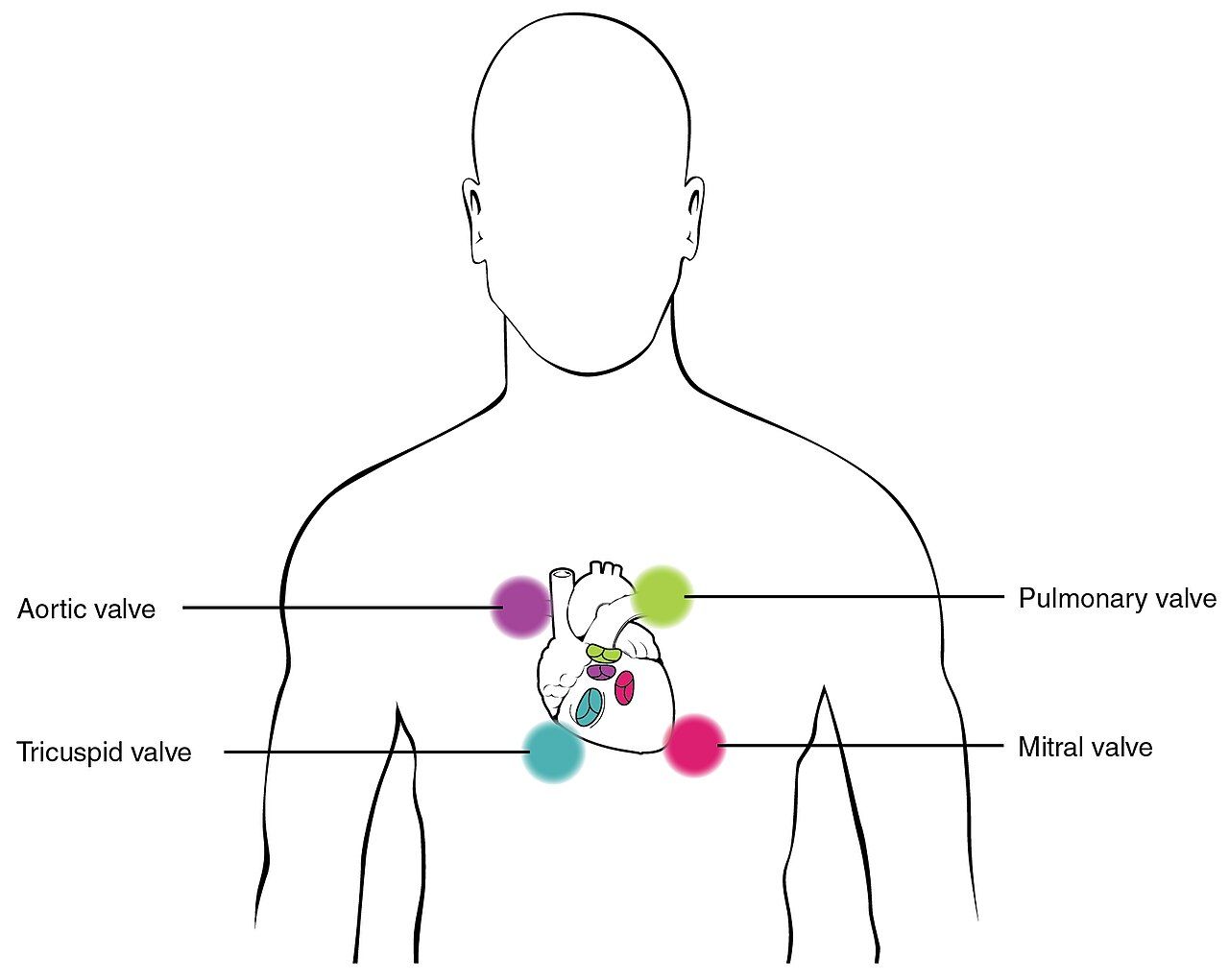
Stand at the patient’s right side and then squat so your eyes are level with the abdomen; a flat contour and a distended one look identical from above and completely different from the side. Describe contour as flat, rounded, scaphoid (sunken, seen in significant weight loss and in dehydration) or protuberant. Then look for symmetry across the midline, because an asymmetric bulge is a mass, a hernia, an enlarged organ or a loop of obstructed bowel until proven otherwise. Ask the patient to lift the head off the pillow: a bulge that becomes more prominent is in the abdominal wall (hernia, diastasis recti), while one that disappears is intra-abdominal.
Inspect the skin systematically. Note scars and map them — every previous abdominal operation is a risk factor for adhesive small-bowel obstruction, and a scar is often the only history a nonverbal patient can give you. Striae that are silvery white are old and expected after pregnancy or weight change; striae that are wide, purple and accompanied by central obesity and a rounded face suggest hypercortisolism. Dilated, tortuous veins radiating from the umbilicus (caput medusae) and spider angiomas suggest portal hypertension. Periumbilical bruising (Cullen sign) and flank bruising (Grey Turner sign) are late signs of retroperitoneal or intraperitoneal bleeding, classically severe hemorrhagic pancreatitis, and they are always an escalation. In deeply pigmented skin neither reads as the textbook blue-purple bruise: look for an area that is darker, duller or violet compared with the surrounding skin and with the same site on the opposite side, use good direct light, and palpate for the tenderness, warmth and induration that go with it.
Watch for movement. Normal respiratory movement is smooth; men breathe more abdominally, women more thoracically. Visible peristaltic waves crossing the abdomen are abnormal in an adult and suggest intestinal obstruction — the bowel is straining against a blockage. In an infant, a wave crossing left to right with projectile, non-bilious vomiting and a firm olive-shaped mass in the right upper quadrant suggests hypertrophic pyloric stenosis. A visible pulsation in the epigastrium may be normal in a thin patient, but a wide, forceful, side-to-side pulsation is not.
| Inspection finding | Expected | Abnormal | What the abnormal suggests |
|---|---|---|---|
| Contour | Flat or rounded, symmetric | Distended, taut, asymmetric bulge, scaphoid | Ascites, obstruction, obesity, mass, organomegaly; scaphoid with severe malnutrition or dehydration |
| Umbilicus | Midline, inverted, no drainage | Everted, displaced, bulging, drainage | Everted with ascites or umbilical hernia; drainage with infection or fistula |
| Skin | Uniform with the patient’s skin tone, silvery striae, healed scars | Purple striae, caput medusae, Cullen or Grey Turner sign, spider angiomas, jaundice | Hypercortisolism, portal hypertension, retroperitoneal bleeding, hepatic disease |
| Movement | Smooth respiratory rise and fall; no visible peristalsis | Visible peristaltic waves; limited movement with splinting | Bowel obstruction, pyloric stenosis in an infant; peritonitis when the patient will not move the abdomen |
| Pulsation | Faint epigastric pulsation in a thin adult | Wide, expansile, laterally pulsating epigastric mass | Abdominal aortic aneurysm — stop, do not palpate deeply, escalate |
Bowel sounds: how long to listen and what to call it
Use the diaphragm of the stethoscope, warmed, and press lightly. Begin in the right lower quadrant, where the ileocecal valve reliably produces sound, then move clockwise: RLQ, RUQ, LUQ, LLQ. For a routine assessment, listen 15 to 30 seconds in each quadrant. Normal bowel sounds are high-pitched, irregular gurgles occurring roughly 5 to 30 times per minute; you should not try to count each one, because they run together. Chart them as present and active in all four quadrants. If you hear nothing in a quadrant, you must listen in that one spot for a full 5 minutes before you may document sounds as absent. “Absent bowel sounds” charted after ten seconds of listening is not an assessment; it is a guess that may prompt a nasogastric tube the patient does not need.
Hypoactive sounds are soft and infrequent (fewer than about 5 per minute). They are expected in the immediate postoperative period and with general anesthesia, opioids, immobility and hypokalemia; they also occur with paralytic ileus and with peritonitis, which are not benign, so never read them apart from the rest of the assessment. Hyperactive sounds are loud, rushing, high-pitched and frequent; you hear them with gastroenteritis, diarrhea, laxatives, gastrointestinal bleeding (blood in the gut is a cathartic) and in early mechanical obstruction. The clinically dangerous pattern is loud, high-pitched, tinkling or rushing sounds proximal to a blockage in a patient with distension and vomiting, which later fade to silence as the bowel fatigues. Hyperactive-then-absent in a distended, vomiting patient is a surgical emergency, not an improvement.
Two sounds are not bowel sounds and are worth naming. Borborygmi are the prolonged, loud, hyperperistaltic rumbles a patient will call “stomach growling” and are usually benign. A succussion splash — a sloshing you elicit by rocking the patient gently at the hips several hours after eating — suggests retained gastric contents from gastric outlet obstruction or gastroparesis.
Vascular sounds and friction rubs
Switch to the bell and listen for vascular sounds, which are lower pitched. A bruit is a blowing, swishing sound of turbulent arterial flow. Listen over the aorta in the epigastrium just left of midline, over each renal artery about 2 to 3 cm above the umbilicus and 2 to 3 cm lateral to it, over the iliac arteries partway between the umbilicus and the inguinal ligament, and over the femoral arteries in the groin. A bruit over the aorta with a widened pulsatile mass supports an aneurysm; a renal artery bruit in a patient with poorly controlled hypertension raises renovascular disease. A venous hum, a soft continuous humming near the umbilicus, suggests increased collateral circulation from portal hypertension. A friction rub — a grating sound over the liver or spleen that varies with respiration — suggests inflammation of the organ’s peritoneal surface, as with a tumor, abscess or infarct, and is rare and always significant.
Auscultation also has a role after abdominal surgery beyond bowel sounds: listen over a new ostomy or drain site if the patient reports a change, and always inspect a dressing and incision line during the same encounter. In pregnancy, fetal heart tones are auscultated with a Doppler over the lower abdomen, expected at 110 to 160 beats per minute; that is a different examination but shares the same real estate.
Auscultate with the diaphragm in all four quadrants starting at the right lower quadrant, use 5 full minutes in one spot before ever charting bowel sounds as absent, then switch to the bell for bruits over the aorta, renal, iliac and femoral arteries.
Section 3Percussion, Palpation and Focused Maneuvers
Percussion: mapping air, fluid and solid organs
Percussion tells you what is under your fingers before you press. Percuss lightly in all four quadrants in the same clockwise pattern. The dominant note over a normal abdomen is tympany, a hollow drum-like note produced by gas in the stomach and intestines. Scattered dullness is expected over the liver, over a full bladder, over a full colon and over the spleen. Generalized dullness where you expect tympany means something has replaced the gas: fluid, stool, a mass or a gravid uterus. Generalized hyperresonance across a distended abdomen suggests gaseous distension from obstruction or ileus.
Measure the liver span in the right midclavicular line. Start well below the umbilicus over tympany and percuss upward until the note changes to dullness; that is the lower border, normally at or just below the right costal margin. Then start in the midclavicular line over lung resonance, around the fourth intercostal space, and percuss downward until dullness begins; that is the upper border, normally in the fifth to seventh intercostal space. Mark both with a skin pencil and measure. The expected adult span at the midclavicular line is 6 to 12 cm; at the midsternal line it is 4 to 8 cm. Spans are larger in tall people and in men and smaller in short people and in women. A span greater than 12 cm is hepatomegaly. Beware two traps: a liver pushed down by chronic obstructive pulmonary disease has a normal span but a palpable edge, and dullness from a right pleural effusion falsely enlarges the span from above.
For the spleen, percuss the lowest left interspace in the anterior axillary line, which is normally tympanitic. Ask the patient to take a deep breath and percuss again; if the note changes to dullness on inspiration, that is a positive Castell sign suggesting splenomegaly. Dullness in Traube space (bounded by the sixth rib, the left midaxillary line and the left costal margin) suggests the same. For the bladder, percuss downward from the umbilicus toward the symphysis pubis; a dull note above the symphysis in a patient who has not voided suggests urinary retention, and a bladder holding roughly 300 mL or more becomes percussible. Confirm with a bladder scanner rather than guessing. For ascites, use shifting dullness: percuss the flanks with the patient supine and mark the fluid level, roll the patient to one side, wait 30 seconds and percuss again; dullness that shifts toward the dependent side is positive. A fluid wave requires an assistant or the patient’s own hand pressed edgewise along the midline while you tap one flank and feel for the impulse on the other; it is specific but insensitive and generally requires a large volume. Physical examination reliably detects ascites at roughly 1500 mL, while ultrasound detects about 100 mL — another reason to measure abdominal girth serially at a marked spot on the skin at the umbilicus, at the same time of day, with the patient supine.
Light and deep palpation
Palpate last, and palpate in two passes. For light palpation, keep the fingers together, hand flat, and depress about 1 to 2 cm with a gentle rotary motion in all four quadrants. Light palpation is a screen for tenderness, surface masses, muscle tone and skin temperature. It is also how you build trust: if you hurt the patient here, the deep pass will be worthless because they will guard. For deep palpation, use the palmar surfaces of the fingers and depress about 4 to 6 cm (some texts teach 5 to 8 cm), using the other hand on top for reinforcement in a large or obese abdomen. Deep palpation delineates organs and masses. Any mass gets described in six terms: location, size in centimeters, shape, consistency, mobility and tenderness.
Do not perform deep palpation when there is a pulsatile epigastric mass or known aortic aneurysm, a suspected abdominal tumor in a child, a transplanted kidney, polycystic kidneys, a recent abdominal surgical site, or an obvious surgical abdomen with rigidity — in that last case you have your answer and further pressing only causes pain. Assess the liver edge by placing your left hand under the right posterior flank at the eleventh and twelfth ribs, pressing upward, and placing your right hand below the right costal margin; ask the patient to inhale deeply and feel for the edge descending against your fingertips. A normal adult liver edge is often not palpable; when felt it is smooth, firm and nontender. A nodular, hard or tender edge is abnormal. The spleen is normally not palpable in an adult; a palpable spleen is enlarged roughly threefold, and you palpate it gently because an enlarged spleen can rupture.
Interpretation of tenderness comes down to one distinction. Voluntary guarding is bilateral, improves with distraction and warm hands, and disappears on expiration. Involuntary guarding — true rigidity — is constant, unaffected by distraction, and is a sign of peritoneal inflammation. A board-like abdomen with rebound tenderness, absent bowel sounds and a patient who will not move is peritonitis until proven otherwise, and it is an emergency.
Focused maneuvers and lifespan variation
| Maneuver | How it is performed | Positive finding | Suggests |
|---|---|---|---|
| Murphy sign | Hook fingers under the right costal margin at the midclavicular line, ask for a deep breath | Patient stops inspiration abruptly from pain | Acute cholecystitis |
| McBurney point tenderness | Palpate one-third of the distance from the right anterior superior iliac spine toward the umbilicus | Sharp localized tenderness | Appendicitis |
| Rovsing sign | Palpate deeply in the left lower quadrant | Pain referred to the right lower quadrant | Appendicitis (peritoneal irritation) |
| Psoas sign | Patient lies on the left side; passively extend the right hip, or have the patient raise the right leg against your hand | Right lower quadrant pain | Retrocecal appendicitis, psoas abscess |
| Obturator sign | Flex the right hip and knee to 90 degrees, then internally rotate the hip | Right lower quadrant or hypogastric pain | Pelvic appendicitis |
| Costovertebral angle (CVA) tenderness | Place one palm over the CVA and strike it with the ulnar surface of the other fist | Sharp flank pain, not just thud | Pyelonephritis, renal calculus |
| Shifting dullness / fluid wave | Percuss flanks supine then in lateral position; or tap one flank and feel the opposite | Dullness shifts to dependent side; impulse transmitted | Ascites |
| Blumberg (rebound) | Press deep and slow, release quickly — perform last | Pain worse on release | Peritoneal irritation |
Infants and children. The infant and toddler abdomen is normally round and protuberant both supine and standing; by the preschool and school-age years the potbelly persists on standing but the abdomen flattens when the child lies supine. A liver edge palpable 1 to 2 cm below the right costal margin is expected in infancy, as is a soft, easily reducible umbilical hernia, which is common in infants of African descent and usually closes without intervention by 2 years of age. Diastasis recti — a midline separation of the rectus muscles — is a benign finding that resolves with growth. Bowel sounds in a newborn occur roughly every 10 to 30 seconds. Flex the child’s knees, use a warm hand, and let a toddler sit on the caregiver’s lap or place your hand over the child’s own hand for the first pass.
Pregnancy. The gravid uterus becomes an abdominal organ at about 12 weeks; fundal height in centimeters approximates gestational age in weeks between roughly 20 and 36 weeks. Progesterone slows gastrointestinal motility, so bowel sounds are diminished and constipation and pyrosis are common. Because the enlarging uterus displaces the appendix upward and laterally, appendicitis in the third trimester may present with right upper quadrant rather than right lower quadrant pain — do not dismiss it. Position a pregnant patient with a wedge under the right hip after about 20 weeks to prevent supine hypotensive syndrome.
Older adults. The abdominal wall is thinner and more lax, so organs are easier to palpate and a mass or aneurysm may be more evident. Salivary flow, esophageal motility and peristalsis decline, and constipation is common but never normal. The most important lesson is presentation: an older adult with peritonitis may have no rigidity, no rebound, and only a low-grade fever, because the abdominal musculature is weaker and the inflammatory response is blunted. New confusion, anorexia, a single fall or tachycardia may be the only sign of an acute abdomen, and an older adult on corticosteroids or with diabetes may be even more silent.
Percuss for tympany, liver span (6 to 12 cm at the midclavicular line) and shifting dullness, then palpate light (1 to 2 cm) before deep (4 to 6 cm) with the painful quadrant last — and never deep-palpate a pulsatile mass, a child’s suspected tumor or a rigid, board-like abdomen.
Section 4Nutritional Assessment and Feeding Safety
Screening, anthropometrics and the numbers that matter
Nutrition screening is a nursing responsibility and, in accredited hospitals, must be completed within 24 hours of admission. A screen is brief and identifies risk; a full nutritional assessment by a registered dietitian follows for anyone who screens positive. Validated tools include the Malnutrition Screening Tool (MST), the Malnutrition Universal Screening Tool (MUST) and, for adults over 65, the Mini Nutritional Assessment Short Form (MNA-SF). All of them ask two things you can ask without any tool at all: Have you lost weight recently without trying? and Have you been eating poorly because of a decreased appetite?
Measure rather than estimate. Height is measured standing without shoes; in a patient who cannot stand, use knee height or recumbent length. Weight is measured with the same scale, at the same time of day, in the same amount of clothing, after voiding. Body mass index is weight in kilograms divided by height in meters squared. It is a screening number, not a diagnosis: it does not distinguish muscle from fat, it underestimates adiposity in older adults who have lost lean mass, and it overestimates risk in muscular athletes. Waist circumference is measured at the level of the iliac crest at the end of a normal expiration, and it adds information BMI cannot, because central adiposity carries the metabolic risk.
Unintentional weight loss is the single most useful bedside nutrition indicator, and it must be calculated, not eyeballed. Percentage of weight change equals usual body weight minus current weight, divided by usual body weight, times 100. In the hospital, remember that a rapid weight change is fluid, not tissue: 1 kg equals approximately 1 L of fluid, and a 2 kg overnight gain in a patient with heart failure is 2 L of retained fluid, not new fat. Also assess intake directly — a 24-hour recall, a 3-day food record, calorie counts, and the percentage of each meal actually eaten. Chart “consumed 25 percent of lunch, refused meat, drank 240 mL milk,” not “poor appetite.”
| Measure | Expected / reference value | Concerning value | What it suggests |
|---|---|---|---|
| BMI (adult) | 18.5–24.9 kg/m² | Under 18.5 underweight; 25.0–29.9 overweight; 30.0–34.9 obesity class I; 35.0–39.9 class II; 40 or above class III | Under-nutrition risk at the low end; cardiometabolic risk at the high end. In adults over 65 a BMI of 23–30 is often the healthier target. |
| Waist circumference | Below 102 cm (40 in) men; below 88 cm (35 in) women | At or above those values | Central adiposity, metabolic syndrome, cardiovascular risk |
| Unintentional weight loss | Stable weight | Over 5 percent in 1 month; over 7.5 percent in 3 months; over 10 percent in 6 months | Significant malnutrition risk; triggers dietitian referral |
| Protein requirement | 0.8 g/kg/day for a healthy adult | Intake below need with wounds, sepsis, burns (needs rise to 1.2–2.0 g/kg/day) | Negative nitrogen balance, impaired wound healing |
| Fluid requirement | Roughly 30 mL/kg/day in a healthy adult | Intake well below need, or restricted intake plus diuretics | Dehydration, constipation, acute kidney injury |
| Fiber | 25 g/day women, 38 g/day men (about 14 g per 1000 kcal) | Chronically low | Constipation, poor glycemic and lipid control |
Physical findings and laboratory data
Malnutrition writes itself on the body, and much of it appears in regions covered by earlier chapters: temporal wasting and hollow cheeks, prominent clavicles and scapulae, loss of subcutaneous fat over the ribs and the orbits, thin dull hair that pulls out easily, spoon-shaped nails (koilonychia) with iron deficiency, angular cheilitis and glossitis with B-vitamin deficiency, poor wound healing, and bilateral dependent edema that is nutrition-related only after cardiac, hepatic and renal causes are excluded. Formal diagnosis of malnutrition uses clinical criteria — insufficient energy intake, weight loss, loss of muscle mass, loss of subcutaneous fat, fluid accumulation and reduced grip strength — not a single laboratory value.
That last point is the one students most often get wrong. Serum albumin (3.5–5.0 g/dL) and prealbumin (15–36 mg/dL) are negative acute-phase reactants. They fall with inflammation, infection, surgery, liver disease and fluid overload regardless of what the patient has eaten, and albumin’s half-life of roughly 20 days makes it useless for tracking short-term nutritional change. A low albumin in a septic patient tells you the patient is inflamed, not that the patient is starving, and a normal albumin does not rule malnutrition out. Use them as severity-of-illness markers, and use intake, weight trend and physical findings for nutrition. Other values worth carrying: hemoglobin 14–18 g/dL in men and 12–16 g/dL in women; hematocrit 42–52 percent and 37–47 percent respectively; sodium 135–145 mEq/L; potassium 3.5–5.0 mEq/L; BUN 10–20 mg/dL; creatinine 0.6–1.2 mg/dL; fasting glucose 70–99 mg/dL.
Watch for refeeding syndrome in anyone who has eaten little for five or more days, in chronic alcohol use, anorexia nervosa or prolonged NPO status. When feeding restarts, insulin drives phosphate, potassium and magnesium into cells and serum levels crash within 12 to 72 hours, producing arrhythmias, respiratory failure and death. Nursing actions are concrete: advance calories slowly per order, obtain baseline and daily phosphorus, potassium and magnesium, monitor cardiac rhythm and daily weights, and report a falling phosphate immediately.
Swallowing, aspiration and enteral feeding
Before any patient with a new stroke, a new neurologic deficit, a recent extubation or a new complaint of choking receives anything by mouth — including water and medications — they need a swallow screen. Keep the patient NPO until the screen is passed. Signs of dysphagia include coughing or throat-clearing during or after swallowing, a wet or gurgly voice afterward, drooling, pocketing food in the cheek, multiple swallows for one bolus, prolonged mealtimes and unexplained weight loss or recurrent pneumonia. Silent aspiration produces none of these signs, which is why a passed bedside screen does not rule out aspiration. Patients who fail a screen — and high-risk patients who pass but go on to show unexplained weight loss, recurrent pneumonia or a wet voice — need a formal speech-language pathology evaluation, which may include an instrumental study such as a videofluoroscopic swallow study or fibreoptic endoscopic evaluation of swallowing (FEES).
Aspiration precautions are nursing work and they are specific: sit the patient fully upright at 90 degrees for meals and keep the head of bed elevated at least 30 to 45 degrees for 30 to 60 minutes afterward; chin-tuck when swallowing if instructed; small bites and single sips; no straws unless approved; alternate solids and liquids; provide oral care at least twice a day, since oral bacteria carried into the lung are what turn aspiration into pneumonia; and never rush or talk to a patient with a mouthful of food. Suction should be available at the bedside for high-risk patients.
For enteral feeding, verification of tube placement is a patient-safety issue with a right answer and a wrong one. An abdominal radiograph is the standard for confirming initial placement of a blind-inserted feeding tube, and nothing may be instilled until it is read. After that, verify at least every 4 hours and before each intermittent feed or medication by combining methods: measure and document the external tube length against the marked exit site, check gastric aspirate pH (a pH of 5.0 or less supports gastric placement, though acid-suppressing medications raise it), and inspect the aspirate. Do not use the auscultatory (air-bolus) method — it is unreliable and has been abandoned. Keep the head of bed at 30 to 45 degrees during continuous feeding unless contraindicated, flush with 30 mL of water before and after medications and every 4 hours during continuous feeds, and give each medication separately in liquid form when possible. Current guidance is that gastric residual volumes should not be checked routinely, and when they are, feeding is generally not held for volumes below 500 mL in the absence of other signs of intolerance — follow your facility protocol, and always report vomiting, new distension or a rise in residual with abdominal pain.
Screen every patient for malnutrition within 24 hours using weight trend and appetite, calculate percentage weight loss rather than estimating it, treat albumin and prealbumin as inflammation markers rather than nutrition markers, and confirm every new feeding tube by radiograph — never by auscultating an air bolus.
Section 5Elimination, Red Flags and Escalation
Bowel elimination, stool characteristics and ostomies
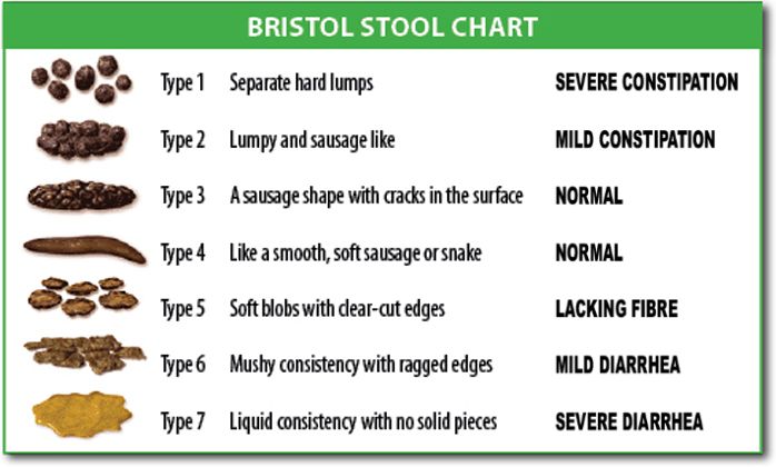
Normal adult bowel frequency ranges widely — from three stools a day to three stools a week — so what matters is the change from the patient’s own baseline. Assess frequency, form, color, odor, effort and continence, plus laxative and opioid use, fluid and fiber intake and mobility. Use the Bristol Stool Form Scale to describe form: type 1 is separate hard lumps, type 2 is lumpy and sausage-shaped, type 3 is sausage-shaped with surface cracks, type 4 is smooth and soft like a sausage or snake (the ideal), type 5 is soft blobs with clear edges, type 6 is fluffy pieces with ragged edges, and type 7 is entirely liquid with no solid pieces. Types 1 and 2 indicate constipation; types 6 and 7 indicate diarrhea. Charting “Bristol type 7 x 4 in 6 hours” means something to the next nurse; “loose stool” does not.
Color and content carry diagnostic weight. Brown is expected. Melena — black, tarry, sticky and foul — means digested blood from an upper gastrointestinal source and is an escalation; black stool from iron or bismuth is dry and not tarry, and you ask before you assume. Hematochezia, bright red blood, points lower. Clay-colored or acholic stool suggests biliary obstruction, especially with dark urine and jaundice. Steatorrhea — greasy, floating, foul stool — suggests fat malabsorption. Currant-jelly stool in an infant with intermittent severe crying and drawn-up legs suggests intussusception and is an emergency. Anyone with new diarrhea and recent antibiotics needs evaluation for Clostridioides difficile: place on contact precautions, wash your hands with soap and water because alcohol-based rub does not kill spores, use a sporicidal (bleach) cleaner for equipment, and do not send a formed specimen.
Assess an ostomy at every shift and at every pouch change. A healthy stoma is beefy red or pink, moist, and slightly raised (budded), and the peristomal skin is intact and matches the patient’s surrounding skin tone. A stoma that is pale, dusky, purple or black is ischemic and must be reported immediately; one that is dry, sunken (retracted) or bleeding beyond a small amount at the mucosa also warrants a call to the wound-ostomy nurse. Bleeding when you clean the surface lightly is common because the stoma is vascular mucosa. Output differs by site: an ileostomy drains continuously with liquid to pasty effluent, typically 500 to 1000 mL per day once it has adapted, and carries a real risk of dehydration and electrolyte loss — sustained output above roughly 1200 mL per day is high-output and must be reported. A descending or sigmoid colostomy produces formed stool on a more predictable schedule. Empty the pouch when it is one-third to one-half full, and expect no stool and no flatus from a new ostomy for the first 24 to 48 hours after surgery; beyond that, report it.
Urinary elimination, retention and fluid balance
Normal urine is pale yellow to amber, clear, with a faint aromatic odor. Expected adult output with normal intake is roughly 1200 to 1500 mL in 24 hours. The number to protect is the hourly minimum: at least 0.5 mL/kg/hour in an adult, which for a 70 kg patient is about 35 mL/hour, or the familiar bedside floor of 30 mL/hour. Children require about 1 mL/kg/hour and infants about 2 mL/kg/hour. Oliguria is under 400 mL in 24 hours or under 0.5 mL/kg/hour sustained for 6 hours; anuria is under 100 mL in 24 hours. Falling urine output is one of the earliest signs of hypoperfusion and one of the first things you report.
Adult bladder capacity is roughly 400 to 600 mL, with the first urge at about 150 to 300 mL. When a patient cannot void, do not reach for a catheter first — scan first. A bladder scanner measures the volume noninvasively; a post-void residual under 50 mL is normal, 50 to 100 mL is borderline and may be acceptable in an older adult, over 100 mL on repeated measurement indicates incomplete emptying, and over 200 mL with discomfort or over 300 to 400 mL is significant retention that is reported and usually catheterized per protocol. Nursing measures come first when the patient is stable: privacy, upright position, running water, warm perineal water, ambulation, and reviewing the medication list for anticholinergics and opioids. Every indwelling catheter is a CAUTI risk — keep the drainage bag below the level of the bladder and off the floor, maintain a closed system, secure the tubing to prevent traction, perform routine perineal care with soap and water, and ask daily whether the catheter can come out. The best CAUTI prevention is removal.
Intake and output requires the same discipline as vital signs. Count everything: oral fluids, ice chips at half their volume, intravenous fluids and flushes, tube feeds and water flushes, urine, emesis, liquid stool, ostomy effluent, drains and measurable wound drainage. Weigh diapers, incontinence pads and dressings when accuracy matters — 1 g equals 1 mL. Pair the I and O record with a daily weight taken at the same time on the same scale, because the weight is the more reliable number. Report a positive or negative balance that does not match the clinical picture rather than simply recording it.
| Parameter | Expected | Abnormal | Suggests |
|---|---|---|---|
| Urine color and clarity | Pale yellow to amber, clear | Dark amber, cloudy, foul, pink or red, tea-colored, orange | Concentration or dehydration; infection; hematuria; myoglobinuria or hepatic disease; phenazopyridine or rifampin |
| Adult hourly output | 0.5 mL/kg/hour or more | Under 0.5 mL/kg/hour for 6 hours | Hypovolemia, hypoperfusion, acute kidney injury, obstruction |
| Post-void residual | Under 50 mL | Over 100 mL repeated; over 300 mL | Incomplete emptying, urinary retention, neurogenic bladder, benign prostatic hyperplasia |
| Stool form | Bristol type 3 or 4 | Type 1–2, or type 6–7 | Constipation with dehydration, opioids, immobility; diarrhea with infection, tube feeds, medication |
| Stool color | Brown | Black tarry, bright red, clay colored, greasy floating | Upper GI bleed, lower GI bleed, biliary obstruction, fat malabsorption |
| Stoma | Beefy red or pink, moist, budded | Pale, dusky, purple, black, retracted | Ischemia or necrosis — report immediately |
Red flags, vital signs, SBAR and delegation
Abdominal emergencies are frequently missed because they present quietly. Escalate now, without waiting for rounds, for any of the following: a rigid, board-like abdomen with involuntary guarding; sudden severe pain, especially if it woke the patient; a distended abdomen with vomiting and absent bowel sounds; a pulsatile epigastric mass, or abdominal or back pain with hypotension; hematemesis or coffee-ground emesis; melena or frank rectal bleeding; Cullen or Grey Turner sign; a dusky or black stoma; wound dehiscence or evisceration; no urine output for 6 to 8 hours or a bladder scan over 400 mL with pain; pain out of proportion to examination findings; and any acute abdominal complaint accompanied by fever, tachycardia and hypotension. If evisceration occurs, do not push the tissue back — cover it with sterile gauze moistened with sterile normal saline, place the patient in low Fowler position with knees flexed, keep them NPO, take vital signs and call the surgical team immediately.
Vital signs frame every one of those findings, and the thresholds shift by age. Compare against the patient’s own baseline first, then against the table.
| Age group | Heart rate (beats/min) | Respiratory rate (breaths/min) | Systolic BP (mmHg) |
|---|---|---|---|
| Newborn (0–1 month) | 100–180 | 30–60 | 60–90 |
| Infant (1–12 months) | 100–160 | 30–53 | 70–100 |
| Toddler (1–3 years) | 90–150 | 22–37 | 80–110 |
| Preschool (3–5 years) | 80–140 | 20–28 | 80–110 |
| School age (6–11 years) | 70–120 | 18–25 | 85–120 |
| Adolescent (12–18 years) | 60–100 | 12–20 | 100–130 |
| Adult | 60–100 | 12–20 | Below 120 systolic and below 80 diastolic is normal; keep mean arterial pressure at 65 or above |
| Older adult | 60–100 | 12–20 | Wider pulse pressure common; a temperature of 37.2 °C (99 °F) may represent fever because baseline temperature is lower and the febrile response is blunted |
Adult oral temperature is 36.5 to 37.5 °C (97.7 to 99.5 °F) and oxygen saturation is 95 to 100 percent on room air. In an acute abdomen, the pattern to fear is a rising heart rate with a narrowing pulse pressure and falling urine output — compensated shock — often before the blood pressure ever drops.
Findings across skin tones require deliberate technique, because the classic color descriptions were written for lightly pigmented skin. Assess jaundice in the sclera near the limbus in good natural light, and confirm on the hard palate, the palms and the soles; carotenemia from a diet high in carrots or squash yellows the palms and soles but spares the sclera. Assess pallor in the conjunctivae, the oral mucosa, the palms, the soles and the nail beds; in richly pigmented skin, pallor may appear as ashen, gray or yellow-brown rather than white. Assess cyanosis in the lips, tongue, oral mucosa, conjunctivae, nail beds, palms and soles; in dark skin it may look gray, ashen or blue-black rather than blue. Assess erythema with your hands as much as your eyes — compare the site to the contralateral side and to surrounding skin, and palpate for warmth, induration, tightness and tenderness, because inflammation in deeply pigmented skin often appears violet, deep brown or simply darker rather than red. This is exactly how early peristomal irritation and early pressure injury get missed.
When you escalate, use SBAR and have the data in hand before you dial. Situation: “This is Maria Ortiz, RN, on 4 West calling about Mr. Daniel Okafor in 412, a 68-year-old man, postoperative day 2 from an open sigmoid colectomy. His abdomen has become rigid and he has new severe pain.” Background: “He has hypertension and type 2 diabetes. He passed no flatus today, had one episode of coffee-ground emesis at 1500, and has had 20 mL of urine over the last 2 hours.” Assessment: “Temperature 38.6 °C, heart rate 122, blood pressure 92/68 down from 138/78, respirations 26, oxygen saturation 94 percent on room air. Abdomen is distended and board-like with involuntary guarding, no bowel sounds in any quadrant after 5 minutes of listening. I think he has peritonitis.” Recommendation: “I need you to come see him now. May I place him NPO, insert a nasogastric tube to low intermittent suction, obtain a stat lactate, CBC and basic metabolic panel, and start a normal saline bolus?” Then read the orders back, document the call with the time, and if you do not get a response, use the chain of command or the rapid response team.
Finally, scope. Unlicensed assistive personnel may measure and record intake and output, weigh patients, obtain and record vital signs on stable patients, assist a stable patient with eating when no dysphagia risk exists, provide perineal and catheter hygiene, empty an established ostomy pouch and drainage bags and record the amounts, collect routine urine and stool specimens, assist with toileting and ambulation, and apply incontinence products. The registered nurse may not delegate assessment (inspection, auscultation, percussion and palpation of the abdomen; interpreting a bladder scan and deciding to catheterize; the initial and ongoing assessment of a stoma), the swallow screen and the first oral intake after it, insertion or verification of a nasogastric or feeding tube, initiation of enteral feeding, evaluation of whether an intervention worked, patient and family teaching, and any care of an unstable patient. The rule that will carry you: assessment, teaching, evaluation, judgment and unstable patients stay with the RN. When you delegate, give the specific task, the expected outcome, the limits, and what to report back and when — “Please empty and measure Mr. Okafor’s ileostomy pouch every 2 hours and tell me immediately if the output is over 200 mL in an hour or if it turns dark.”
A rigid abdomen, absent bowel sounds with vomiting and distension, coffee-ground emesis, melena, a dusky stoma, a pulsatile mass or urine output under 0.5 mL/kg/hour are escalations right now — communicated in SBAR with vital signs, and never handed off to unlicensed personnel, because assessment, teaching, evaluation and unstable patients stay with the RN.
The chapter in one breath
The abdomen is examined in a sequence that exists nowhere else in the body: inspect, auscultate, percuss, palpate — because pressing first changes the bowel sounds you are about to record. Set the patient up supine with knees flexed and bladder empty, examine the painful quadrant last, and save any rebound test for the very end. Map findings to the four quadrants so your language survives handoff. Auscultate with the diaphragm starting in the right lower quadrant, expect 5 to 30 irregular gurgles a minute, and listen a full 5 minutes before charting sounds as absent; then switch to the bell for bruits. Percuss for tympany, a liver span of 6 to 12 cm at the midclavicular line, and shifting dullness. Palpate light at 1 to 2 cm before deep at 4 to 6 cm, and never deep-palpate a pulsatile mass or a rigid abdomen. Nutrition rests on weight trend, intake, BMI and physical findings — not on albumin. Elimination rests on Bristol type, stool color, urine output of at least 0.5 mL/kg/hour and post-void residual. And the whole chapter ends where nursing always ends: recognize the red flag, hand the story over in SBAR, and keep assessment, teaching, evaluation and unstable patients with the RN.
Words worth knowing
| Term | What it means |
|---|---|
| Ascites | Accumulation of free fluid in the peritoneal cavity; detected clinically by shifting dullness or a fluid wave, usually only above about 1500 mL. |
| Borborygmi | Loud, prolonged, hyperperistaltic gurgling audible without a stethoscope; usually benign. |
| Bristol Stool Form Scale | Seven-type classification of stool form, from type 1 (separate hard lumps) to type 7 (entirely liquid); types 3 and 4 are ideal. |
| Bruit | Blowing or swishing sound of turbulent arterial flow, heard with the bell over the aorta, renal, iliac or femoral arteries. |
| Caput medusae | Dilated tortuous veins radiating from the umbilicus, a sign of portal hypertension. |
| Cullen sign / Grey Turner sign | Periumbilical bruising and flank bruising respectively, indicating retroperitoneal or intraperitoneal bleeding. |
| Guarding, voluntary vs involuntary | Voluntary guarding is anticipatory muscle tensing that relaxes with distraction; involuntary guarding (rigidity) persists and signals peritoneal inflammation. |
| Hematochezia / melena | Bright red rectal bleeding from a lower gastrointestinal source; black, tarry, foul stool from digested blood in an upper source. |
| Liver span | Vertical distance between the upper and lower borders of hepatic dullness at the right midclavicular line; 6 to 12 cm in an adult. |
| Murphy sign | Abrupt cessation of inspiration when the examiner presses beneath the right costal margin; suggests acute cholecystitis. |
| McBurney point | Point one-third of the way from the right anterior superior iliac spine to the umbilicus; classic site of appendiceal tenderness. |
| Oliguria / anuria | Urine output under 400 mL in 24 hours or under 0.5 mL/kg/hour for 6 hours; anuria is under 100 mL in 24 hours. |
| Post-void residual (PVR) | Volume remaining in the bladder immediately after voiding; under 50 mL is normal, over 100 mL repeated indicates incomplete emptying. |
| Prealbumin / albumin | Serum proteins (15–36 mg/dL and 3.5–5.0 g/dL) that fall with inflammation; markers of illness severity, not of nutritional intake. |
| Refeeding syndrome | Dangerous drop in phosphate, potassium and magnesium when nutrition restarts after prolonged starvation; monitor labs and cardiac rhythm. |
| Rovsing sign | Pain referred to the right lower quadrant when the left lower quadrant is palpated; supports appendicitis. |
| Scaphoid abdomen | Sunken, concave abdominal contour seen with marked weight loss or dehydration. |
| Stoma | Surgically created opening of bowel or ureter onto the abdominal wall; expected to be beefy red or pink, moist and budded. |
| Tympany | Loud, drum-like percussion note over gas-filled bowel; the dominant note of a normal abdomen. |
| Visceral vs parietal pain | Visceral pain is dull, crampy and poorly localized from a stretched hollow organ; parietal pain is sharp, well localized and worse with movement, from peritoneal inflammation. |