The High-Risk Newborn
Most newborns make the transition from placenta to air on their own. A minority cannot, and the nursing work of the first minutes, hours and days decides how much of that minority survives intact. A high-risk newborn is one whose gestational age, growth, birth events, physiology or environment puts them at greater-than-usual risk of illness, injury or death — the 34-week late preterm baby who looks deceptively term, the macrosomic infant of a parent with diabetes whose glucose falls within ninety minutes, the meconium-stained post-term newborn whose pulmonary vasculature will not relax, the baby whose bilirubin is climbing faster than the nomogram allows. This chapter moves the way the bedside moves: mechanism first, then what you assess, then what you do, then what goes wrong if you are slow. Every number here is per kilogram, per hour or per week of gestation, because in this population a decimal point is a diagnosis.
By the end of this chapter you can
- Classify a newborn by gestational age and by birth weight, and distinguish small for gestational age, appropriate for gestational age and large for gestational age from intrauterine growth restriction.
- Sequence the Neonatal Resuscitation Program algorithm through the Golden Minute, including the corrective ventilation steps (MR SOPA), compression ratio and the weight-based dose of epinephrine.
- Differentiate respiratory distress syndrome, transient tachypnoea of the newborn, meconium aspiration syndrome and persistent pulmonary hypertension of the newborn by history, examination and radiograph.
- Calculate weight-based volumes and doses, including the 4–2–1 maintenance-fluid rule, the glucose infusion rate and a neonatal antibiotic dose, and check every result against the maximum adult dose.
- Screen, interpret and treat neonatal hypoglycaemia and hyperbilirubinaemia using hour-specific thresholds, and describe the progression to acute bilirubin encephalopathy and kernicterus.
- Recognise early-onset and late-onset neonatal sepsis and necrotising enterocolitis from their earliest, least dramatic signs.
- State the single correct first action for congenital diaphragmatic hernia, oesophageal atresia with tracheo-oesophageal fistula, myelomeningocele, gastroschisis and a ductal-dependent cardiac lesion.
- Apply non-pharmacologic Eat, Sleep, Console care to the newborn experiencing withdrawal, and describe therapeutic hypothermia for hypoxic-ischaemic encephalopathy.
- Sequence the obstetric emergency responses — postpartum haemorrhage, magnesium sulfate toxicity, cord prolapse, shoulder dystocia and uterine rupture — that decide which newborn you receive.
- Escalate deterioration using SBAR, act on a safeguarding concern at the threshold of reasonable suspicion, and delegate within scope, retaining assessment, teaching, evaluation and the unstable patient for the registered nurse.
Section 1Who Counts as High Risk: Age, Growth and the Birth Itself
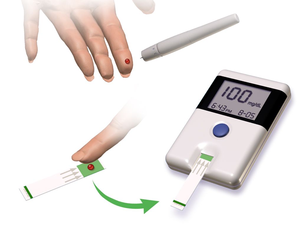
Gestational age and birth weight are two different questions
Ask them separately, because the answers do not travel together. Gestational age is counted in completed weeks from the first day of the last menstrual period, and it predicts organ maturity: lung surfactant, gut motility, hepatic conjugation of bilirubin, thermal control, the integrity of the germinal matrix. Birth weight predicts a different set of problems — substrate reserve, surface-area-to-mass ratio, and the mechanical difficulty of the birth itself. A 1,900 g newborn may be a well-grown 33-week infant or a growth-restricted 39-week infant, and those two babies need almost opposite anticipatory care.
Preterm means birth before 37 completed weeks. Within that, late preterm covers 34 0/7 to 36 6/7 weeks, moderate preterm 32 0/7 to 33 6/7, very preterm below 32 weeks, and extremely preterm below 28 weeks. Early term is 37 0/7 to 38 6/7, full term 39 0/7 to 40 6/7, late term 41 0/7 to 41 6/7, and post-term 42 0/7 weeks and beyond. Low birth weight is under 2,500 g, very low birth weight under 1,500 g, and extremely low birth weight under 1,000 g. When the dating is uncertain, physical and neuromuscular maturity are estimated with the New Ballard Score, which should be performed within 12 hours of birth for infants under 26 weeks and remains valid up to 96 hours in more mature infants, and which can assess infants as immature as 20 weeks.
The late preterm newborn is the trap of this section. At 35 weeks a baby often weighs 2,400 g, feeds a little, and looks like a small term infant to a tired family and a busy unit. Physiologically the baby is not term: brain mass is roughly two-thirds of term brain mass, hepatic uridine diphosphate glucuronosyltransferase activity is low, brown fat stores are thin, and the suck-swallow-breathe sequence is immature. Late preterm infants are several times more likely than term infants to develop hypoglycaemia, hyperbilirubinaemia requiring phototherapy, temperature instability, apnoea, feeding difficulty and readmission after discharge. Treat every 34-to-36-week newborn as a surveillance patient, not a well baby.
| Category | Definition | What it predicts at the bedside |
|---|---|---|
| Late preterm | 34 0/7 – 36 6/7 weeks | Hypoglycaemia, jaundice, cold stress, poor feeding, apnoea, readmission |
| Very preterm | Less than 32 weeks | RDS, intraventricular haemorrhage, patent ductus arteriosus, NEC, retinopathy |
| Extremely preterm | Less than 28 weeks | All of the above plus insensible water loss, BPD, long-term neurodevelopmental risk |
| Post-term | 42 0/7 weeks or more | Meconium aspiration, placental insufficiency, hypoglycaemia, birth trauma, polycythaemia |
| Low birth weight | Less than 2,500 g | Thermal instability, hypoglycaemia, feeding immaturity |
| Very low birth weight | Less than 1,500 g | Ventilatory support, central access, parenteral nutrition, ROP screening |
| Extremely low birth weight | Less than 1,000 g | Massive transepidermal water loss, humidified isolette, extreme fragility |
Growth: SGA, LGA and the two shapes of restriction
Plot the weight on a gestational-age growth curve. Below the 10th percentile is small for gestational age, between the 10th and 90th is appropriate for gestational age, and above the 90th is large for gestational age. Small for gestational age is a statistical statement about size; intrauterine growth restriction is a pathological statement about a fetus that did not achieve its own growth potential. Most growth-restricted fetuses are small for gestational age, but a constitutionally small baby with healthy parents may be perfectly well, and a genetically large fetus can be growth restricted while still plotting at the 25th percentile.
Symmetric restriction — head, length and weight all reduced proportionally — usually begins early and reflects an intrinsic problem: chromosomal abnormality, congenital infection from cytomegalovirus, toxoplasmosis, rubella or syphilis, or early exposure to alcohol. Asymmetric restriction spares the head while the abdomen and weight fall away, and it points to late uteroplacental insufficiency from chronic hypertension, pre-eclampsia, pre-gestational vascular disease, smoking or high-altitude hypoxia. The asymmetric baby has burned the hepatic glycogen and the subcutaneous fat, which is exactly why the loose-skinned, wide-eyed, scaphoid-abdomened newborn becomes hypoglycaemic and hypothermic within the first two hours.
Large for gestational age is most often the newborn of a birthing parent with diabetes. Fetal hyperinsulinaemia in response to maternal hyperglycaemia produces macrosomia, organomegaly and, occasionally, an interventricular septum thick enough to obstruct outflow. At birth the placental glucose supply stops but the pancreatic insulin does not, so glucose can fall steeply in the first 30 to 90 minutes. These newborns also carry risks of birth injury, hypocalcaemia, polycythaemia, hyperbilirubinaemia and respiratory distress syndrome even at term, because insulin antagonises cortisol-driven surfactant maturation.
The birth events that hand you a compromised newborn
The high-risk newborn is often created in the ten minutes before delivery, so the newborn nurse must understand the obstetric emergency sequences. In every one of them the correct first action is the counterintuitive one, and getting it wrong changes the baby you receive. Postpartum haemorrhage: massage the fundus first, before the oxytocin, before the bladder, before the laboratory tube — uterine atony is the cause in most cases and a contracted uterus is the treatment. Magnesium sulfate toxicity in a patient being treated for pre-eclampsia: deep tendon reflexes disappear before respiratory depression, so absent reflexes are the early warning, not a late one; stop the infusion, and the antidote is calcium gluconate 1 g intravenously over 3 minutes. Magnesium crosses the placenta, so the newborn may arrive hypotonic, hyporeflexic and hypoventilating, and needs ventilatory support rather than naloxone.
Umbilical cord prolapse: relieve the pressure of the presenting part on the cord with a gloved hand and place the birthing parent in knee-chest or steep Trendelenburg position while preparing for immediate caesarean birth. Never attempt to replace the cord into the uterus, and never clamp it; keep any exposed loop warm and moist. Shoulder dystocia: McRoberts hyperflexion of the maternal hips and suprapubic pressure, in that order, and never fundal pressure, which drives the anterior shoulder harder into the symphysis and risks uterine rupture. The newborn from a shoulder dystocia gets a careful examination for clavicular fracture, brachial plexus injury and asphyxia. Uterine rupture announces itself as sudden severe abdominal pain, loss of the fetal station, an abnormal fetal heart rate pattern with prolonged deceleration or bradycardia, and maternal signs of hypovolaemia; the response is immediate surgical birth, and the newborn arrives acidotic.
The newborn of a pregnancy complicated by pre-eclampsia is commonly growth restricted, preterm, and transiently neutropenic and thrombocytopenic.
| Obstetric emergency | First action — and the tempting wrong one | What arrives at the warmer |
|---|---|---|
| Postpartum haemorrhage | Massage the fundus FIRST; not the oxytocin bolus first | Usually a well newborn; the emergency is the parent, so protect bonding and feeding once stable |
| Magnesium sulfate toxicity | Stop infusion; calcium gluconate 1 g IV; NOT naloxone | Hypotonic, hyporeflexic, hypoventilating newborn needing PPV, not an antidote |
| Cord prolapse | Lift the presenting part off the cord, knee-chest or Trendelenburg; do NOT reduce the cord | Acidotic, possibly bradycardic newborn; full resuscitation team |
| Shoulder dystocia | McRoberts plus suprapubic pressure; NEVER fundal pressure | Clavicle fracture, Erb palsy, asphyxia; check for asymmetric Moro |
| Uterine rupture | Immediate surgical birth and volume resuscitation | Severe acidosis, candidate for therapeutic hypothermia |
Gestational age, birth weight and the events of the birth are three independent risk axes, and the newborn who looks least sick — the late preterm infant — is the one most often discharged too soon.
Section 2The Golden Minute: Resuscitation and Respiratory Distress
The Golden Minute and the numbers that drive it
Roughly one newborn in ten needs some assistance to begin breathing and about one in a hundred needs extensive resuscitation. Preparation is therefore an assessment skill: before every birth, ask the four antenatal questions — what is the gestational age, is the amniotic fluid clear, how many babies are expected, and are there additional risk factors — and agree the umbilical cord management plan. Preheat the room to 23–25 °C, turn on the radiant warmer, and check the equipment. For a vigorous newborn, delay cord clamping for 30 to 60 seconds; do not delay if immediate resuscitation is required or the placental circulation is disrupted by abruption, praevia or cord avulsion.
Three rapid questions at birth decide everything: term gestation, good tone, breathing or crying. Three yes answers mean the baby stays skin-to-skin with routine care. Any no answer moves the baby to the warmer for the initial steps — warm, dry, stimulate, position the airway in the neutral sniffing position, and clear secretions only if they obstruct. Then assess respirations and heart rate. If the newborn is apnoeic or gasping, or the heart rate is under 100 beats per minute, begin positive-pressure ventilation within the first 60 seconds of life. This is the Golden Minute, and it is a deadline, not an aspiration.
Ventilate at 40 to 60 breaths per minute. The first and most reliable sign of effective ventilation is a rising heart rate; chest rise is the second. If the heart rate does not rise, run the corrective steps in order — MR SOPA: Mask readjust, Reposition the head, Suction mouth then nose, Open the mouth, Pressure increase in increments, and Alternative airway by endotracheal tube or laryngeal mask. A laryngeal mask is an appropriate rescue airway from about 34 weeks and roughly 1,500 to 2,000 g. If the heart rate remains under 60 after 30 seconds of ventilation that visibly moves the chest, preferably through an advanced airway, start chest compressions with two thumbs on the lower third of the sternum, depressing one-third of the anterior-posterior chest diameter, in a 3:1 ratio — 90 compressions and 30 breaths, 120 events, every minute — and increase the oxygen to 100 per cent.
Reassess after 60 seconds of coordinated compressions and ventilation, using cardiac monitor leads for the most accurate heart rate. If the heart rate is still under 60, give epinephrine. Volume expansion with 10 mL/kg of normal saline or O-negative blood over 5 to 10 minutes is reserved for suspected blood loss with poor response, and is given more slowly in preterm infants because rapid volume shifts contribute to intraventricular haemorrhage. Naloxone is not recommended in the delivery room, and neither is routine sodium bicarbonate; both are classic distractors from the actual intervention, which is ventilation. If there is no detectable heart rate after 20 minutes of adequate resuscitation, the team discusses discontinuation with the family.
Oxygen is a drug: saturation targets by minute of life
A healthy newborn does not saturate 95 per cent at birth and should not be pushed there. Fetal preductal saturation is around 60 per cent and climbs over ten minutes as the lungs fill and pulmonary vascular resistance falls. Place the pulse oximeter probe preductally on the right hand or wrist. Begin resuscitation of newborns 35 weeks or more in 21 per cent oxygen, and those under 35 weeks in 21 to 30 per cent, titrating to the target range. Hyperoxia in the preterm infant fuels retinopathy of prematurity and bronchopulmonary dysplasia; hypoxia sustains pulmonary vasoconstriction. The target is a corridor, not a ceiling.
| Minutes after birth | Target preductal SpO2 |
|---|---|
| 1 minute | 60–65 per cent |
| 2 minutes | 65–70 per cent |
| 3 minutes | 70–75 per cent |
| 4 minutes | 75–80 per cent |
| 5 minutes | 80–85 per cent |
| 10 minutes | 85–95 per cent |
The Apgar score is assigned at 1 and 5 minutes and, if the 5-minute score is under 7, every 5 minutes to 20 minutes. It scores heart rate, respiratory effort, muscle tone, reflex irritability and colour from 0 to 2 each. It describes the transition and it belongs in the record, but it never dictates resuscitation — by the time the 1-minute score is calculated, ventilation should already be underway. For meconium-stained amniotic fluid, routine intubation and tracheal suctioning is no longer recommended, even for a non-vigorous newborn; perform the initial steps and, if the baby is not breathing effectively, begin positive-pressure ventilation.
Telling the respiratory syndromes apart
Respiratory distress in the newborn shows itself as tachypnoea above 60 breaths per minute, nasal flaring, intercostal and subcostal retractions, expiratory grunting, cyanosis and, in the deteriorating infant, apnoea. Grunting is the sound of a baby closing the glottis in expiration to hold open alveoli that want to collapse; it is a compensation, and when it stops without improvement elsewhere, the baby is tiring. Silverman-Andersen or a similar respiratory distress score converts these observations into a trackable number.
Respiratory distress syndrome is surfactant deficiency. It belongs to the preterm infant, is worsened by maternal diabetes, caesarean birth without labour, asphyxia and cold stress, and is prevented by antenatal betamethasone 12 mg intramuscularly given twice, 24 hours apart, between 24 and 34 weeks, and in selected patients up to 36 6/7 weeks. The radiograph shows a diffuse ground-glass reticulogranular pattern with air bronchograms and low lung volumes. Treatment is early continuous positive airway pressure at 5 to 6 cm H2O and, when required, exogenous surfactant — poractant alfa 200 mg/kg (2.5 mL/kg) as an initial dose with 100 mg/kg repeats, or beractant 100 mg/kg (4 mL/kg) up to four doses. Improvement can be abrupt, so lower the ventilator pressures and the oxygen promptly to avoid air leak and hyperoxia.
Transient tachypnoea of the newborn is retained fetal lung fluid, most often in a term or late preterm baby born by caesarean without labour. Respiratory rate can reach 100 to 120, but oxygen requirement is modest and the baby usually needs nothing more than time, warmth and, in a very tachypnoeic newborn, a pause in oral feeding to protect the airway. It resolves within 24 to 72 hours. Meconium aspiration syndrome affects post-term and growth-restricted infants; meconium obstructs airways in a ball-valve fashion, inactivates surfactant and causes chemical pneumonitis, producing coarse patchy infiltrates with hyperinflation, a high risk of pneumothorax and a high risk of persistent pulmonary hypertension.
Persistent pulmonary hypertension of the newborn is failure of the pulmonary vasculature to relax, so blood shunts right to left across the ductus arteriosus and foramen ovale. The signature finding is labile, profound hypoxaemia with a preductal-to-postductal saturation difference greater than 10 per cent between the right hand and either foot. Management is meticulous oxygenation and ventilation, correction of acidosis, minimal handling because agitation worsens shunting, inhaled nitric oxide typically at 20 parts per million, and extracorporeal membrane oxygenation when maximal therapy fails. A sudden deterioration with asymmetric chest movement and a shifted point of maximal impulse is a pneumothorax until proven otherwise, and needs immediate needle decompression.
| Condition | Typical newborn | Radiograph | Core management |
|---|---|---|---|
| Respiratory distress syndrome | Preterm; worse with maternal diabetes or asphyxia | Ground-glass, air bronchograms, low volumes | CPAP 5–6 cm H2O, surfactant, wean oxygen briskly |
| Transient tachypnoea | Term or late preterm, caesarean without labour | Fluid in fissures, perihilar streaking | Warmth, oxygen if needed, time; resolves 24–72 h |
| Meconium aspiration | Post-term or growth restricted, stained fluid | Patchy infiltrates plus hyperinflation | Ventilation, watch for air leak and PPHN |
| PPHN | Term, after asphyxia, MAS or sepsis | Often clearer than the hypoxaemia suggests | Oxygen, gentle ventilation, correct acidosis (no deliberate hyperventilation or alkali therapy), minimal handling, iNO, ECMO |
Apnoea of prematurity is a pause of more than 20 seconds, or a shorter pause with bradycardia under 100 or desaturation, in an infant under 37 weeks. It reflects an immature respiratory centre, but the first response is never to assume immaturity — new apnoea in a previously stable preterm infant is sepsis, necrotising enterocolitis, intraventricular haemorrhage, hypoglycaemia, seizure, anaemia or reflux until those are excluded. Treatment is gentle tactile stimulation, positioning, and caffeine citrate: a loading dose of 20 mg/kg intravenously over 30 minutes, then 5 to 10 mg/kg once daily. Bronchopulmonary dysplasia is defined by a continuing need for supplemental oxygen at 36 weeks postmenstrual age and is the price of oxygen, pressure and time; it is minimised by gentle ventilation, vitamin A, caffeine and avoidance of fluid overload.
Ventilation of the lungs is the intervention that resuscitates a newborn; if the heart rate is not rising, fix the ventilation with MR SOPA before reaching for compressions or drugs.
Section 3Warmth, Sugar and Fluid: The Metabolic Newborn
Cold stress is a metabolic emergency, not a comfort issue
Newborns lose heat by four routes: conduction to cold surfaces, convection to moving air, evaporation from wet skin and mucosa, and radiation to nearby cold objects such as a window or an incubator wall. They lose it fast because their surface-area-to-mass ratio is large, their subcutaneous fat is thin and their vasomotor control is immature. They cannot shiver. Instead they perform non-shivering thermogenesis by metabolising brown adipose tissue at the nape of the neck, between the scapulae, in the axillae and around the kidneys and adrenals — stores that the preterm and growth-restricted newborn largely do not have.
The consequences cascade in a predictable order. Heat production consumes oxygen and glucose, so cold stress produces hypoxia and hypoglycaemia. Anaerobic metabolism generates lactic acid, and metabolic acidosis causes pulmonary vasoconstriction, which reduces pulmonary perfusion and inhibits surfactant production, which worsens the hypoxia. A cold baby is also a jaundiced baby, because free fatty acids released from brown fat compete with bilirubin for albumin binding sites. Target axillary temperature is 36.5 to 37.5 °C. Below 36.5 °C is cold stress; below 36.0 °C is moderate hypothermia and warrants escalation.
Prevention is nursing work: dry the newborn immediately and remove the wet linen, put a hat on, use skin-to-skin contact with the birthing parent as the default warming device for stable infants, and for infants under 32 weeks place the baby without drying into a polyethylene wrap or bag with a thermal mattress and humidified warmed gases. Preheat surfaces, keep the isolette away from external walls, and cluster cares to limit the time the portholes are open. Rewarm a hypothermic newborn slowly, about 0.5 °C per hour, because rapid rewarming provokes apnoea and vasodilatory hypotension.
Hypoglycaemia: who, when and how much
Glucose falls physiologically in the first one to two hours of life in every newborn and then recovers as counter-regulation and feeding begin. Screening is targeted, not universal: the newborn of a parent with any form of diabetes, the large-for-gestational-age newborn, the small-for-gestational-age or growth-restricted newborn, the late preterm and preterm newborn, and any newborn who is cold, septic, asphyxiated, polycythaemic or symptomatic. Screen at approximately one hour of age, roughly 30 minutes after the first feed, then before feeds every three hours through the first 12 to 24 hours.
Signs are non-specific and easy to attribute to something else: jitteriness, high-pitched or weak cry, poor feeding, hypotonia, lethargy, temperature instability, tachypnoea, apnoea, cyanosis, diaphoresis and, late, seizure. Any point-of-care result must be confirmed on a laboratory plasma sample, but treatment starts on the bedside value — do not wait for the laboratory to treat a symptomatic baby.
Common operational thresholds in the first 24 hours: from birth to 4 hours of age, a value under 25 mg/dL (1.4 mmol/L) after a feed, or any symptomatic value, calls for intravenous dextrose; 25 to 40 mg/dL calls for a feed and a recheck in one hour. From 4 to 24 hours, treat under 35 mg/dL (1.9 mmol/L) intravenously and feed-and-recheck between 35 and 45 mg/dL. The general goal is a plasma glucose of at least 45 mg/dL (2.5 mmol/L) before routine feeds, rising toward 60 mg/dL after 48 hours. Buccal 40 per cent dextrose gel at 0.5 mL/kg, given with a feed, treats many asymptomatic episodes and keeps parent and baby together.
When intravenous treatment is needed, give a minibolus of 2 mL/kg of 10 per cent dextrose — that is 200 mg/kg of glucose — and follow immediately with a continuous infusion, because a bolus alone provokes rebound insulin secretion. Start the infusion at a glucose infusion rate of 5 to 8 mg/kg/min. The formula is worth memorising: glucose infusion rate in mg/kg/min equals the dextrose percentage multiplied by the rate in mL/hr, divided by the weight in kilograms multiplied by 6. A 3 kg newborn on 10 per cent dextrose at 12 mL/hr receives (10 × 12) ÷ (3 × 6) = 120 ÷ 18 = 6.7 mg/kg/min. Persistent hypoglycaemia requiring more than 8 to 10 mg/kg/min beyond 48 hours suggests hyperinsulinism and needs endocrine evaluation.
Weight-based dosing: the arithmetic and the ceiling
Children are not small adults, and the only safe way to express a paediatric dose is per kilogram of actual measured weight in kilograms, taken on admission, not reported by a caregiver in pounds. Write the calculation out, have a second nurse verify it independently for high-alert drugs, and then perform the step that is skipped most often: compare the calculated dose against the maximum adult dose. A weight-based formula that is correct for a 12 kg toddler becomes an overdose in a 70 kg adolescent because the milligrams-per-kilogram relationship stops scaling.
Worked example. Acetaminophen is dosed at 15 mg/kg per dose. For a 14 kg child: 14 × 15 = 210 mg. For a 62 kg adolescent: 62 × 15 = 930 mg, which is under the 1,000 mg adult single-dose maximum, so it stands. For an 84 kg adolescent: 84 × 15 = 1,260 mg, which exceeds the maximum — the dose is capped at 1,000 mg, and the daily total must stay under 75 mg/kg/day and under the 4,000 mg adult daily maximum. The same discipline applies to ibuprofen at 10 mg/kg per dose with a 400 mg per dose ceiling, and to amoxicillin at 45 to 90 mg/kg/day divided, capped at the adult daily maximum. If the calculated dose exceeds the adult dose, the calculation is not wrong — the ceiling is right.
Neonatal antibiotic arithmetic follows the same pattern. Ampicillin for suspected early-onset sepsis is 50 mg/kg per dose (100 mg/kg per dose when meningitis is suspected). A 3.2 kg newborn: 3.2 × 50 = 160 mg per dose. Gentamicin is 4 to 5 mg/kg per dose with the interval set by postmenstrual age — commonly every 24 hours at 35 weeks or more, and every 36 to 48 hours below that, because renal clearance is slower. For the same newborn at 4 mg/kg: 3.2 × 4 = 12.8 mg. Draw a trough before the next dose when therapy extends beyond 48 hours, and monitor for nephrotoxicity and ototoxicity.
Volumes follow the same per-kilogram discipline but not the same numbers at every age, and this is the pair most often confused. In the delivery room and in the neonatal unit, volume expansion is 10 mL/kg of isotonic crystalloid or O-negative blood over 5 to 10 minutes, given more slowly still in the preterm infant because rapid volume shifts contribute to intraventricular haemorrhage. Beyond the newborn period, the paediatric fluid resuscitation bolus for shock is 20 mL/kg of isotonic crystalloid — normal saline or lactated Ringer’s — given rapidly, with reassessment of heart rate, perfusion, mental state and liver edge after each bolus before the next is given. Reduce it to 10 mL/kg and give it more cautiously in diabetic ketoacidosis, in suspected cardiogenic shock and in severe acute malnutrition. Reassessment between boluses is part of the dose, not an optional extra.
Feeding the preterm gut, and necrotising enterocolitis
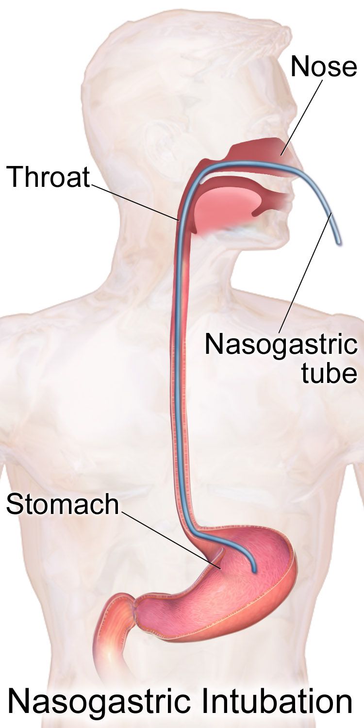
Coordinated sucking, swallowing and breathing matures at about 32 to 34 weeks, so infants below that are fed by orogastric or nasogastric tube while non-nutritive sucking on a pacifier during gavage builds the reflex. Feeding readiness is assessed by cues — rooting, hand-to-mouth activity, stable respiratory rate, sustained alert state — not by the clock and never by labelling a baby a poor feeder. Describe what you observe: this infant sustains four to five sucks before pausing to breathe and desaturates to 88 per cent during bursts. That sentence generates a plan; a label does not.
Human milk is the preferred enteral nutrition and is protective against necrotising enterocolitis; when the birthing parent’s own milk is unavailable, pasteurised donor human milk is preferred for very low birth weight infants. Preterm infants need fortification for protein, calcium and phosphorus to support bone mineralisation. Trophic or gut-priming feeds of 10 to 20 mL/kg/day stimulate mucosal maturation and hormone release without providing meaningful nutrition, and are advanced steadily rather than in leaps.
Necrotising enterocolitis is inflammatory and ischaemic injury to the bowel, usually presenting in the second or third week of life in a preterm infant whose feeds are advancing. The early signs are quiet: increased gastric residuals, feeding intolerance, abdominal distension with visible loops, a shiny or discoloured abdominal wall, bilious emesis, blood in the stool, and a generalised deterioration with apnoea, temperature instability and lethargy. The abdominal radiograph shows pneumatosis intestinalis, and free air means perforation. Management is immediate nil by mouth, orogastric tube to low intermittent suction for decompression, intravenous fluids and parenteral nutrition, broad-spectrum antibiotics, blood cultures, serial abdominal radiographs and girths, and surgical consultation. Do not palpate the abdomen repeatedly and never take a rectal temperature in a newborn with suspected necrotising enterocolitis.
Warmth, glucose and fluid are one physiological system: a cold newborn burns glucose and oxygen to make heat, and every dose or volume given must be per kilogram, written out, and checked against the maximum adult dose.
Section 4Bilirubin, Blood and Infection
Why every newborn turns a little yellow, and when it stops being normal
Newborns have a high haematocrit, a red cell lifespan of only 70 to 90 days, and an immature hepatic conjugating enzyme, uridine diphosphate glucuronosyltransferase. Haem breakdown yields unconjugated, fat-soluble, albumin-bound bilirubin that the liver must conjugate before it can be excreted in bile. What reaches the gut is partly deconjugated by beta-glucuronidase and reabsorbed through the enterohepatic circulation, which is why delayed or inadequate feeding raises bilirubin and why stooling lowers it. Physiologic jaundice therefore appears after 24 hours of age, peaks on day three to five in the term infant and day five to seven in the preterm infant, and resolves without treatment.
Jaundice is pathologic when it appears in the first 24 hours of life, when total serum bilirubin rises faster than 0.2 mg/dL per hour or more than 5 mg/dL per day, when the direct (conjugated) fraction exceeds 1 mg/dL or 20 per cent of the total, or when visible jaundice persists beyond two weeks in a term infant. Jaundice within 24 hours is haemolysis, sepsis or both until proven otherwise, and it never waits for morning rounds. A raised direct fraction is never phototherapy-responsive and points toward biliary atresia, which is time-critical surgery, or toward sepsis, metabolic disease or parenteral-nutrition cholestasis.
Distinguish two feeding-related patterns without blaming anyone. Suboptimal-intake jaundice appears on day two to four when a chestfeeding dyad has not yet established volume; the treatment is more effective and more frequent feeding, 8 to 12 times in 24 hours, with lactation support — not the substitution of water or dextrose, which worsens it. Breast-milk jaundice appears later, around day five to ten, persists for weeks, and occurs in a thriving, well-gaining baby; feeding continues.
| Feature | Physiologic | Pathologic |
|---|---|---|
| Onset | After 24 hours of age | Within the first 24 hours |
| Rate of rise | Under 0.2 mg/dL per hour | Over 0.2 mg/dL per hour or over 5 mg/dL per day |
| Direct fraction | Under 1 mg/dL | Over 1 mg/dL or over 20 per cent of total |
| Duration | Resolves by day 7–10 (term) | Persists beyond 2 weeks (term) or 3 weeks (preterm) |
| Baby | Feeding, stooling, gaining | Lethargic, poor feeding, pale stools or dark urine, unwell |
Phototherapy, exchange transfusion and the brain
Every newborn is screened before discharge with a transcutaneous bilirubin or serum bilirubin, and the result is plotted against the exact age in hours on a curve chosen for gestational age and neurotoxicity risk factors — not compared to a single remembered number. Neurotoxicity risk factors include gestational age under 38 weeks, isoimmune haemolytic disease, glucose-6-phosphate dehydrogenase deficiency, sepsis, albumin under 3.0 g/dL and significant clinical instability in the preceding 24 hours; any of these lowers the treatment threshold. Once phototherapy has begun, transcutaneous readings are unreliable and serum values are used.
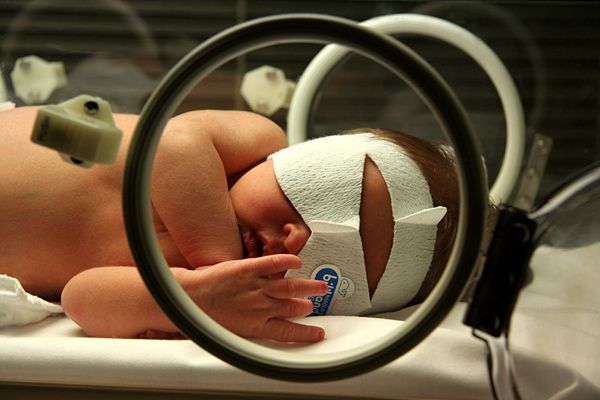
Phototherapy uses narrow-band blue light near 460 to 490 nm to photoisomerise bilirubin in the skin into water-soluble forms excreted in bile and urine without conjugation. Nursing care is specific: expose the maximum skin surface with only a nappy, apply opaque eye shields and check the eyes each shift for discharge or corneal abrasion, keep the prescribed light-to-skin distance, monitor temperature because both hyperthermia and cold stress occur, and increase attention to hydration — insensible losses rise and stools become loose and green. Continue feeding; do not interrupt phototherapy for prolonged periods, and never substitute water. Expect a transient bronze discolouration if the direct fraction is raised.
Exchange transfusion, usually double-volume at about 160 mL/kg through an umbilical venous catheter, is reserved for bilirubin at or above the exchange threshold or for any sign of acute bilirubin encephalopathy. In isoimmune haemolytic disease, intravenous immunoglobulin 0.5 to 1 g/kg may reduce the need for exchange. Acute bilirubin encephalopathy progresses through a recognisable sequence: lethargy, hypotonia and poor feeding first; then a high-pitched cry, irritability, retrocollis and opisthotonos, and fever; then apnoea, seizures, coma and death. The chronic sequel is kernicterus — athetoid cerebral palsy, sensorineural hearing loss, upward gaze palsy and dental enamel dysplasia — and it is permanent, which is why the bilirubin curve is treated as a time-critical value.
Haemolysis, polycythaemia and anaemia
Rh incompatibility arises when an Rh-negative birthing parent carries an Rh-positive fetus and becomes sensitised, usually at a previous birth or bleeding event; maternal IgG anti-D then crosses the placenta and destroys fetal red cells, causing anaemia, hepatosplenomegaly, hydrops in severe cases, and rapid postnatal jaundice. Prevention is Rho(D) immune globulin 300 micrograms intramuscularly at 28 weeks and again within 72 hours of birth of an Rh-positive newborn, with additional dosing guided by a Kleihauer-Betke test after a large fetomaternal bleed. ABO incompatibility — a group O parent with a group A or B newborn — is more common, generally milder, and can occur in a first pregnancy. A positive direct antiglobulin (Coombs) test on the newborn confirms antibody-coated red cells.
Polycythaemia is a central venous haematocrit above 65 per cent, seen after delayed cord clamping, in growth restriction, in infants of parents with diabetes, at altitude and in twin-to-twin transfusion. Hyperviscosity produces a plethoric, ruddy baby with sluggish capillary refill, respiratory distress, hypoglycaemia, hyperbilirubinaemia, poor feeding and, at worst, thrombosis. Treatment of a symptomatic infant is a partial exchange transfusion replacing blood with normal saline. At the other end, anaemia of prematurity reflects low erythropoietin, short red cell survival and phlebotomy losses, and is managed by minimising blood draws, iron supplementation and restrictive transfusion thresholds.
Sepsis: early, late and almost silent
Early-onset sepsis presents in the first 72 hours and is acquired vertically, most often from group B Streptococcus, Escherichia coli or Listeria. Risk factors are maternal group B streptococcal colonisation without adequate intrapartum antibiotic prophylaxis, rupture of membranes for 18 hours or more, intraamniotic infection with maternal fever of 38 °C or higher, and preterm labour. Adequate prophylaxis means penicillin G, or ampicillin, given at least four hours before birth. Late-onset sepsis appears after 72 hours and is usually healthcare-associated or environmental — coagulase-negative staphylococci, Staphylococcus aureus, Gram-negative organisms and Candida — and central lines are the dominant risk.
The newborn does not localise infection and rarely mounts a convincing fever; the classic presentation is that the baby simply is not right. Temperature instability, and more often hypothermia than hyperthermia, poor feeding, lethargy, hypotonia, tachypnoea or apnoea, tachycardia or bradycardia, mottling, delayed capillary refill, hypoglycaemia or hyperglycaemia, jaundice within 24 hours and irritability are the vocabulary. Neonatal meningitis does not produce nuchal rigidity; look for a bulging fontanelle, a high-pitched shrill cry, seizures and opisthotonos. Investigation is blood culture before antibiotics, complete blood count with differential including the immature-to-total neutrophil ratio (0.2 or higher is concerning), C-reactive protein trends and lumbar puncture when meningitis is suspected. Antibiotics are started empirically within the first hour of suspicion and narrowed when cultures return.
Jaundice in the first 24 hours and a newborn who is subtly not right are both emergencies: plot bilirubin against the hour-specific curve, and draw the blood culture and start antibiotics before the baby looks septic.
Section 5Injury, Anomaly, Withdrawal and the Family
Birth injury and hypoxic-ischaemic encephalopathy
Three scalp swellings must be told apart, because one of them is a resuscitation. Caput succedaneum is oedema of the presenting scalp, present at birth, soft, pitting, and it crosses suture lines; it resolves within days. Cephalohaematoma is subperiosteal bleeding that appears hours after birth, is firm, and does not cross suture lines; it resolves over weeks and raises the bilirubin load as it breaks down. Subgaleal haemorrhage is bleeding into the potential space between the aponeurosis and the periosteum, it crosses suture lines and shifts with position, it feels boggy and fluctuant, and it can hold 20 to 40 per cent of the newborn’s blood volume. It is associated with vacuum-assisted birth and it is a haemorrhagic shock emergency: measure occipitofrontal circumference hourly, watch for tachycardia, pallor, falling haematocrit and hypotension, and escalate at once.
A clavicular fracture after shoulder dystocia presents with crepitus, a lump, crying on movement of the arm and an asymmetric Moro reflex; treatment is gentle handling and pinning the sleeve to the shirt. Erb-Duchenne palsy from C5 to C6 stretch produces an arm held adducted and internally rotated with the forearm pronated — the waiter’s tip position — an absent Moro on that side, and an intact grasp reflex, which is the finding that distinguishes it from a lower plexus injury. Facial nerve palsy after forceps shows as asymmetric crying with an eye that will not close on the affected side.
Safeguarding belongs in this section, because birth injury is where nurses learn to read an injury against a mechanism. Every injury must be consistent with both the history given and the child’s developmental stage. A cephalohaematoma after a vacuum-assisted birth is consistent; a bruise, a torn frenulum, a rib or long-bone fracture, a retinal haemorrhage or a burn in an infant who is not yet mobile is not. Use the milestones as the yardstick: an infant rolls at about 4 to 6 months, sits unsupported at about 6 months, crawls at about 8 to 9 months, pulls to stand and cruises at about 9 to 12 months, and walks alone at about 12 to 15 months — so bruising in a pre-mobile infant is a red flag until explained. A history that changes between tellings, an unexplained delay in seeking care, or an injury that outruns what the child can actually do raises the same duty. Mandatory reporting is a legal obligation, not a clinical judgement call. The threshold is reasonable suspicion, not proof, not certainty and not a completed diagnosis; you report to the statutory child-protection authority yourself rather than passing the concern to someone else to file, you inform the treating clinician, you keep the infant safe in the meantime, and you document objectively — what you saw, what you measured, and what you were told, in quotation marks where it was said. Investigating, interrogating the family or deciding whether the concern is founded is not the nurse’s role.
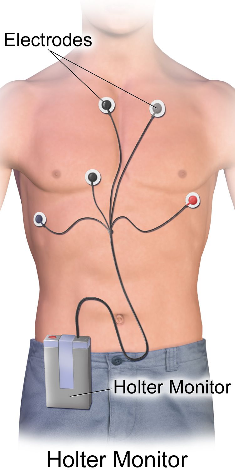
Hypoxic-ischaemic encephalopathy follows a sentinel event such as cord prolapse, abruption or uterine rupture. Candidacy for therapeutic hypothermia typically requires 36 weeks or more of gestation and about 1,800 g or more, plus evidence of perinatal acidosis such as a cord pH of 7.0 or less or a base deficit of 12 mmol/L or more, or an Apgar of 5 or less at 10 minutes or continued resuscitation at 10 minutes, together with moderate or severe encephalopathy on a structured neurological examination. Cooling to a core temperature of 33.5 °C must begin within 6 hours of birth and continues for 72 hours, followed by rewarming at no more than 0.5 °C per hour. Nursing during cooling means expecting sinus bradycardia in the 80 to 100 range as normal, monitoring amplitude-integrated EEG for subclinical seizures, watching for coagulopathy, thrombocytopenia, hypotension and subcutaneous fat necrosis, and above all preventing hyperthermia, which worsens injury.
Congenital anomalies: the first action is the whole answer
Congenital diaphragmatic hernia is the emergency that punishes reflexes. The abdominal contents herniate into the chest, so the baby has a scaphoid abdomen, a barrel chest, absent breath sounds and audible bowel sounds on the affected side, and often mediastinal shift. Do not give bag-mask positive-pressure ventilation: it fills the herniated stomach and bowel with gas and compresses the lung further. Intubate immediately, place a large-bore orogastric tube to low suction, ventilate gently, position with the affected side down, keep the baby nil by mouth, and transfer to a surgical and extracorporeal membrane oxygenation centre.
Oesophageal atresia with tracheo-oesophageal fistula presents with copious frothy oral secretions, choking, coughing and cyanosis with the first feed, and an orogastric tube that will not pass. Keep the baby nil by mouth, place a sump catheter in the upper pouch on continuous low suction, elevate the head of the bed 30 to 45 degrees to reduce reflux of gastric acid through the fistula into the trachea, and arrange surgery. Myelomeningocele demands prone or side-lying positioning to keep pressure off the sac, a sterile non-adherent dressing moistened with warm sterile normal saline, a strictly latex-free environment because of the high rate of latex sensitisation, daily occipitofrontal circumference measurement for hydrocephalus, and surgical closure typically within 24 to 72 hours.
Gastroschisis and omphalocele both need the exposed viscera covered immediately with a sterile saline-moistened dressing and a bowel bag, a nil-by-mouth order, orogastric decompression, side-lying positioning to protect the mesenteric blood supply, and a very high fluid requirement — often two to three times maintenance for gastroschisis because evaporative and third-space losses are extreme. Choanal atresia produces a newborn who is pink while crying and cyanotic at rest, relieved by an oral airway. A ductal-dependent congenital heart lesion deteriorates as the ductus closes over the first hours to days; the treatment is prostaglandin E1 at 0.01 to 0.05 micrograms/kg/min, and the nurse must anticipate its predictable side effects of apnoea, hypotension and fever with intubation equipment at the bedside. Critical congenital heart disease screening by pulse oximetry is performed at 24 hours or later in the right hand and either foot, and a pass requires 95 per cent or higher in either limb with a difference of 3 per cent or less.
The newborn experiencing withdrawal
Neonatal opioid withdrawal, still often called neonatal abstinence syndrome, is a physiological consequence of in-utero exposure, not a moral event, and the family deserves language that says so: a parent with a substance use disorder in treatment, a newborn experiencing withdrawal. Timing depends on the drug half-life — short-acting opioids typically 24 to 48 hours, methadone and buprenorphine 48 to 72 hours and sometimes as late as day five to seven, selective serotonin reuptake inhibitors within one to two days. Signs cluster in four systems: central nervous (high-pitched persistent cry, tremors, hypertonia, irritability, poor sleep, exaggerated Moro, seizures), autonomic (sweating, mottling, nasal stuffiness, frequent sneezing and yawning, low-grade fever), gastrointestinal (uncoordinated suck, regurgitation, loose stools, excoriated buttocks, poor weight gain) and respiratory (tachypnoea).
Care is non-pharmacologic first, and it is nursing care rather than pharmacy care. Room the baby in with the parent, maximise skin-to-skin contact, dim the lights and cluster the cares, swaddle in flexion, offer a pacifier, feed small volumes frequently with a high-calorie formula or human milk, and support chestfeeding when the parent is engaged in treatment and there is no contraindication such as untreated HIV or ongoing illicit polysubstance use. Protect the skin of the buttocks with a barrier ointment and change nappies promptly. The Eat, Sleep, Console approach evaluates function rather than tallying symptoms: can the newborn eat at least an ounce or chestfeed well, sleep undisturbed for at least an hour, and be consoled within ten minutes? If not, optimise the non-pharmacologic care first, then consider medication. Where a scored approach is used, the Finnegan tool commonly triggers treatment at three consecutive scores of 8 or more, or two scores of 12 or more. When medication is needed, oral morphine at approximately 0.04 to 0.05 mg/kg per dose every three to four hours is typical, weaned by protocol, with phenobarbital or clonidine as adjuncts.
Priorities, delegation and going home
A newborn compensates with tachycardia and vasoconstriction and keeps the blood pressure normal until very late, so hypotension is a pre-arrest finding, not an early one, and the deterioration you are actually meant to catch is the compensated one — rising heart rate, rising respiratory rate, a narrowing pulse pressure, capillary refill over 2 seconds, cool mottled peripheries and any change in level of consciousness. Read the newborn’s own ranges rather than an adult range: a term newborn breathes 30 to 60 times a minute, runs a heart rate of roughly 100 to 205 awake, and is hypotensive below a systolic of 60 mmHg. Priority then follows airway, breathing and circulation, and the acute problem outranks the chronic one: the preterm infant who has just become apnoeic is assessed before the stable infant whose phototherapy is overdue, and the newborn with grunting and retractions is assessed before the newborn with a rising but sub-threshold bilirubin.
Delegation in the neonatal unit follows the same rule as everywhere else, and the high-risk newborn stretches it hard. The registered nurse retains assessment, teaching, evaluation and any unstable patient. That means the admission and shift assessments, interpretation of a bilirubin or glucose result, the decision to begin or stop a feed, phototherapy assessment, discharge teaching about safe sleep and car seats, the first feed of an at-risk newborn, and every aspect of the care of an infant on respiratory support, receiving therapeutic hypothermia or being resuscitated. Assistive personnel may weigh and measure, bathe a stable newborn, change nappies and record output, stock the bed space, transport a stable infant with a nurse, and hold or bottle-feed a stable term newborn after the nurse has assessed and specified the plan. Licensed practical nurse scope varies by jurisdiction and by employer, and unit policy governs.
Discharge from a neonatal unit is a teaching event, not a paperwork event. Confirm the newborn metabolic screen was collected at 24 to 48 hours and repeated when required, the hearing screen was performed, critical congenital heart disease screening passed, and immunisations are given by chronological age — a preterm infant is vaccinated on the calendar, not on corrected age — while developmental milestones are judged by corrected age until about two years. The routine schedule is hepatitis B at birth, then diphtheria-tetanus-acellular pertussis, inactivated polio, Haemophilus influenzae type b, pneumococcal conjugate and rotavirus vaccines beginning at 2 months and repeated at 4 and 6 months according to the product used, with measles-mumps-rubella and varicella at 12 to 15 months. Doses are never reduced or split for a small infant. The single accepted exception to the calendar is the birth dose of hepatitis B vaccine in an infant under 2,000 g whose birthing parent is hepatitis B surface antigen negative, which is deferred to 1 month of age or to discharge; if that status is positive or unknown, both the vaccine and hepatitis B immune globulin are given within 12 hours of birth whatever the weight. Preterm infants under 37 weeks need a car seat tolerance screen of 90 to 120 minutes before discharge. Teach safe sleep without exception: supine, alone, on a firm flat surface, no soft bedding, room-sharing without bed-sharing, and no smoking. Arrange follow-up for bilirubin and weight within 24 to 48 hours, and ensure the family knows exactly who to call, for what, and at what number.
Finally, the family is the patient too. Support parental presence and unrestricted access, teach kangaroo care and let parents provide it as care rather than as a reward, involve them in cares and in decision-making, and protect lactation with early and frequent expression when the baby cannot yet feed. Name the baby, not the diagnosis or the bed number. When an infant is dying or has a life-limiting condition, palliative care is offered alongside, not instead of, active treatment: symptom control, memory-making, sibling inclusion, cultural and spiritual practices honoured, and bereavement follow-up planned before the family leaves.
For every high-risk newborn there is one correct first action, and it is usually the one that overrides instinct — intubate rather than bag, cool rather than warm, console rather than medicate, and always assess the family alongside the infant.
The chapter in one breath
A newborn becomes high risk through gestational age, growth, the events of the birth, or the physiology of the first hours — and the late preterm infant, who looks well, is the one most often underestimated. Resuscitation is a timed sequence: initial steps, then positive-pressure ventilation within the Golden Minute if the newborn is apnoeic or the heart rate is under 100, MR SOPA when the heart rate will not rise, 3:1 compressions when it stays under 60 despite chest-moving ventilation, and epinephrine 0.02 mg/kg intravenously only after that. Saturation targets climb minute by minute from 60–65 per cent at one minute to 85–95 per cent at ten. Warmth protects glucose, glucose protects the brain, and every volume and dose is per kilogram, written out and checked against the adult maximum — with 4–2–1 for maintenance beyond the newborn period and mL/kg/day within it. Jaundice before 24 hours, a bilirubin climbing faster than the hour-specific curve, and a baby who is simply not right are all emergencies. Congenital anomalies each have one counterintuitive first action. Hypotension in a newborn is a pre-arrest sign rather than an early one, and an injury that does not fit the history or the child’s developmental stage is reported on reasonable suspicion, not on proof. Throughout, the language is family-centred, the escalation is SBAR, and assessment, teaching, evaluation and the unstable patient stay with the registered nurse.
Words worth knowing
| Term | What it means |
|---|---|
| Late preterm | Birth at 34 0/7 to 36 6/7 weeks; physiologically immature despite a term-like appearance. |
| Small for gestational age | Birth weight below the 10th percentile for gestational age; a size statement, not a diagnosis. |
| Intrauterine growth restriction | Failure of a fetus to achieve its own growth potential; symmetric (early, intrinsic) or asymmetric (late, placental). |
| Golden Minute | The first 60 seconds of life, within which initial steps are completed and positive-pressure ventilation begun if needed. |
| MR SOPA | Corrective ventilation steps: Mask, Reposition, Suction, Open mouth, Pressure increase, Alternative airway. |
| Surfactant | Phospholipid-protein complex that lowers alveolar surface tension; deficient in respiratory distress syndrome. |
| Persistent pulmonary hypertension | Failure of pulmonary vascular resistance to fall, causing right-to-left shunting and a preductal-postductal saturation gap over 10 per cent. |
| Bronchopulmonary dysplasia | Chronic lung disease defined by continued oxygen need at 36 weeks postmenstrual age. |
| Non-shivering thermogenesis | Heat production by metabolism of brown adipose tissue; the newborn’s only means of generating heat. |
| Neutral thermal environment | The ambient conditions in which oxygen consumption and metabolic demand are minimal, keeping axillary temperature 36.5–37.5 °C. |
| Glucose infusion rate | Milligrams of glucose per kilogram per minute; (dextrose per cent × mL/hr) ÷ (kg × 6). |
| 4–2–1 rule | Hourly maintenance fluid: 4 mL/kg for the first 10 kg, 2 mL/kg for the next 10 kg, 1 mL/kg thereafter; not for the first days of life. |
| Necrotising enterocolitis | Ischaemic-inflammatory bowel injury of the preterm infant; pneumatosis intestinalis on radiograph, treated with nil by mouth, decompression and antibiotics. |
| Kernicterus | Permanent bilirubin-induced neurological damage: athetoid cerebral palsy, sensorineural hearing loss, upward gaze palsy, enamel dysplasia. |
| Direct antiglobulin test | Coombs test detecting antibody bound to newborn red cells; positive in Rh and ABO isoimmune haemolysis. |
| Subgaleal haemorrhage | Bleeding beneath the scalp aponeurosis that crosses sutures and can hold 20–40 per cent of blood volume; a shock emergency. |
| Therapeutic hypothermia | Cooling to 33.5 °C within 6 hours of birth for 72 hours in moderate to severe hypoxic-ischaemic encephalopathy at 36 weeks or more. |
| Eat, Sleep, Console | Function-based assessment of the newborn experiencing withdrawal; non-pharmacologic care is optimised before medication is considered. |
| Corrected age | Chronological age minus the weeks of prematurity; used to judge development to about two years, but never to delay immunisations. |