Cardiac Disorders: Coronary Artery Disease and Heart Failure
Two problems account for an enormous share of the patients you will care for on a medical floor, and they are the same disease at two different stages. Coronary artery disease starves myocardium of oxygen; when a coronary artery closes, muscle dies. Dead muscle does not contract, so the pump weakens, pressure backs up behind it, fluid moves into the lungs and the legs, and the patient returns months later with heart failure. Understanding that continuity means you never have to memorise two unrelated lists. In this chapter you build the mechanism first — supply versus demand, plaque rupture, remodelling, neurohormonal compensation — and then layer on the assessment findings, the diagnostics with real numbers, the drug classes by what they actually do, and the priority reasoning that decides what you handle first when four patients need you at once. Chest pain and shortness of breath are the two complaints that will define your shift. Learn to think through both.
By the end of this chapter you can
- Explain myocardial oxygen supply and demand, the formation and rupture of an atherosclerotic plaque, and how ischaemia, injury and infarction produce their distinct findings.
- Differentiate stable angina, unstable angina, NSTEMI, STEMI and vasospastic angina by presentation, 12-lead ECG and troponin, using correct troponin release timing.
- Interpret cardiac diagnostics with stated values — high-sensitivity troponin, CK-MB, BNP, lipid panel, ejection fraction — and localise an infarct to a coronary territory from the leads involved.
- Prioritise the first ten minutes of an acute coronary syndrome, critique the MONA mnemonic, and state the contraindications to nitrates and the correct indication for oxygen.
- Compare reperfusion strategies — PCI and fibrinolysis — against the door-to-balloon and door-to-needle clocks, and deliver safe post-catheterisation nursing care.
- Describe the pathophysiology of HFrEF and HFpEF, distinguish left-sided from right-sided failure, and apply NYHA class and ACC/AHA stage.
- Apply guideline-directed medical therapy for heart failure by class, with the monitoring parameters that matter, including potassium, renal function and digoxin levels.
- Teach daily weights, the 2 to 3 pound per day or 5 pound per week rule, sodium and fluid limits, and the discharge priorities that prevent readmission.
- Recognise cardiac red flags, escalate using SBAR, defend delegation decisions to LPN and UAP, and anticipate the atypical presentation of ischaemia and failure in the older adult.
Section 1Coronary Perfusion, Plaque and the Anginas
Supply, demand and the arithmetic of ischaemia
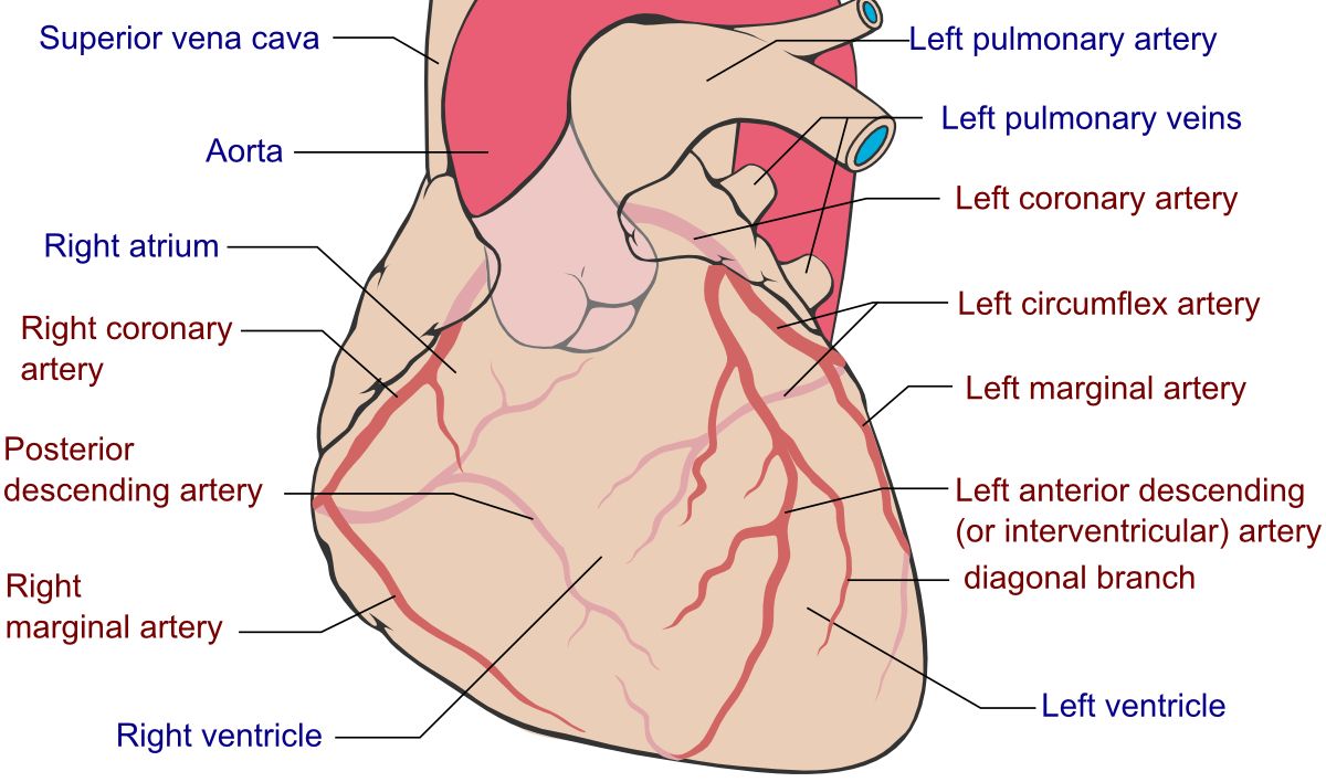
The myocardium is the most oxygen-greedy tissue in the body and it has almost no reserve. Most tissues extract roughly a quarter of the oxygen delivered to them and can extract more when demand rises. Cardiac muscle already extracts about seventy-five percent at rest. That single fact drives the whole chapter: when the heart needs more oxygen, the only way to get it is to increase coronary blood flow. If the artery cannot dilate — because a plaque holds the lumen fixed — demand outruns supply and the tissue becomes ischaemic.
Supply depends on three things. First, the diameter of the coronary artery, which is where plaque does its damage. Second, the oxygen-carrying capacity of the blood, which is why a haemoglobin of 7 g/dL (normal roughly 12 to 16 g/dL in women and 14 to 18 g/dL in men) can precipitate angina in a patient whose arteries have not changed at all. Third, diastolic time. The coronaries fill during diastole, when the aortic valve has closed and the myocardium is relaxed. Tachycardia shortens diastole disproportionately, so a heart rate of 150 both raises demand and cuts supply at the same moment. This is the single most useful piece of physiology in cardiac nursing, and it explains why rate control is therapeutic, why beta-blockers reduce angina, and why the patient in atrial fibrillation with a rapid ventricular response develops chest pain.
Demand rises with heart rate, contractility, and wall tension (preload and afterload). Anything that increases those — exertion, cold, a heavy meal, emotional stress, fever, hyperthyroidism, hypertension, cocaine — can tip a fixed lesion into symptoms. Note that a plaque generally has to obstruct about seventy percent of the lumen before it limits flow during exertion, and about ninety percent before it limits flow at rest. That is why patients feel entirely well for decades while the disease builds.
Anatomy matters for prediction. The left anterior descending artery feeds the anterior wall, the anterior two-thirds of the septum and much of the left ventricle — occlusion here is the “widow-maker” and the infarct most likely to cause heart failure and cardiogenic shock. The left circumflex feeds the lateral wall. The right coronary artery feeds the inferior wall, the right ventricle and, in most people, the SA and AV nodes — which is why inferior infarcts come with bradycardia and heart block. Since the coronaries perfuse from the epicardial surface inward, the innermost layer, the subendocardium, is the watershed and dies first. Partial-thickness subendocardial damage produces ST depression; full-thickness transmural damage produces ST elevation.
Building a plaque and the risk factors that actually change outcomes
Atherosclerosis begins with endothelial injury from shear stress at branch points, hypertension, tobacco toxins, hyperglycaemia or oxidised LDL. The damaged endothelium becomes sticky and permeable. LDL particles slip into the intima and oxidise. Monocytes follow, transform into macrophages, gorge on the oxidised lipid and become foam cells, which pile up as a fatty streak. Smooth muscle migrates in and lays down a fibrous cap over a soft, inflammatory, lipid-rich core. The plaque is now a two-part structure, and how it behaves depends on which part predominates.
A stable plaque has a thick fibrous cap and a small lipid core. It narrows the lumen slowly and produces predictable exertional angina. A vulnerable plaque has a thin cap over a large, inflamed lipid core — and here is the point students miss: vulnerable plaques are often not the tightest lesions. Roughly two-thirds of infarctions arise from plaques that were obstructing less than fifty percent of the lumen. When the thin cap erodes or tears, the thrombogenic core is exposed to flowing blood. Platelets adhere, activate and aggregate, the coagulation cascade fires, and a thrombus forms in minutes. Partial occlusion produces unstable angina or NSTEMI; complete, sustained occlusion produces STEMI. That is the whole mechanism of acute coronary syndrome in one paragraph, and every drug you will give — aspirin, P2Y12 inhibitors, heparin, fibrinolytics — targets one step in it.
Non-modifiable risk factors are age (men 45 and older, women 55 and older or post-menopausal), male sex until menopause equalises the risk, family history of premature coronary disease (a first-degree male relative under 55 or female relative under 65), and ethnicity. The modifiable ones are where nursing lives: tobacco use, hypertension, diabetes (a coronary risk equivalent), dyslipidaemia, obesity with central adiposity, physical inactivity, an atherogenic diet, excess alcohol, chronic kidney disease and untreated obstructive sleep apnoea. Metabolic syndrome — three or more of waist circumference above 40 inches in men or 35 inches in women, triglycerides 150 mg/dL or higher, HDL below 40 mg/dL in men or 50 mg/dL in women, blood pressure 130/85 mmHg or higher, and fasting glucose 100 mg/dL or higher — bundles several of them.
Know the lipid targets, because you will teach them. A standard fasting panel reports total cholesterol (desirable below 200 mg/dL), LDL (optimal below 100 mg/dL, and below 70 mg/dL for patients with established disease or diabetes), HDL (protective at 60 mg/dL or higher, a risk factor below 40) and triglycerides (normal below 150 mg/dL). Modern practice puts less weight on chasing a single LDL number and more on statin intensity matched to risk, but the numbers still anchor patient teaching. Smoking cessation is the single highest-yield intervention you can deliver: risk of a coronary event begins falling within a year of quitting.
Pearl. Do not reassure a patient because a stress test or a catheterisation last year showed “only thirty percent blockage.” Degree of stenosis predicts angina; cap stability predicts infarction. The lesion that kills is frequently the one that was never flow-limiting. Treat new or changing chest pain on its own merits regardless of prior imaging.
Stable angina — and the two anginas that are not stable
Stable angina is the symptom of a fixed lesion under load. It is predictable: the same amount of exertion produces the same substernal pressure, tightness or squeezing, lasting three to five minutes, relieved by rest or by sublingual nitroglycerin. Patients often describe it with a fist over the sternum rather than a fingertip. It may radiate to the left arm, both arms, the jaw, the neck, the epigastrium or between the shoulder blades. There is no myocardial necrosis, so troponin stays normal and the resting ECG is frequently normal — ST depression appears only during the episode or during stress testing.
Unstable angina is the same pain that has broken its pattern: new onset, occurring at rest, lasting longer, occurring with less exertion than before, or no longer fully relieved by rest or nitroglycerin. Mechanistically this is plaque rupture with a partially occluding thrombus. It is an acute coronary syndrome, it is admitted, and it carries a real short-term risk of progressing to infarction. Troponin is negative — and that negative troponin is precisely what distinguishes unstable angina from NSTEMI, not the severity of the pain.
Vasospastic (Prinzmetal or variant) angina is a different animal. It results from coronary spasm rather than fixed obstruction, occurs at rest and characteristically between midnight and early morning, may occur in relatively young patients without classic risk factors, and produces transient ST elevation that resolves when the spasm breaks. Calcium channel blockers and nitrates are the treatment; non-selective beta-blockers can worsen it by leaving alpha-mediated vasoconstriction unopposed. Cocaine and methamphetamine cause the same picture in a much more dangerous form, and in cocaine-associated chest pain benzodiazepines and nitrates come before beta-blockers.
Chronic stable angina is managed with the goal of shifting the supply-demand balance. Nitrates dilate veins to cut preload and dilate coronaries. Beta-blockers lower heart rate, contractility and blood pressure, and lengthen diastolic filling time. Calcium channel blockers relax vascular smooth muscle and, in the case of the non-dihydropyridines diltiazem and verapamil, also slow the rate. Ranolazine is added for refractory angina without meaningful effect on heart rate or blood pressure. Aspirin and a high-intensity statin address plaque stability rather than symptoms. Cardiac rehabilitation, weight management, glycaemic control and blood pressure control complete the plan.
| Type | Trigger and pattern | ECG during pain | Troponin | Priority |
|---|---|---|---|---|
| Stable angina | Predictable exertion, relieved by rest or nitroglycerin within 5 min | Often normal; may show ST depression | Normal | Outpatient optimisation |
| Unstable angina | New, at rest, longer, or worse with less effort | ST depression or T-wave inversion, or normal | Normal | Admit, ACS pathway |
| NSTEMI | Rest pain, often prolonged | ST depression, T-wave inversion; no ST elevation | Elevated | Admit, antithrombotics, angiography usually within 24–72 h |
| STEMI | Sudden, severe, unrelieved, with diaphoresis and dread | ST elevation in 2 contiguous leads, or new LBBB pattern | Elevated (do not wait for it) | Immediate reperfusion |
| Vasospastic angina | At rest, often 12 a.m. to early morning, cyclical | Transient ST elevation that resolves | Usually normal | Calcium channel blocker plus nitrate |
Myocardium extracts nearly all the oxygen it is offered at rest, so ischaemia is always a flow problem — and because coronaries fill in diastole, anything that shortens diastole raises demand and cuts supply in the same beat.
Section 2Acute Coronary Syndrome: Reading the Presentation
One ruptured plaque, three diagnoses
Acute coronary syndrome is an umbrella covering unstable angina, non-ST-elevation myocardial infarction and ST-elevation myocardial infarction. All three begin with the same event — plaque disruption and thrombosis — and differ only by how completely and for how long the artery is occluded. Think of it as a spectrum of one disease, because the initial nursing response is identical no matter which of the three it turns out to be.
The classic presentation is crushing substernal chest pressure, often described as an elephant on the chest, lasting more than twenty minutes, unrelieved by rest or nitroglycerin, accompanied by diaphoresis, nausea, dyspnoea, weakness and a sense of impending doom. Radiation to the left arm, jaw, neck, back or epigastrium is common. Skin is often cool, ashen and clammy because sympathetic outflow shunts blood centrally. Blood pressure may be elevated from catecholamines or low if the pump is failing.
Now unlearn the assumption that this is universal. Up to a third of infarcts present without chest pain. Women more often report fatigue that preceded the event by days or weeks, dyspnoea, indigestion, nausea, anxiety, and pain between the shoulder blades or in the jaw. Patients with diabetes may have autonomic neuropathy that blunts the pain signal entirely — the silent MI. Older adults commonly present with dyspnoea, sudden confusion or delirium, syncope, a fall, weakness or new-onset heart failure rather than with pain, and delirium in an older adult should always raise the possibility of an acute cardiac or infectious event. Do not require chest pain before you obtain an ECG.
One further distinction shapes treatment. Type 1 MI is the plaque-rupture event described above and it is what reperfusion therapy is designed for. Type 2 MI is myocardial injury from supply-demand mismatch without plaque rupture — severe anaemia, sepsis, sustained tachydysrhythmia, hypotension, hypertensive crisis, respiratory failure. Troponin rises in both. A patient in septic shock with a troponin of 250 ng/L usually needs the sepsis treated, not the catheterisation lab. Knowing that the number has more than one explanation keeps you from anchoring.
The 12-lead ECG: territory, timing and the leads you forget
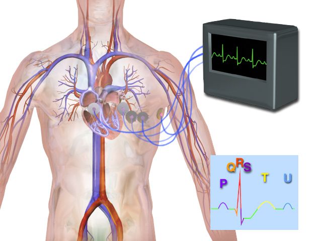
A 12-lead ECG must be obtained and interpreted within ten minutes of a patient reporting ischaemic symptoms — in the emergency department, on your floor, anywhere. That ten-minute standard is a national quality measure, and on the NCLEX “obtain a 12-lead ECG” is nearly always among the first actions in a chest-pain stem, alongside aspirin and continuous cardiac monitoring.
Read the tracing in three layers. Ischaemia shows as ST depression or symmetric T-wave inversion and is reversible. Injury shows as ST elevation and represents tissue that is dying but salvageable — this is the finding that starts the clock. Infarction shows as a pathologic Q wave, which develops over hours to days and is permanent, the electrical scar. Diagnostic ST elevation is measured at the J point in two anatomically contiguous leads: at least 1 mm (0.1 mV) in all leads except V2 and V3, where the threshold is 2 mm in men aged 40 and older, 2.5 mm in men under 40, and 1.5 mm in women. A new left bundle branch block with an ischaemic presentation is managed as a STEMI equivalent.
Localisation lets you predict the complications before they happen, which is what separates a nurse who documents from a nurse who anticipates. Two lead groups deserve special attention. First, posterior infarction produces no ST elevation on a standard 12-lead; it shows as tall R waves and ST depression in V1 and V2 — a mirror image — and requires posterior leads V7 to V9 to confirm. Second, and more important at the bedside, whenever you see an inferior STEMI in II, III and aVF, obtain a right-sided ECG with V4R, because up to half of these involve the right ventricle.
| Territory | Leads with changes | Usual artery | Anticipate |
|---|---|---|---|
| Septal | V1, V2 | LAD (septal branches) | Bundle branch block, septal rupture |
| Anterior | V3, V4 | LAD | Large infarct, pump failure, cardiogenic shock |
| Lateral | I, aVL, V5, V6 | Left circumflex or diagonal | Often combined with anterior |
| Inferior | II, III, aVF | Right coronary (usually) | Bradycardia, AV block, RV involvement |
| Right ventricular | V4R (right-sided leads) | Proximal right coronary | Preload dependence — hypotension with nitrates |
| Posterior | Tall R plus ST depression V1–V2; confirm V7–V9 | Circumflex or right coronary | Easily missed entirely |
Pearl. Inferior MI with right ventricular involvement is the classic trap. The failing right ventricle depends on preload to push blood through the lungs. Give nitroglycerin or morphine — both venodilators — and the preload drops out from under it, producing sudden, profound hypotension. The management is the opposite of what you reflexively expect: hold the nitrate and give a fluid bolus. If the patient with an inferior MI becomes hypotensive after sublingual nitroglycerin, stop the nitrate, lay them flat, raise the legs, open the intravenous fluids and call the provider.
Troponin, the other markers, and the rest of the workup
Cardiac troponin I and T are structural proteins of the contractile apparatus and they are the diagnostic standard because they are essentially specific to cardiac muscle. Timing is what students get wrong, so learn it precisely. With conventional assays troponin begins to rise about three to six hours after the onset of injury, peaks at twelve to twenty-four hours, and remains detectable for seven to fourteen days. That long tail makes troponin excellent for diagnosing an event the patient sat on at home for two days, and poor for detecting a re-infarction a few days later. High-sensitivity troponin assays detect a rise within one to three hours and are used in serial 0-hour and 1-hour, or 0-hour and 2-hour, rule-out protocols. Cut-points are assay-specific and often sex-specific — the 99th-percentile upper reference limit for high-sensitivity troponin T is commonly around 14 ng/L, with separate thresholds for men and women. Always use your own laboratory’s reference range, and interpret the delta — the change between draws — not a single value.
Two consequences follow. First, a normal troponin drawn one hour into chest pain does not rule out infarction; serial sampling is mandatory, and a patient who is discharged on a single negative troponin has been failed. Second, you never wait for a troponin result to activate reperfusion in a STEMI. The ECG, not the laboratory, triggers the catheterisation lab. CK-MB retains a narrow role: it rises in four to six hours, peaks at eighteen to twenty-four hours and normalises within forty-eight to seventy-two hours, so a second rise after it has normalised is useful evidence of re-infarction. Myoglobin rises earliest but is so non-specific that it has largely been abandoned.
The rest of the workup rounds out the picture. Order a CBC (haemoglobin, because anaemia worsens ischaemia), a basic metabolic panel with potassium 3.5 to 5.0 mEq/L and magnesium 1.5 to 2.5 mg/dL because both derangements provoke ventricular dysrhythmias, creatinine 0.6 to 1.2 mg/dL before contrast exposure, coagulation studies before anticoagulation, a lipid panel within twenty-four hours, a glucose and A1C, and a BNP if failure is suspected. Echocardiography quantifies the ejection fraction (normal 55 to 70 percent) and shows regional wall-motion abnormalities that correspond to the infarcted territory, plus complications such as acute mitral regurgitation or a pericardial effusion.
For patients who are not in an acute syndrome, non-invasive testing sorts out chest pain. Exercise stress testing is preferred when the patient can walk; pharmacologic stress with adenosine, regadenoson or dipyridamole is substituted when they cannot. Nursing preparation matters and is frequently tested: nothing by mouth for four to six hours, no caffeine for twelve to twenty-four hours (caffeine blocks adenosine and invalidates the test), hold beta-blockers if instructed, wear walking shoes, and hold theophylline. Adenosine-class agents are contraindicated in bronchospastic disease because they can provoke severe bronchoconstriction — dobutamine stress is used instead. Coronary CT angiography and coronary calcium scoring are increasingly used for risk stratification, and cardiac catheterisation with angiography remains the definitive anatomic test.
The ECG within ten minutes decides whether the patient goes to the catheterisation lab now; troponin confirms and quantifies afterward, so a single early negative troponin rules nothing out and a positive troponin is never the reason to delay reperfusion in a STEMI.
Section 3Managing ACS: The First Ten Minutes and the Next Ten Days
The immediate bundle, and what is wrong with MONA
When a patient reports chest pain, your actions overlap rather than queue. Stay with the patient and call for help. Place them in semi-Fowler position, obtain a full set of vital signs including bilateral blood pressures, apply continuous cardiac monitoring with defibrillator available, obtain the 12-lead ECG within ten minutes, establish two large-bore intravenous lines, draw troponin and the baseline labs, and give aspirin 162 to 325 mg chewed (non-enteric-coated, chewed so it is absorbed in minutes) unless there is a true allergy or active bleeding. Then treat pain and arrange reperfusion.
You were probably taught MONA — morphine, oxygen, nitroglycerin, aspirin. It is a useful memory hook and a poor treatment order, and current practice has revised nearly every letter. Learn the critiques, because they are now tested directly.
Aspirin comes first, not last. It is the intervention with the clearest mortality benefit and it takes seconds to give. Oxygen is not routine. Supplemental oxygen in a normoxic patient causes coronary vasoconstriction and confers no benefit; large trials showed no reduction in mortality or infarct size. Give oxygen only if the SpO2 is below 90 percent (some protocols use 94 percent) or the patient is dyspnoeic or in respiratory distress, and titrate to that target rather than running high flow reflexively.
Nitroglycerin is given as 0.4 mg sublingually, repeated every five minutes for up to three doses, with blood pressure checked before each dose; hold it and reassess for a systolic pressure below 90 mmHg or a drop of more than 30 mmHg from baseline, for heart rate below 50 or above 100, and for suspected right ventricular infarction. The absolute contraindication you must never miss is recent phosphodiesterase-5 inhibitor use: sildenafil or vardenafil within 24 hours, tadalafil within 48 hours. The combination causes refractory, potentially fatal hypotension. Ask every patient of any age and any sex, privately and without embarrassment, and remember that these drugs are also prescribed for pulmonary hypertension. Teach outpatients to take one tablet at the first sign of pain, sit or lie down first because of the hypotension and headache, and call emergency services if the pain is unimproved five minutes after the first dose rather than finishing all three at home. Tablets are stored in the original dark glass bottle away from heat and light and replaced every six months once opened.
Morphine has moved from routine to selective. It relieves pain, reduces sympathetic drive and venodilates, which lowers preload — helpful in pulmonary oedema, harmful in right ventricular infarction. It also slows gastric emptying and measurably delays absorption of oral P2Y12 inhibitors, and observational data raised concern about outcomes in NSTEMI. Reserve it for pain that persists despite nitrates and anti-ischaemic therapy, give 2 to 4 mg intravenously, and monitor blood pressure, respiratory rate and level of consciousness. Never treat ongoing ischaemic pain with analgesia alone: pain that continues means muscle that is still dying, and the answer is reperfusion.
Reperfusion: PCI, fibrinolysis and the clock
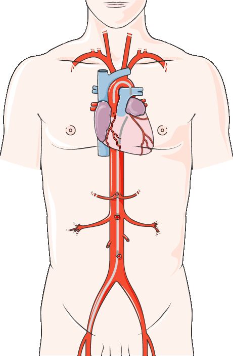
Time is muscle. Necrosis begins within twenty to thirty minutes of complete occlusion and is largely complete by six hours, so every minute of delay is myocardium that will not come back. Primary percutaneous coronary intervention is the preferred strategy: a catheter is advanced from the radial or femoral artery, the thrombosed segment is crossed, a balloon expands the lesion and a stent holds it open. The benchmark is first medical contact to device within 90 minutes at a PCI-capable facility, or within 120 minutes when the patient must be transferred.
If PCI cannot be delivered within 120 minutes, fibrinolytic therapy is given, with a door-to-needle time of 30 minutes and greatest benefit within the first twelve hours of symptom onset. Alteplase, reteplase and tenecteplase convert plasminogen to plasmin and dissolve the clot. Screening is a nursing responsibility. Absolute contraindications include any prior intracranial haemorrhage, known cerebral vascular malformation or intracranial malignancy, ischaemic stroke within three months, active bleeding or a bleeding diathesis, suspected aortic dissection, and significant closed head or facial trauma within three months. Relative contraindications include uncontrolled hypertension above 180/110 mmHg, recent major surgery or internal bleeding, pregnancy, current anticoagulant use and prolonged CPR. After administration, monitor neurologic status closely, avoid unnecessary punctures and invasive lines, hold pressure longer at any site, watch for signs of reperfusion (relief of pain, resolution of ST elevation, and reperfusion dysrhythmias such as accelerated idioventricular rhythm, which is usually transient and benign), and observe for bleeding — a sudden change in level of consciousness or a new headache is intracranial haemorrhage until proven otherwise.
Post-catheterisation nursing is high-yield and reliably tested. Assess the access site and the distal pulses, colour, temperature, capillary refill and sensation of the affected extremity on the standard schedule — typically every fifteen minutes for the first hour, then tapering. With femoral access keep the leg straight, maintain bed rest with the head of the bed no higher than about 30 degrees, and do not let the patient flex the hip. With radial access a compression band is used, the wrist is kept straight, and the patient may sit up much sooner. Watch for haematoma, and for the complication students forget: retroperitoneal bleeding, which presents as flank or back pain, hypotension, tachycardia and a falling haematocrit with an unremarkable-looking groin. Contrast is nephrotoxic, so hydrate, monitor urine output and creatinine, and follow institutional policy on holding metformin for 48 hours after contrast in patients with renal impairment because of lactic acidosis risk. Report chest pain after PCI immediately — it may signal acute stent thrombosis.
| Class | Examples | Why it is given | Nursing monitoring |
|---|---|---|---|
| Antiplatelet — COX inhibitor | Aspirin 162–325 mg chewed, then 81 mg daily | Blocks thromboxane A2, prevents platelet aggregation | Bleeding, GI upset, tinnitus; take with food; lifelong |
| Antiplatelet — P2Y12 inhibitor | Clopidogrel, ticagrelor, prasugrel | Blocks ADP-mediated aggregation; mandatory after stenting | Bleeding; hold before surgery per protocol; prasugrel contraindicated with prior stroke or TIA; ticagrelor causes dyspnoea |
| Anticoagulant | Unfractionated heparin, enoxaparin, bivalirudin, fondaparinux | Stops clot propagation | UFH: aPTT 1.5–2.5 times control; platelets for HIT; enoxaparin 1 mg/kg subcutaneous twice daily, do not expel the air bubble, do not aspirate or massage |
| Nitrate | Sublingual, topical or intravenous nitroglycerin | Venodilation lowers preload; dilates coronaries | Blood pressure before every dose; headache; hold with PDE5 inhibitor use or RV infarct; IV titration is an RN function |
| Beta-blocker | Metoprolol, carvedilol, bisoprolol | Lowers rate, contractility, demand; reduces reinfarction and dysrhythmia | Hold for HR below 60 or SBP below 100; contraindicated in acute decompensated failure, cardiogenic shock, high-grade block, active bronchospasm; masks hypoglycaemia except sweating; never stop abruptly |
| ACE inhibitor / ARB | Lisinopril, enalapril; losartan, valsartan | Limits ventricular remodelling; start within 24 h especially with anterior MI or EF under 40 percent | Potassium, creatinine, blood pressure; dry cough; angioedema; contraindicated in pregnancy |
| High-intensity statin | Atorvastatin 40–80 mg, rosuvastatin 20–40 mg | Stabilises plaque beyond LDL lowering; start in hospital for everyone | Baseline and symptom-driven liver enzymes; report unexplained muscle pain, weakness or dark urine (rhabdomyolysis); avoid grapefruit juice with the CYP3A4-metabolised statins — atorvastatin, simvastatin and lovastatin — while rosuvastatin and pravastatin are not affected |
| Glycoprotein IIb/IIIa inhibitor | Eptifibatide, tirofiban, abciximab | Final common pathway of aggregation; used selectively around PCI | Profound bleeding risk; monitor platelets and puncture sites |
Pearl. Dual antiplatelet therapy after a drug-eluting stent is not optional and it is not a refill the patient can skip until payday. Stopping a P2Y12 inhibitor early is a leading cause of acute stent thrombosis, which carries mortality far higher than the original infarction. Before discharge, confirm in words that the patient has the prescription, can afford it, and understands to call the cardiologist — not to simply stop — if any dentist or surgeon asks them to hold it.
Complications, priorities and communicating the deterioration
Complications cluster by time, and knowing the timeline lets you anticipate. In the first hours, dysrhythmias dominate and ventricular fibrillation is the leading cause of pre-hospital death — which is why continuous monitoring, patent intravenous access and an available defibrillator are non-negotiable. Inferior infarcts bring bradycardia and AV block. Within the first day to week, pump failure appears: acute heart failure, pulmonary oedema, and cardiogenic shock when more than roughly forty percent of the left ventricle is lost, presenting as hypotension, cool mottled skin, oliguria, altered mentation and a rising lactate.
Mechanical complications typically appear between day two and day seven, as necrotic tissue softens before scar forms. Papillary muscle rupture produces sudden acute mitral regurgitation — a new loud holosystolic murmur at the apex with abrupt flash pulmonary oedema. It follows an inferior infarct most often, because the posteromedial papillary muscle has only one blood supply, from the posterior descending branch of the right coronary artery. Ventricular septal rupture produces a new harsh murmur with a thrill and rapid decompensation. Free wall rupture produces sudden pulseless electrical activity from tamponade and is usually fatal. Any new murmur in a post-MI patient is an emergency, not a charting detail. Later still come ventricular aneurysm, mural thrombus with embolic stroke, and Dressler syndrome — an autoimmune pericarditis one to eight weeks out with pleuritic pain that improves sitting forward, fever, a friction rub and diffuse ST elevation, treated with anti-inflammatories rather than reperfusion.
Priority reasoning is what the NCLEX rewards. Apply ABC first: airway and breathing problems and lethal rhythms outrank everything. Then unstable over stable, acute over chronic, actual over potential. Given four post-MI patients, you see the one with new dyspnoea and crackles before the one whose pain is relieved and vital signs are stable, and you see the patient with a run of ventricular tachycardia before either. Red flags that mean call now: chest pain unrelieved after three sublingual nitroglycerin doses; new or worsening ST elevation; systolic pressure below 90 mmHg with signs of poor perfusion; heart rate below 50 or a new high-grade block; sustained ventricular tachycardia or any new ventricular ectopy with instability; a new murmur; sudden severe dyspnoea with pink frothy sputum; altered mentation; and signs of bleeding after fibrinolysis or PCI.
Escalate using SBAR, and rehearse it until it is automatic. Situation: “Dr. Reyes, this is Jordan Park, RN, on 4 West. Mr. Alvarez in 412, day one after an inferior STEMI, has new chest pain rated 8 out of 10 that has not responded to three sublingual nitroglycerin.” Background: “He had a right coronary stent yesterday and is on aspirin, ticagrelor and a heparin infusion.” Assessment: “Blood pressure is 88 over 54, heart rate 48 and sinus bradycardic, SpO2 93 percent on room air, skin cool and diaphoretic. The repeat 12-lead shows new ST elevation in II, III and aVF and I have obtained a right-sided lead.” Recommendation: “I have held the nitroglycerin, started a 250 mL fluid bolus and I need you to see him now. Should I activate the catheterisation lab?”
Recovery teaching begins before discharge. Refer every patient to cardiac rehabilitation. Teach that activity is resumed gradually, that sexual activity may generally be resumed when the patient can climb two flights of stairs or walk a block without symptoms, that nitroglycerin must never be combined with erectile dysfunction drugs, and that isometric straining, heavy lifting and extremes of temperature increase cardiac work. Address smoking cessation at every contact, review a heart-healthy dietary pattern, and make sure the patient can state which symptoms mean call 911 rather than wait for the clinic to open.
In a STEMI the ECG opens the artery and the clock — 90 minutes to a device or 30 minutes to a needle — while aspirin comes before everything, oxygen only if the saturation is below 90 percent, and nitrates never with a phosphodiesterase-5 inhibitor or a right ventricular infarct.
Section 4Heart Failure: When the Pump Cannot Keep Up
Forward failure, backward failure, and compensation that turns on the patient
Heart failure is a clinical syndrome in which the ventricle cannot fill or eject sufficiently to meet metabolic demand at normal filling pressures. Two consequences follow from that one sentence and they generate every sign and symptom. Forward failure — inadequate cardiac output — produces fatigue, exercise intolerance, weakness, cool extremities, oliguria and, in the older adult, confusion. Backward failure — congestion behind the failing chamber — produces the fluid findings. If you can name which ventricle is failing, you can predict the assessment.
The common causes are coronary artery disease and prior infarction (dead muscle does not contract), long-standing hypertension (the ventricle hypertrophies against afterload and eventually stiffens and dilates), valvular disease, cardiomyopathies, dysrhythmias, myocarditis, and high-output states such as thyrotoxicosis or severe anaemia. Precipitants of decompensation in a previously stable patient are worth memorising because they are largely preventable and dominate the readmission data: dietary sodium indiscretion, medication non-adherence, new atrial fibrillation, infection, anaemia, uncontrolled hypertension, NSAIDs (which cause sodium and water retention), thiazolidinediones, excess intravenous fluid, and myocardial ischaemia.
The cruelty of heart failure is that the compensations are helpful for weeks and destructive for years. When cardiac output falls, the sympathetic nervous system fires: heart rate and contractility rise and vessels constrict, which maintains blood pressure but raises both afterload and myocardial oxygen demand and shortens diastolic filling. Reduced renal perfusion activates the renin-angiotensin-aldosterone system: angiotensin II vasoconstricts and aldosterone retains sodium and water, which restores preload but worsens congestion. Antidiuretic hormone retains free water and produces dilutional hyponatraemia (sodium normally 135 to 145 mEq/L). Over months these signals drive ventricular remodelling — hypertrophy, chamber dilation, fibrosis and a shift to a spherical, inefficient shape.
Once you see that, modern therapy stops looking like a random list. Every disease-modifying drug in heart failure blocks one of those compensations: ACE inhibitors, ARBs and ARNIs block RAAS; beta-blockers block sympathetic drive; mineralocorticoid receptor antagonists block aldosterone; SGLT2 inhibitors act through metabolic and renal pathways that reduce congestion and remodelling. Diuretics, by contrast, relieve symptoms without changing the trajectory of the disease. That distinction — symptom relief versus mortality benefit — explains why the patient who “feels fine” still must take four medications.
HFrEF, HFpEF and the left-right split
Classify by ejection fraction, the percentage of end-diastolic volume ejected each beat; normal is 55 to 70 percent. HFrEF (reduced, 40 percent or less) is a systolic problem: the ventricle is dilated and weak and cannot eject. It follows infarction and dilated cardiomyopathy, and it is the phenotype with proven drug therapy. HFpEF (preserved, 50 percent or higher) is a diastolic problem: the ventricle is stiff and non-compliant and cannot fill, so filling pressures rise and back up even though the squeeze looks normal on echo. HFpEF is typical of older women with long-standing hypertension, diabetes, obesity and atrial fibrillation, and it accounts for roughly half of all heart failure. A patient can be drowning in pulmonary congestion with an ejection fraction of 60 percent, and telling a family “the pump is fine” on that basis is a common and damaging error. Ejection fraction between 41 and 49 percent is termed mildly reduced.
Left-sided failure backs up into the pulmonary circulation. Expect dyspnoea on exertion, orthopnoea (quantified by how many pillows the patient sleeps on), paroxysmal nocturnal dyspnoea (waking at two in the morning gasping and needing to sit up or open a window, caused by reabsorption of interstitial fluid when recumbent), bibasilar crackles that do not clear with coughing, a dry hacking nocturnal cough, frothy or pink-tinged sputum in pulmonary oedema, fatigue, nocturia, an S3 gallop, tachycardia and a laterally displaced point of maximal impulse. The S3 is worth chasing with the bell at the apex in the left lateral position: in an adult it is one of the earliest and most specific signs of volume overload.
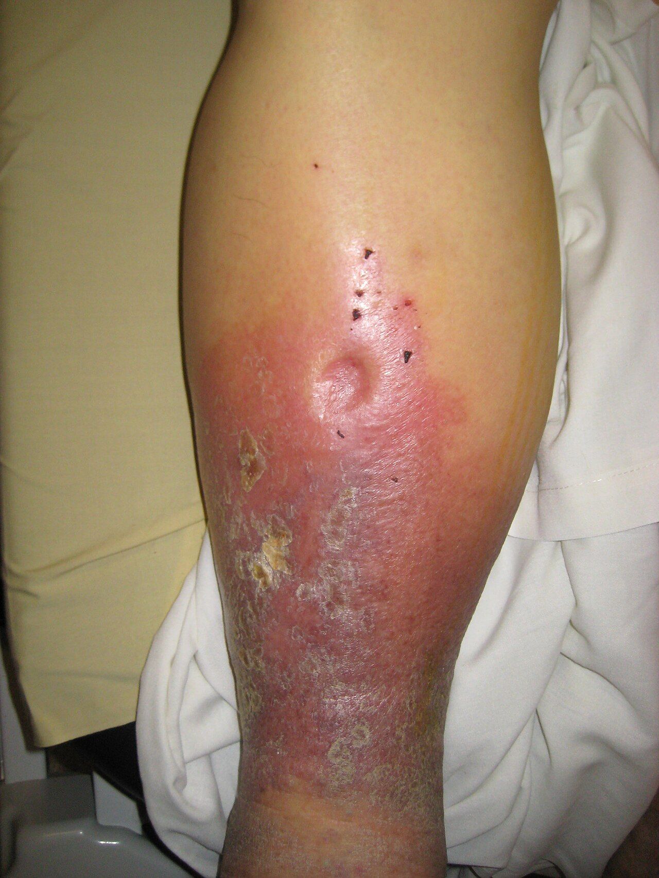
Right-sided failure backs up into the systemic venous circulation. Expect jugular venous distension, hepatojugular reflux, dependent pitting oedema (ankles in an ambulatory patient, sacrum in a bedbound one — always check the sacrum), weight gain, hepatomegaly and right upper quadrant fullness or tenderness, splenomegaly, ascites, anorexia, nausea and early satiety from gut congestion. The most common cause of right-sided failure is left-sided failure; when it arises from pulmonary disease alone it is called cor pulmonale. Most hospitalised patients have findings on both sides.
| Left-sided (pulmonary congestion) | Right-sided (systemic congestion) |
|---|---|
| Dyspnoea on exertion, orthopnoea, paroxysmal nocturnal dyspnoea | Jugular venous distension, hepatojugular reflux |
| Bibasilar crackles, wheeze (“cardiac asthma”) | Dependent pitting oedema of ankles or sacrum |
| Dry nocturnal cough; pink frothy sputum if acute | Weight gain, ascites, abdominal girth |
| S3 gallop, tachycardia, displaced apical impulse | Hepatomegaly, right upper quadrant tenderness |
| Fatigue, nocturia, restlessness and confusion | Anorexia, nausea, early satiety from gut oedema |
Diagnostics: BNP, imaging and the numbers you must know
B-type natriuretic peptide is released by ventricular myocytes in response to stretch, so it rises with volume overload. Learn the interpretation, not just the number. A BNP below 100 pg/mL makes heart failure an unlikely explanation for acute dyspnoea; 100 to 400 pg/mL is a grey zone requiring clinical correlation; above 400 pg/mL strongly supports it, and values above 1,000 are common in decompensation. For NT-proBNP, a level below 300 pg/mL effectively rules out acute heart failure, and the confirming thresholds are age-adjusted — roughly above 450 pg/mL under age 50, above 900 between 50 and 75, and above 1,800 over 75. BNP is most useful for answering the question that actually arises at 3 a.m.: is this dyspnoeic patient in failure or having a COPD exacerbation?
Three caveats separate a nurse who reports a number from one who interprets it. Renal impairment and atrial fibrillation raise BNP independently of failure. Obesity lowers it, so a value of 250 pg/mL in a patient with a BMI of 45 may represent significant congestion. And sacubitril/valsartan raises BNP because the neprilysin inhibitor blocks BNP degradation — in a patient on that drug, follow NT-proBNP instead, which is unaffected. Trend the value against the patient’s own baseline rather than against the reference range.
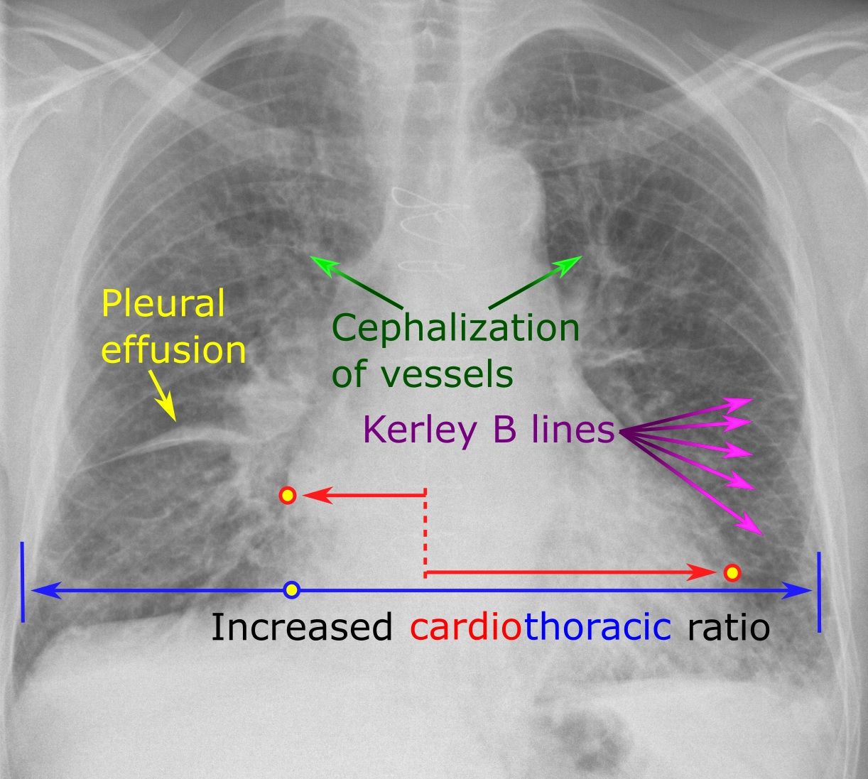
The rest of the workup: chest radiography shows cardiomegaly, cephalisation of pulmonary vessels, Kerley B lines, interstitial and then alveolar oedema, and pleural effusions. Echocardiography is the central test — it gives ejection fraction, chamber sizes, wall motion, diastolic function and valve assessment. ECG identifies infarction, hypertrophy, atrial fibrillation and bundle branch block (a QRS of 150 ms or more with left bundle branch block, in a symptomatic patient whose ejection fraction remains 35 percent or below on optimal therapy, is what qualifies a patient for resynchronisation therapy). Laboratory monitoring is relentless in these patients: potassium, sodium, magnesium, BUN 10 to 20 mg/dL and creatinine 0.6 to 1.2 mg/dL (a rising ratio suggests poor renal perfusion), liver enzymes with hepatic congestion, thyroid studies, CBC for anaemia and iron studies, and a digoxin level where relevant. Right heart catheterisation is reserved for uncertain cases, where the numbers to recognise are CVP 2 to 6 mmHg, pulmonary artery wedge pressure 6 to 12 mmHg and cardiac output 4 to 8 L/min.
Two classification systems coexist and both appear on examinations. The New York Heart Association functional classes describe symptoms and can move up and down: class I, no limitation; class II, slight limitation with ordinary activity; class III, marked limitation with less-than-ordinary activity; class IV, symptoms at rest. The ACC/AHA stages describe structural progression and only move forward: stage A, at risk with no structural disease; stage B, structural disease without symptoms; stage C, structural disease with current or prior symptoms; stage D, refractory disease requiring advanced interventions. Staging drives therapy; NYHA class drives the conversation about how the patient is actually living.
Pearl. The scale is a better fluid monitor than intake and output records, and it is cheaper than a BNP. One litre of retained fluid is about 2.2 pounds, and roughly five to ten pounds of fluid accumulate before crackles or visible oedema appear. By the time you hear the lungs change, the patient has been retaining for days. Weigh every heart failure patient daily, at the same time, on the same scale, in the same amount of clothing, after voiding and before breakfast — and chart it where the next nurse will actually see the trend.
Name the failing side and you can predict the findings — left backs up into the lungs, right backs up into the body — and remember that a normal ejection fraction never rules out heart failure, because half of these patients have a stiff ventricle that cannot fill.
Section 5Treating Heart Failure and Teaching the Patient to Live With It
Guideline-directed medical therapy: four pillars and the drugs around them
For HFrEF, four drug classes each independently reduce mortality, and the current standard is to start all four early at low doses and titrate upward rather than maximising one before adding the next. Students should be able to name them cold. First, a renin-angiotensin blocker: an ARNI (sacubitril/valsartan) is preferred, with an ACE inhibitor or ARB as the alternative. Second, an evidence-based beta-blocker — only carvedilol, metoprolol succinate (the extended-release salt) and bisoprolol have been shown to work, so metoprolol tartrate is not a substitute. Third, a mineralocorticoid receptor antagonist: spironolactone or eplerenone. Fourth, an SGLT2 inhibitor: dapagliflozin or empagliflozin, which benefit patients with and without diabetes. Beyond the four, hydralazine plus isosorbide dinitrate is added for self-identified Black patients on optimal therapy and for those who cannot take RAAS blockade, ivabradine is added when the sinus rate remains at 70 or above, and digoxin reduces hospitalisation without reducing mortality. Loop diuretics are used at the lowest effective dose for congestion; they make patients feel better but do not extend life. For HFpEF, the evidence base is thinner: control blood pressure, control rate and rhythm in atrial fibrillation, diurese for congestion, treat obesity and sleep apnoea, and use an SGLT2 inhibitor, with an MRA considered.
Three safety points recur on examinations and in practice. Switching from an ACE inhibitor to sacubitril/valsartan requires a 36-hour washout to avoid angioedema, and the two are never given together. Beta-blockers are started when the patient is euvolaemic and stable, not during an acute decompensation, and are titrated slowly because they transiently worsen symptoms before they help. And potassium is the recurring hazard of the whole regimen: loop diuretics drive it down while ACE inhibitors, ARBs, ARNIs, MRAs and potassium supplements drive it up. Never assume the direction — look at the actual value. And when potassium must be replaced, it is given orally or as a dilute intravenous infusion on a pump, never as an intravenous push and never as a concentrated bolus — undiluted intravenous potassium stops the heart.
| Class and examples | Effect | Key nursing monitoring and teaching |
|---|---|---|
| Loop diuretic — furosemide, bumetanide, torsemide | Blocks reabsorption in the loop of Henle; rapid symptom relief | Daily weight, strict intake and output, orthostatic blood pressure, potassium 3.5–5.0 mEq/L, sodium, magnesium, BUN and creatinine, hearing. Give IV push slowly (no faster than about 20 mg/min) — rapid administration causes ototoxicity. Dose in the morning so the patient is not up all night. |
| Thiazide — hydrochlorothiazide, metolazone | Distal tubule; metolazone potentiates a loop in diuretic resistance | Profound potassium and sodium loss when combined; give metolazone about 30 min before the loop; watch for hypovolaemia |
| ACE inhibitor — lisinopril, enalapril, captopril | Blocks angiotensin II; reduces afterload and remodelling | Potassium, creatinine and blood pressure; dry persistent cough (a reason to switch, not to stop unsupervised); angioedema is an emergency; first-dose hypotension; avoid salt substitutes containing potassium chloride; contraindicated in pregnancy |
| ARB — losartan, valsartan, candesartan | Same axis, no cough | As above without the cough; still causes hyperkalaemia |
| ARNI — sacubitril/valsartan | Adds neprilysin inhibition; superior to ACE inhibitor in HFrEF | 36-hour washout from an ACE inhibitor; hypotension, hyperkalaemia, renal function; raises BNP — monitor NT-proBNP instead |
| Beta-blocker — carvedilol, metoprolol succinate, bisoprolol | Blocks sympathetic overdrive; reverses remodelling over months | Apical pulse for a full minute before each dose, hold for HR below 60 or SBP below 90 per parameters; start low and go slow; warn that fatigue is temporary; never stop abruptly (rebound ischaemia); masks hypoglycaemia except diaphoresis |
| MRA — spironolactone, eplerenone | Blocks aldosterone; potassium-sparing; mortality benefit | Hyperkalaemia is the danger — check potassium and creatinine at baseline, at one week and periodically; generally avoid if potassium is above 5.0 mEq/L or eGFR is below 30; no potassium supplements or salt substitutes; spironolactone causes gynaecomastia |
| SGLT2 inhibitor — dapagliflozin, empagliflozin | Reduces hospitalisation and death in HFrEF and HFpEF | Genital mycotic and urinary infections, volume depletion, euglycaemic ketoacidosis; hold before major surgery per protocol; teach perineal hygiene and adequate fluids |
| Cardiac glycoside — digoxin | Positive inotrope, negative chronotrope; symptom and hospitalisation benefit only | Apical pulse one full minute; hold and notify if below 60. Therapeutic level 0.5–2.0 ng/mL, with 0.5–0.9 ng/mL targeted in heart failure. Hypokalaemia potentiates toxicity. Toxicity: anorexia, nausea, vomiting, visual halos or yellow-green vision, confusion, bradycardia and dysrhythmias. Antidote is digoxin immune Fab. |
| Vasodilator — hydralazine plus isosorbide dinitrate | Reduces preload and afterload | Headache, orthostatic hypotension, reflex tachycardia; rise slowly |
| Inotropes — dobutamine, milrinone | Short-term support in low-output states | Continuous monitoring, dysrhythmias, hypotension with milrinone; titration is an RN function in a monitored bed |
Acute decompensation, pulmonary oedema and cardiogenic shock
Acute decompensated heart failure is the most common reason these patients are admitted, and flash pulmonary oedema is its most frightening form: extreme dyspnoea, severe anxiety and air hunger, tachypnoea, tachycardia, diffuse crackles, wheezing, cyanosis and pink frothy sputum from plasma forced into the alveoli. The patient will not lie down, and you should not make them. Interventions, in order: sit the patient upright with the legs dependent (this pools blood peripherally and immediately reduces preload — it is the fastest intervention you own and requires no order), apply oxygen and titrate to saturation, obtain vital signs and an ECG, establish intravenous access, and prepare for an intravenous loop diuretic, nitroglycerin infusion for afterload and preload reduction if the blood pressure tolerates it, and non-invasive positive pressure ventilation (CPAP or BiPAP), which pushes fluid out of the alveoli and often prevents intubation. Morphine, once routine here, is now used sparingly. Insert a urinary catheter if precise output measurement is required, and monitor potassium closely as diuresis proceeds.
Cardiogenic shock is failure of the pump severe enough to compromise tissue perfusion: systolic pressure below 90 mmHg or a sustained fall of more than 30 mmHg, urine output below 0.5 mL/kg/hour, cool mottled extremities, weak thready pulses, altered mentation and a rising serum lactate (normal roughly 0.5 to 2.2 mmol/L). Mortality is high and the patient belongs in intensive care. Management combines inotropic support (dobutamine or milrinone), vasopressor support (norepinephrine is generally first-line), careful volume assessment, treatment of the underlying cause — usually urgent revascularisation when the cause is infarction — and mechanical support such as an intra-aortic balloon pump or a percutaneous ventricular assist device. Your role on the floor is early recognition and fast escalation, because the window is short.
For advanced, refractory disease, options include an implantable cardioverter-defibrillator for primary prevention of sudden death when the ejection fraction remains 35 percent or below after at least three months of optimal therapy, cardiac resynchronisation therapy for a wide QRS with left bundle branch block, a left ventricular assist device as a bridge to transplant or as destination therapy, and cardiac transplantation. Palliative care is not a failure of treatment and belongs in the conversation earlier than most teams introduce it — symptom burden in advanced heart failure rivals that of many cancers, and goals-of-care discussions should include device deactivation planning.
Daily weights, sodium, delegation and the discharge conversation
Heart failure has one of the highest thirty-day readmission rates in medicine, and most of those readmissions are preventable with teaching that the patient actually retains. Lead with the scale. Teach the patient to weigh every morning, after voiding, before eating, in similar clothing, on the same scale, and to write it down. The action threshold you must teach in these exact terms: call the provider for a gain of 2 to 3 pounds in 24 hours, or 5 pounds in a week. That gain is fluid, not fat, and it precedes symptoms by days. Many patients will also have a diuretic action plan permitting an extra dose within defined limits — confirm the patient can state theirs.
Diet is the second pillar. Sodium is limited to roughly 2 to 3 grams per day, and teaching that works is concrete: read labels for sodium per serving, avoid canned soups, deli meats, frozen dinners, pickled and smoked foods, cheese and restaurant meals, cook with herbs and citrus instead of salt, and do not switch to a salt substitute without asking, because most contain potassium chloride and are dangerous with an ACE inhibitor, ARB or MRA. Fluid is restricted to about 1.5 to 2 litres per day in advanced disease or hyponatraemia; teach the patient to measure it, spread it across the day, count ice chips and gelatine, and manage thirst with hard candy, chewing gum, frequent mouth care and cold water rinses. Add daily activity as tolerated, alcohol moderation or abstinence, smoking cessation, annual influenza and appropriate pneumococcal and COVID-19 vaccination (infection is a leading trigger of decompensation), and a plan for medication adherence including a pill organiser and a realistic conversation about cost.
Delegation is tested constantly and misunderstood constantly. The UAP may obtain and record daily weights, measure and record intake and output, take vital signs on stable patients, assist with hygiene and repositioning, ambulate a stable patient and report findings back to you. The LPN or LVN may administer most oral and many routine medications within their state scope, reinforce teaching that the RN has already provided, monitor an established stable patient, and perform many procedures. The RN retains initial and ongoing assessment, initial patient teaching, evaluation of the response to therapy, care of the unstable patient, and titration of intravenous vasoactive infusions including nitroglycerin, dobutamine and heparin. The rule that will carry you through most items: you may delegate a task, never the assessment, the teaching, the evaluation or the unstable patient — and you remain accountable for the outcome of anything you delegate.
Older adults deserve a final paragraph because their presentation and their pharmacology both differ. Heart failure in the older adult may present as confusion, delirium, falls, anorexia, weakness or functional decline long before the classic dyspnoea, and a sudden change in mental status warrants a cardiac and infectious workup rather than a sedative. Reduced lean body mass and declining renal clearance raise digoxin levels at unchanged doses, so toxicity presents insidiously as nausea, anorexia and confusion. Diuretics plus vasodilators produce orthostatic hypotension and falls — teach patients to rise in stages and check orthostatic vital signs. Polypharmacy is the rule, and NSAIDs taken over the counter for arthritis are a frequent and hidden cause of decompensation. Ask about them by name. Red flags in heart failure that mean call now: weight gain of 2 to 3 pounds in a day or 5 in a week; new or worsening orthopnoea or paroxysmal nocturnal dyspnoea; resting dyspnoea, pink frothy sputum or a falling oxygen saturation; chest pain; syncope, palpitations or a new irregular rhythm; systolic pressure below 90 with poor perfusion; urine output below 0.5 mL/kg/hour; and any new confusion.
Pearl. When a heart failure patient tells you they “could not sleep last night and had to sit in the recliner,” that is not a comfort complaint — it is orthopnoea, and it is a documented worsening of congestion. Quantify it (“how many pillows, and how many last month?”), weigh the patient, listen to the bases, look at the sacrum and the neck veins, and report the change. The sentences patients volunteer in passing are frequently the most valuable data you will collect all shift.
Heart failure is managed with four mortality-reducing drug classes, a diuretic for symptoms, and a patient who weighs themselves every morning and calls for 2 to 3 pounds in a day or 5 pounds in a week — and the teaching that prevents the readmission is the intervention only the RN can deliver.
The chapter in one breath
Cardiac muscle extracts nearly all the oxygen it receives at rest, so ischaemia is always a flow problem, and because the coronaries fill during diastole, tachycardia raises demand and cuts supply simultaneously. Atherosclerosis builds a lipid core under a fibrous cap; a stable cap produces predictable exertional angina, while a thin, inflamed cap ruptures and thromboses, producing unstable angina, NSTEMI or STEMI depending on how completely the artery closes. The 12-lead ECG within ten minutes decides the pathway: ST elevation in two contiguous leads sends the patient to reperfusion within 90 minutes of first medical contact, or fibrinolysis within 30 minutes of arrival when PCI is unavailable. Troponin rises in three to six hours and stays up for one to two weeks, so serial draws are mandatory and no single early value rules out infarction. Aspirin comes first, oxygen only below 90 percent saturation, and nitrates never with a phosphodiesterase-5 inhibitor or a right ventricular infarct. Infarcted muscle does not contract, so the same patient returns with heart failure — left failure floods the lungs, right failure floods the body, and half of these patients have a preserved ejection fraction. Four drug classes reduce mortality, diuretics only relieve symptoms, and the daily weight the patient takes at home is the earliest warning either of you will get.
Words worth knowing
| Term | What it means |
|---|---|
| Acute coronary syndrome | Umbrella term for unstable angina, NSTEMI and STEMI — all caused by plaque rupture with thrombosis, differing by degree and duration of occlusion. |
| Angina pectoris | Chest pain from reversible myocardial ischaemia. Stable if predictable and relieved by rest or nitroglycerin; unstable if new, at rest, or worsening. |
| Atherosclerosis | Progressive accumulation of lipid, foam cells and fibrous tissue in the arterial intima, forming a plaque that narrows the lumen and may rupture. |
| BNP / NT-proBNP | Peptides released with ventricular stretch. BNP under 100 pg/mL makes heart failure unlikely; NT-proBNP under 300 pg/mL rules out acute failure. Sacubitril/valsartan raises BNP but not NT-proBNP. |
| Cardiogenic shock | Pump failure severe enough to impair tissue perfusion — hypotension, oliguria, cool mottled skin, altered mentation, rising lactate. Requires intensive care. |
| Door-to-balloon time | Interval from first medical contact to PCI device deployment in STEMI; goal 90 minutes, or 120 minutes with interfacility transfer. |
| Dressler syndrome | Autoimmune pericarditis one to eight weeks after MI, with pleuritic pain relieved by sitting forward, fever, friction rub and diffuse ST elevation. |
| Ejection fraction | Percentage of end-diastolic volume ejected per beat; normal 55 to 70 percent. Reduced at 40 percent or below, preserved at 50 percent or above. |
| Guideline-directed medical therapy | The four mortality-reducing drug classes in HFrEF: ARNI or ACE inhibitor/ARB, evidence-based beta-blocker, mineralocorticoid receptor antagonist, and SGLT2 inhibitor. |
| HFpEF | Heart failure with preserved ejection fraction — a stiff, non-compliant ventricle that cannot fill; typical of older adults with hypertension, diabetes and obesity. |
| Orthopnoea | Dyspnoea when lying flat, relieved by sitting up; quantified by the number of pillows required. A sensitive sign of pulmonary congestion. |
| Paroxysmal nocturnal dyspnoea | Waking abruptly at night gasping for air, caused by reabsorption of interstitial fluid during recumbency. |
| Phosphodiesterase-5 inhibitor | Sildenafil, vardenafil, tadalafil. Nitrates are absolutely contraindicated within 24 hours (48 for tadalafil) because of catastrophic hypotension. |
| Preload | Ventricular stretch at end-diastole, determined by venous return. Nitrates and diuretics lower it; the right-ventricular infarct patient depends on it. |
| Remodelling | Chronic hypertrophy, dilation, fibrosis and change in ventricular shape driven by sympathetic and RAAS activation; the target of ACE inhibitors, ARNIs and beta-blockers. |
| S3 gallop | Extra low-pitched sound just after S2, best heard with the bell at the apex; in an adult it is an early and relatively specific sign of volume overload. |
| STEMI | Full-thickness infarction with ST elevation in two contiguous leads; a reperfusion emergency diagnosed by ECG, never delayed for troponin results. |
| Troponin | Cardiac-specific contractile protein. Conventional assays rise in three to six hours, peak at twelve to twenty-four hours and persist seven to fourteen days; serial draws are required. |
| Type 2 myocardial infarction | Myocardial injury from supply-demand mismatch (sepsis, anaemia, tachydysrhythmia, hypotension) without plaque rupture; treat the underlying cause. |