Infectious Disease and Antimicrobial Stewardship
Volume I taught you to recognise sepsis and start the hour-one bundle. This chapter is the rest of the story: the patient who is infected but not yet septic, the culture that comes back at 0300 with a name and a susceptibility panel, and the four to six weeks of therapy that follow. You will learn to build a diagnostic picture before the first dose rather than after it, to read antimicrobials by mechanism so that the monitoring parameters stop being memorised trivia, and to hold the line on stewardship — the discipline of giving the narrowest effective drug for the shortest effective time. You will meet the resistant organisms by name, the device bundles that prevent most of what we treat, and four infections that fill med-surg beds: cellulitis, necrotising soft-tissue infection, osteomyelitis and infective endocarditis. The chapter closes with tuberculosis, HIV and viral hepatitis — chronic communicable illness, where the nursing is adherence, stigma and public health as much as pharmacology.
By the end of this chapter you can
- Explain how pathogen invasion and the host inflammatory response generate fever, leukocytosis, local erythema, induration and organ-specific findings, and why the older adult may present with delirium or hypothermia instead.
- Collect blood cultures and other specimens correctly and interpret WBC, bandemia, absolute neutrophil count, CRP, ESR, procalcitonin and lactate against real reference values.
- Classify antimicrobials by mechanism — beta-lactams, glycopeptides, lipopeptides, oxazolidinones, aminoglycosides, fluoroquinolones, macrolides, tetracyclines, sulfonamides, nitroimidazoles — and state the monitoring each demands.
- Apply the core actions of antimicrobial stewardship: culture before therapy, de-escalation, IV-to-PO conversion, defined duration, penicillin-allergy delabelling and refusal to treat asymptomatic bacteriuria.
- Differentiate MRSA, VRE, ESBL, CRE, Candida auris and Clostridioides difficile, and select the correct transmission-based precautions and hand-hygiene method for each.
- Prioritise care in necrotising fasciitis, neutropenic fever and septic embolisation using ABC, unstable-over-stable and acute-over-chronic reasoning, and escalate with a complete SBAR.
- Teach the patient on RIPE antitubercular therapy, antiretroviral therapy and direct-acting antivirals what to expect, what to report and why adherence is non-negotiable.
- Delegate appropriately, retaining assessment, teaching, evaluation and the unstable patient for the registered nurse.
Section 1Recognising Infection and Building the Diagnostic Picture
Why infection looks the way it does
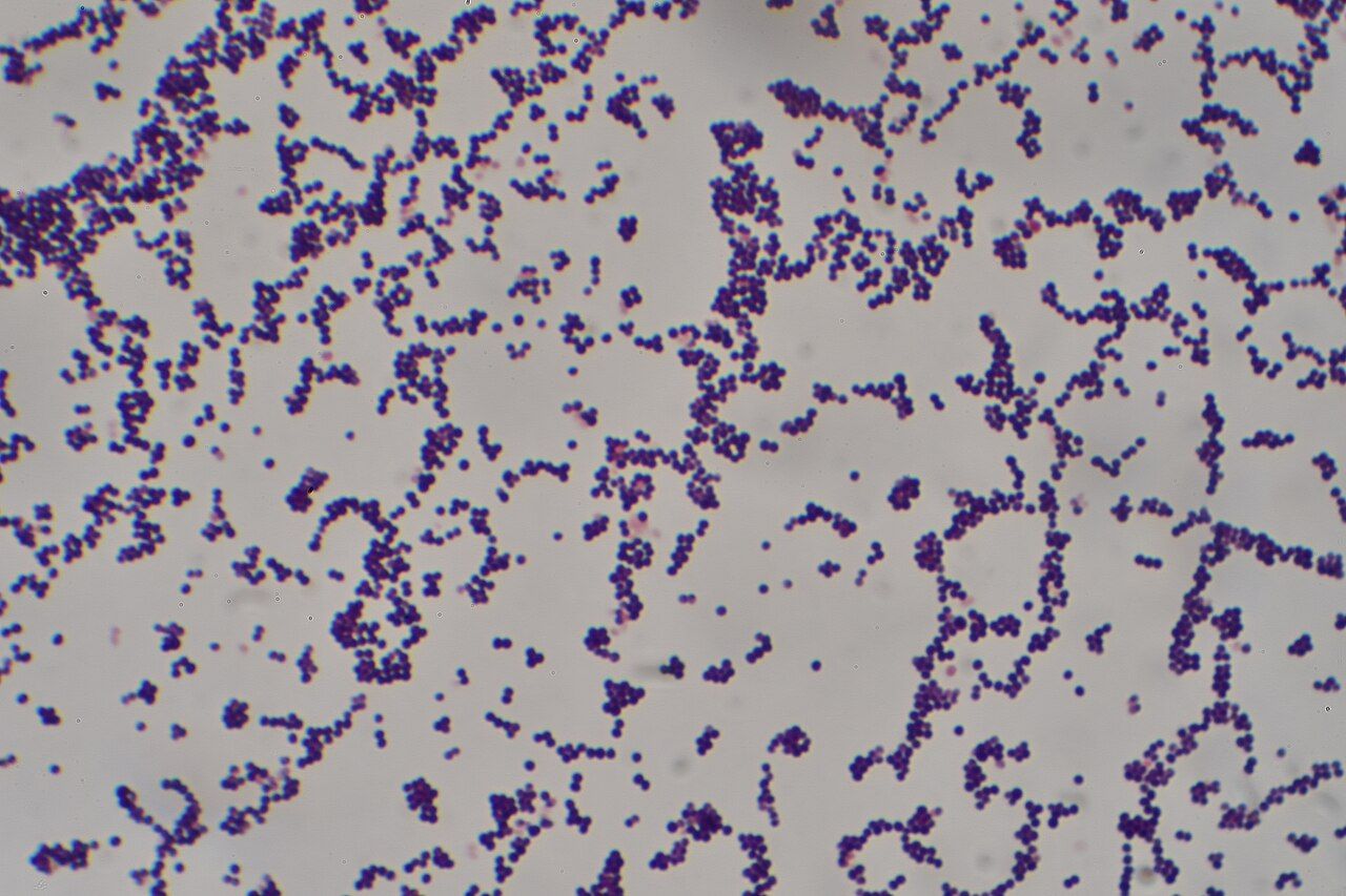
An infection begins when a pathogen breaches a barrier — skin, mucosa, an intravascular catheter, an endotracheal tube — and multiplies faster than local defences can clear it. Bacterial cell-wall components such as lipopolysaccharide and lipoteichoic acid are recognised by tissue macrophages, which release interleukin-1, interleukin-6 and tumour necrosis factor. Those cytokines do three things you can see at the bedside. They reset the hypothalamic thermostat upward, producing fever and the shivering, vasoconstricted, cold-feeling phase that precedes it. They dilate local capillaries and increase permeability, producing the erythema, warmth, swelling and pain of a cellulitic limb or an inflamed wound. And they drive the liver to make acute-phase reactants, which is why C-reactive protein climbs within hours.
The same cytokines pull neutrophils out of the marrow. A mature neutrophil takes days to build, so when demand outstrips supply the marrow ships immature band forms — the left shift. Bands above roughly 10 percent are a meaningful sign of bacterial stress even when the total white count is normal. In overwhelming infection the marrow can fail to keep pace and the white count falls instead of rising; a white count of 2,800/mm³ in an ill patient is more worrying, not less, than one of 18,000/mm³.
Local findings tell you the compartment. Dysuria and suprapubic tenderness point to bladder; costovertebral angle tenderness adds kidney. Productive cough with focal crackles and bronchial breath sounds points to lung. A new murmur with peripheral emboli points to valve. Pain that is far worse than the skin looks points to fascia. Learning to pair the systemic response with the local sign is what turns “the patient has a fever” into a working hypothesis you can hand to a provider.
The focused infection assessment, and how the older adult differs
Assess in this order: airway and work of breathing, then perfusion, then the source. Take a full set of vital signs including an accurate temperature by a consistent route, and note the respiratory rate yourself — tachypnoea is the earliest and most-ignored sign of deterioration. Look for tachycardia, a widening pulse pressure, a falling systolic pressure, mottled knees, delayed capillary refill and cool extremities. Check mentation against the patient’s documented baseline, not against your impression of “normal for their age.” Then hunt for the source with your hands and eyes: every line site, every drain, every wound dressing, the perineum and sacrum, between the toes, under pendulous skin folds, the mouth. Infections hide where nobody looks.
Fever is defined as a temperature of 38.0 °C (100.4 °F) or higher, but in the older adult that threshold misses a great deal. Baseline temperature falls with age, so a resident whose usual oral temperature is 36.1 °C is febrile at 37.8 °C. Accepted geriatric criteria are a single oral reading above 37.8 °C (100 °F), repeated readings above 37.2 °C (99 °F) oral or 37.5 °C (99.5 °F) rectal, or a rise of 1.1 °C (2 °F) over the patient’s own baseline. Up to a third of older adults with serious bacterial infection are afebrile, and some are frankly hypothermic, which carries a worse prognosis.
More often the presentation is functional rather than febrile. New confusion or a change in the character of established dementia, a fall, incontinence in a previously continent patient, anorexia, or simply “she is not herself” from a family member are legitimate infection findings in this population and should trigger a full assessment. The corollary matters just as much: confusion alone in an older adult with a positive urine culture and no urinary symptoms is not pyelonephritis. Treating that patient’s asymptomatic bacteriuria exposes them to C. difficile and resistance while the real cause — dehydration, a new opioid, hypoxia, pain, constipation — goes unaddressed.
Cultures, markers and what the numbers mean
Cultures come before antimicrobials whenever it can be done without meaningful delay. In suspected sepsis, obtain them within the first minutes and do not let collection push the first antibiotic dose beyond 45 to 60 minutes. Draw two sets of blood cultures from two separate venepuncture sites, 8 to 10 mL into each bottle, using scrupulous skin antisepsis with chlorhexidine-alcohol and a friction scrub with adequate dry time. Volume is the single biggest determinant of yield; a skimpy bottle is a false negative waiting to happen. Do not routinely draw through an existing catheter — the exception is suspected catheter-related bloodstream infection, where one peripheral set plus one set from the line lets the laboratory compare time to positivity.
Sputum should be deep and purulent, not saliva; the laboratory will reject a specimen with abundant squamous epithelial cells. Wound cultures come from cleansed viable tissue or from aspirated pus, never from a swab dragged across old slough, which grows colonisers and earns the patient an unnecessary antibiotic. Stool is tested for C. difficile only when it is unformed — testing a formed stool detects colonisation and mislabels the patient. Urine is collected clean-catch midstream, or from the sampling port of a freshly changed catheter, never from the drainage bag.
| Test | Reference range | What it tells you at the bedside |
|---|---|---|
| White blood cells | 4,500–11,000/mm³ | Elevation suggests bacterial stress; leukopenia in an ill patient is ominous, not reassuring |
| Neutrophils / bands | Segs 55–70 percent; bands under 5 percent | Bands over 10 percent equal a left shift — the marrow is being emptied |
| Absolute neutrophil count | Over 1,500/mm³ | ANC equals WBC times (segs plus bands) divided by 100; under 500/mm³ is severe neutropenia |
| C-reactive protein | Under 10 mg/L (under 1 mg/dL) | Rises within 6–8 hours, falls quickly with response; good for trending therapy |
| Erythrocyte sedimentation rate | Under 15 mm/hr men, under 20 mm/hr women; rises with age (roughly age÷2 in men, [age+10]÷2 in women) | Slow to move; useful for tracking bone and joint infection over weeks |
| Procalcitonin | Under 0.25 ng/mL low; over 0.5 ng/mL supports bacterial sepsis | Helps justify stopping antibiotics; a falling value supports de-escalation |
| Lactate | Under 2.0 mmol/L | Over 2 is tissue hypoperfusion; over 4 is high risk — repeat within 2–4 hours |
| Blood cultures | No growth at 5 days | Two sets, two sites, before antibiotics; single-bottle coagulase-negative staph is usually contaminant |
One combination of numbers outranks everything else on the list. A single oral temperature of 38.3 °C (101 °F), or 38.0 °C (100.4 °F) sustained for an hour, in a patient whose absolute neutrophil count is under 500/mm³ — or under 1,000/mm³ and predicted to fall — is neutropenic fever, and it is an emergency. Without neutrophils the patient cannot make pus, a visible infiltrate or much erythema, so fever may be the only sign while the bloodstream is already seeded. Draw blood cultures, then give a broad-spectrum antipseudomonal beta-lactam — cefepime, piperacillin-tazobactam or meropenem — within 60 minutes. Do not wait for a source, an imaging study or a rising white count. Avoid rectal temperatures, suppositories and enemas in neutropenia, because mucosal trauma seeds the blood.
Imaging follows the compartment: chest radiograph for pulmonary findings, ultrasound for abscess and for gallbladder, computed tomography for intra-abdominal and deep neck and retroperitoneal collections, magnetic resonance imaging for bone and spine, echocardiography for valve. Remember the governing principle of infection management: source control beats drug selection. An abscess that is not drained, a necrotic limb that is not debrided, an obstructed ureter that is not stented and an infected catheter that is not removed will not be cured by any antibiotic, at any dose, for any duration.
Build the diagnostic picture — systemic response, local source, adequate cultures, correct markers — before the first dose, because after the first dose the cultures may never give you a name.
Section 2Antimicrobial Therapy by Mechanism
Cell-wall agents and the penicillin-allergy label
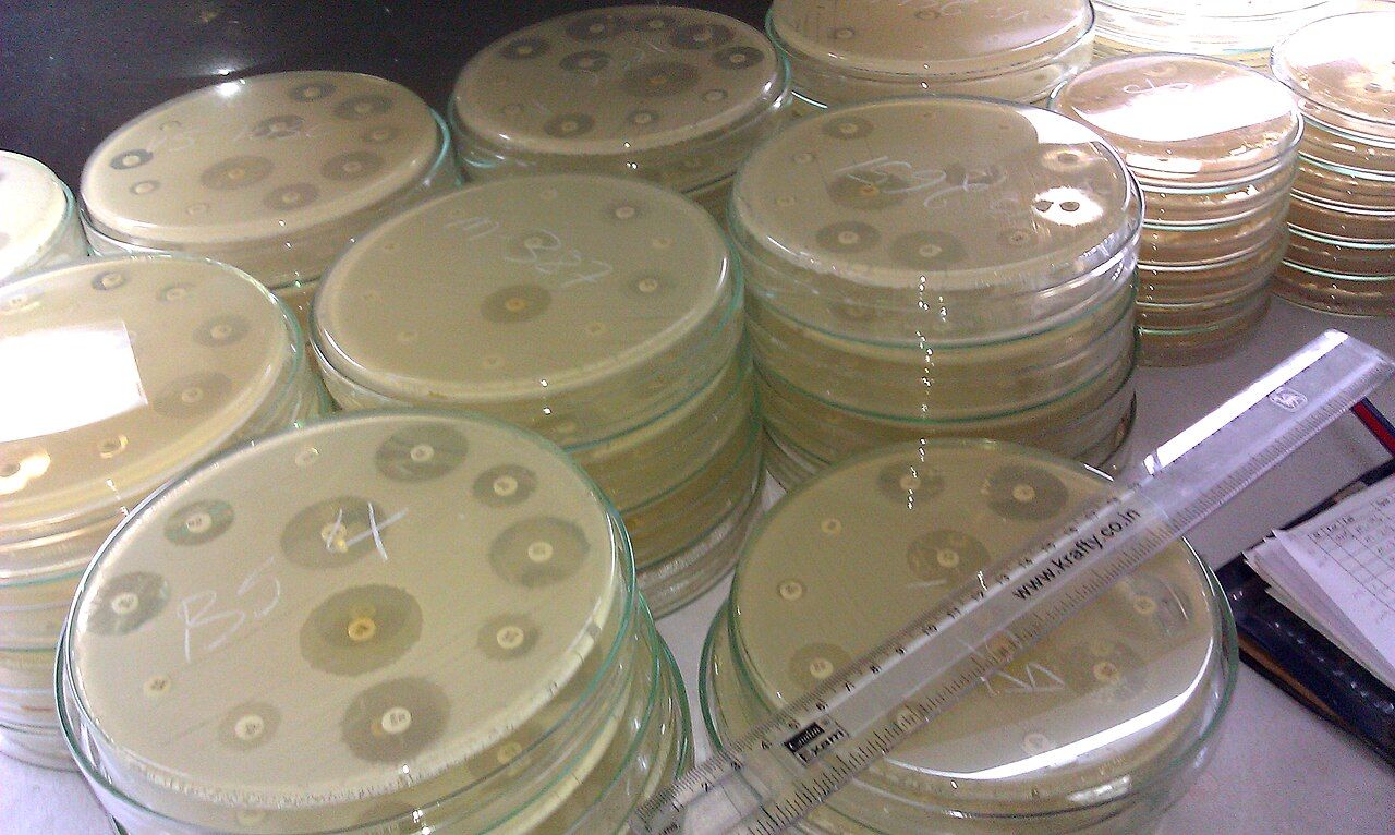
Beta-lactams — penicillins, cephalosporins, carbapenems and the monobactam aztreonam — bind penicillin-binding proteins and stop cross-linking of the bacterial cell wall, so the organism lyses as it divides. They are bactericidal, remarkably safe for the kidney and liver, and remain the backbone of therapy for streptococci, methicillin-susceptible Staphylococcus aureus, most Enterobacterales and anaerobes. Their killing is time-dependent: what matters is the fraction of the dosing interval that the free drug concentration stays above the organism’s minimum inhibitory concentration. That is why piperacillin-tazobactam and cefepime are increasingly given as extended three- to four-hour infusions and why a late dose is a real clinical event, not a documentation problem.
Know the practical hazards. Cefepime and high-dose beta-lactams accumulate in renal impairment and cause encephalopathy, myoclonus and non-convulsive seizures — new confusion on cefepime with a rising creatinine is a drug effect until proven otherwise. Ceftriaxone causes biliary sludging and must never be co-administered with calcium-containing solutions in neonates. Piperacillin-tazobactam combined with vancomycin raises the risk of acute kidney injury above either drug alone. All beta-lactams disrupt colonic flora and predispose to C. difficile.
Roughly one in ten hospitalised patients carries a penicillin-allergy label, yet fewer than one in a hundred has a true IgE-mediated allergy, and about 80 percent of those who once did lose sensitivity after ten years. The label is not harmless: it pushes patients onto vancomycin, fluoroquinolones and clindamycin, which raise resistance, C. difficile, surgical site infection and cost. Your job is to take a real history. What happened, at what age, how soon after the dose, and what was done about it? A childhood rash, gastrointestinal upset, a family history or “my mother told me” is an intolerance or an unverifiable story, not an allergy. Hives within an hour, angio-oedema, bronchospasm, hypotension or anaphylaxis is an allergy. Blistering, mucosal involvement or skin sloughing describes a severe cutaneous reaction — Stevens-Johnson syndrome or toxic epidermal necrolysis — and that patient must never be rechallenged with the class. Cross-reactivity between penicillins and modern cephalosporins is under 2 percent and is driven by side-chain similarity, not by the beta-lactam ring itself.
The gram-positive workhorses: vancomycin, daptomycin, linezolid
Vancomycin is a glycopeptide that blocks cell-wall precursor assembly and remains first-line intravenous therapy for MRSA. It is poorly absorbed orally, which is a defect everywhere except the colon — and that is precisely why oral vancomycin treats C. difficile while intravenous vancomycin does not. Dosing is now guided by an area-under-the-curve target of 400 to 600 mg·h/L; where trough monitoring is still used, the goal is 15 to 20 mcg/mL for serious MRSA infection, with the trough drawn within 30 minutes before the fourth dose. Monitor serum creatinine at least twice weekly, more often with concurrent nephrotoxins, and watch urine output.
Vancomycin infusion reaction — the flushing, pruritus and erythema of the face, neck and upper torso formerly called red man syndrome — is direct histamine release from rapid infusion, not an allergy. It is a rate problem: infuse at no faster than 1 gram per hour, extend the time, and pretreat with an antihistamine. Do not let a documented infusion reaction be miscoded as an allergy and cost the patient the drug.
Daptomycin is a lipopeptide that depolarises the gram-positive cell membrane. It is excellent for MRSA bacteraemia, right-sided endocarditis and skin infection, and it is inactivated by pulmonary surfactant, so it is never used for pneumonia — a favourite examination point and a real prescribing error. Check creatine kinase at baseline and weekly, and report muscle pain or weakness. Linezolid is an oxazolidinone that blocks the 50S ribosome, is nearly 100 percent orally bioavailable, and covers MRSA and VRE. It causes reversible myelosuppression, especially thrombocytopenia, so draw a baseline CBC and then a weekly CBC in every patient regardless of the expected duration, watching more closely in pre-existing myelosuppression, renal impairment or concurrent marrow-suppressive drugs; the risk rises steeply beyond about two weeks. Beyond about four weeks it can cause peripheral and optic neuropathy. Because it is a weak monoamine oxidase inhibitor, combining it with SSRIs, SNRIs, tramadol or triptans risks serotonin syndrome — agitation, hyperthermia, clonus, hyper-reflexia.
Ribosomal, DNA-active and anaerobic agents
Aminoglycosides — gentamicin, tobramycin, amikacin — bind the 30S subunit and are bactericidal against gram-negative aerobes. Their killing is concentration-dependent with a long post-antibiotic effect, which is why extended-interval dosing (about 5 to 7 mg/kg every 24 hours for gentamicin) gives better killing with less toxicity than the old three-times-daily schedule. Nephrotoxicity is usually reversible; ototoxicity is often not. Ask daily about tinnitus, a sense of fullness, hearing change, vertigo and unsteadiness, and document a baseline. Aminoglycosides also potentiate neuromuscular blockade and are avoided in myasthenia gravis.
Fluoroquinolones inhibit DNA gyrase and topoisomerase, have superb oral bioavailability, and carry a boxed warning worth reciting to patients: tendinitis and Achilles tendon rupture, peripheral neuropathy, central nervous system effects from insomnia to psychosis, exacerbation of myasthenia gravis, plus QT prolongation, dysglycaemia, aortic aneurysm and dissection, and a high C. difficile risk. Antacids, sucralfate, calcium, iron, zinc and multivitamins chelate the drug and can abolish absorption entirely — give the fluoroquinolone at least 2 hours before, or 6 hours after, any of them. Macrolides as a class prolong the QT interval — check an ECG when other QT-prolonging drugs are on board. Cytochrome inhibition, however, is not a class property: erythromycin and clarithromycin are potent CYP3A4 inhibitors with clinically important interactions (statins, with a risk of rhabdomyolysis, plus colchicine, tacrolimus, warfarin and carbamazepine), while azithromycin has minimal CYP inhibition and is deliberately chosen over clarithromycin when interaction risk matters — its signature hazard is QT prolongation. Tetracyclines and doxycycline cause photosensitivity and oesophagitis — take with a full glass of water, sit upright for 30 minutes, and use sunscreen. Trimethoprim-sulfamethoxazole causes hyperkalaemia, a creatinine rise that is partly artefactual, rash, and marrow suppression, and it potentiates warfarin. Metronidazole covers anaerobes and protozoa, causes a metallic taste and peripheral neuropathy, and produces a disulfiram-like reaction — teach absolute avoidance of alcohol, including mouthwash and cough syrup, during therapy and for three days after.
| Class / example | Mechanism | Nursing priority and monitoring |
|---|---|---|
| Penicillins (piperacillin-tazobactam) | Cell-wall cross-link inhibition | Time-dependent — give on time, extended infusion; allergy history; renal dosing |
| Cephalosporins (cefazolin, ceftriaxone, cefepime) | Cell-wall | Cefepime neurotoxicity in renal failure; ceftriaxone and calcium incompatibility in neonates |
| Carbapenems (meropenem, ertapenem) | Cell-wall, very broad | Reserve agents; seizure risk (imipenem); stewardship review at 48–72 hours |
| Glycopeptide (vancomycin IV) | Cell-wall precursor binding | AUC or trough monitoring, creatinine; infuse no faster than 1 g/hr (at least 60 min per gram) |
| Lipopeptide (daptomycin) | Membrane depolarisation | Never for pneumonia; weekly creatine kinase; report muscle pain |
| Oxazolidinone (linezolid) | 50S ribosome | Baseline then weekly CBC in every patient — risk rises steeply beyond 2 weeks; serotonin syndrome with SSRIs; oral equals IV |
| Aminoglycosides (gentamicin) | 30S ribosome | Levels, creatinine, hearing and balance; concentration-dependent dosing |
| Fluoroquinolones (levofloxacin) | DNA gyrase | Tendon rupture, QT, neuropathy, C. difficile; separate from cations |
| Macrolides (azithromycin, clarithromycin, erythromycin) | 50S ribosome | QT prolongation across the class; GI upset; erythromycin and clarithromycin — not azithromycin — are strong CYP3A4 inhibitors (statins, colchicine, tacrolimus, warfarin) |
| Tetracycline (doxycycline) | 30S ribosome | Photosensitivity, oesophagitis; upright with water; avoid in pregnancy and under age 8 |
| Sulfonamide (TMP-SMX) | Folate synthesis | Potassium, creatinine, CBC, rash; warfarin interaction; push fluids |
| Nitroimidazole (metronidazole) | DNA strand breakage | No alcohol during and 3 days after; metallic taste; neuropathy |
| Echinocandin (micafungin) | Fungal cell wall | First-line for candidaemia; few interactions; LFTs |
| Amphotericin B (liposomal) | Fungal membrane sterols | Infusion reactions, nephrotoxicity, hypokalaemia and hypomagnesaemia — replace and monitor |
| Acyclovir IV | Viral DNA polymerase | Crystal nephropathy — hydrate generously, infuse over at least 1 hour |
Every antimicrobial class carries one signature toxicity that flows directly from its mechanism — learn the mechanism and the monitoring parameter names itself.
Section 3Stewardship, Resistance and Healthcare-Associated Infection
What stewardship asks of the bedside nurse
Antimicrobial stewardship is the coordinated effort to give the right drug, at the right dose, by the right route, for the right duration — and to give none at all when none is needed. It exists because resistance is a shared resource problem: every unnecessary course selects for organisms that will one day infect someone else. The formal programme belongs to pharmacy, infectious diseases and microbiology, but the levers that actually move are at the bedside, and most of them are nursing actions.
Six of them are yours. Get adequate cultures before the first dose. Get the first dose in on time — delayed administration is the most common failure in an otherwise correct plan. Take an accurate allergy history and push for delabelling when the story is not an allergy. Prompt the 48- to 72-hour antibiotic time-out: once the culture and susceptibilities are back, ask the team whether the broad empiric regimen can be narrowed to a targeted one, which is de-escalation. Prompt IV-to-PO conversion when the patient has been afebrile for 24 to 48 hours, is haemodynamically stable, has a working gut and is improving — levofloxacin, moxifloxacin, linezolid, metronidazole, doxycycline, fluconazole and TMP-SMX are so well absorbed that the oral route is therapeutically equivalent and removes an intravenous line. Finally, know the intended stop date. Modern evidence supports shorter courses than tradition: five days for most community-acquired pneumonia that is responding, seven days for uncomplicated gram-negative bacteraemia and for most hospital-acquired pneumonia, five to seven days for uncomplicated pyelonephritis.
Refuse two temptations. Do not send cultures reflexively from every warm patient — a positive result from a colonised site generates treatment nobody needed. And do not treat asymptomatic bacteriuria, which is common in older adults and universal in long-term catheters; the only accepted exceptions are pregnancy and before a urologic procedure that will traumatise mucosa.
The resistant organisms, by name
MRSA carries the mecA gene and an altered penicillin-binding protein, so every beta-lactam except ceftaroline fails; treat with vancomycin, daptomycin or linezolid. VRE, usually Enterococcus faecium, is treated with linezolid or daptomycin. ESBL-producing E. coli and Klebsiella destroy penicillins and cephalosporins, so serious infection needs a carbapenem. CRE defeats the carbapenems too and requires newer agents such as ceftazidime-avibactam, meropenem-vaborbactam or cefiderocol. Pseudomonas aeruginosa is intrinsically resistant to much of the formulary and thrives in water, ventilator circuits and moist wounds. Candida auris is a yeast that resists multiple antifungal classes, persists on surfaces for weeks, and requires contact precautions plus a sporicidal-grade disinfectant proven against it — ordinary quaternary-ammonium wipes are not adequate.
Placement and equipment matter as much as drugs. Resistant organisms travel on hands, gowns, stethoscopes, blood-pressure cuffs, glucometers and workstations on wheels. Use dedicated single-patient equipment, disinfect anything shared between patients, and observe contact-time on the wipe label — the surface must stay visibly wet for the full stated dwell time or the disinfection did not happen.
Clostridioides difficile: the infection we cause
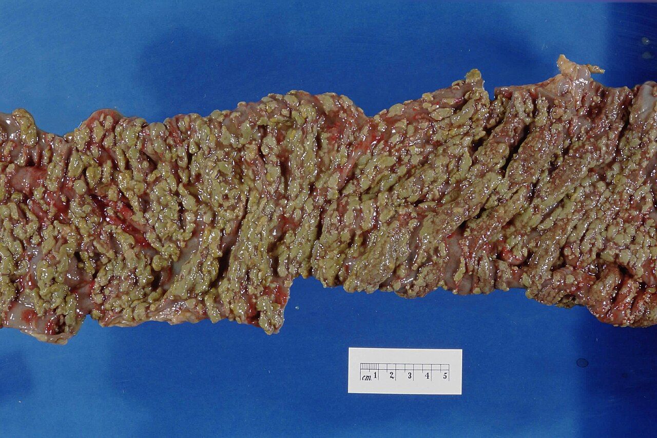
Antibiotics strip the colonic anaerobes that normally suppress C. difficile; spores germinate and release toxins A and B, which disrupt the cytoskeleton of colonocytes and produce watery diarrhoea, cramping, fever, leukocytosis and, in the classic case, pseudomembranes. Clindamycin, fluoroquinolones, cephalosporins and carbapenems are the highest-risk classes; age over 65, prolonged hospitalisation, proton pump inhibitors and enteral feeding add risk. Test only unformed stool, ideally three or more loose stools in 24 hours with no laxative, and never test for cure — the assay stays positive long after the patient is well.
Treat an initial episode with oral vancomycin 125 mg four times daily for 10 days or fidaxomicin 200 mg twice daily for 10 days; metronidazole is now reserved for when neither is available. Fulminant disease — hypotension, shock, ileus or megacolon — is treated with high-dose oral or nasogastric vancomycin 500 mg four times daily plus intravenous metronidazole, with rectal vancomycin instillation added if there is ileus, and surgical consultation. Watch for the most dangerous transition in the disease: a patient whose diarrhoea suddenly stops while the abdomen becomes distended, tympanitic and quiet has not improved — that is toxic megacolon, and it is an emergency.
Isolation is contact precautions in a private room with dedicated equipment. Wash hands with soap and water; alcohol-based rub does not kill spores. Clean with a sporicidal agent such as a bleach-based product. Continue precautions for at least 48 hours after diarrhoea resolves — many facilities continue until discharge. Antimotility agents such as loperamide are avoided in acute disease because retaining toxin worsens colitis. Review the proton pump inhibitor: many are continued out of habit and can be stopped.
Precautions and the device bundles
Standard precautions apply to every patient, always: hand hygiene at the five moments, gloves for anything wet, and gown, mask and eye protection whenever splash is possible. Transmission-based precautions are layered on top. Contact means gown and gloves for MRSA, VRE, ESBL, CRE, C. difficile, scabies and draining wounds. Droplet means a surgical mask for influenza, pertussis, Neisseria meningitidis, group A streptococcal pharyngitis and mumps, with the patient masked during transport. Airborne means a fit-tested N95 or a powered air-purifying respirator and a negative-pressure airborne infection isolation room with the door closed, for tuberculosis, measles, varicella and disseminated zoster; only immune staff should enter measles and varicella rooms.
Most of what we treat, we introduced. The prevention bundles are short and they work. CLABSI: hand hygiene, maximal sterile barriers at insertion, chlorhexidine-alcohol skin prep with full dry time, avoid the femoral site, scrub the hub for at least 15 seconds with friction, daily chlorhexidine bathing, and above all a daily out-loud review of whether the line is still needed. CAUTI: insert only for a true indication, use aseptic technique, keep a closed system, secure the tubing, keep the bag below the level of the bladder and off the floor, and remove the catheter at the earliest opportunity — duration is the dominant risk factor. VAP: head of bed 30 to 45 degrees, oral care with toothbrushing, subglottic secretion drainage, daily sedation interruption paired with a spontaneous breathing trial, and early mobility. Surgical site infection: prophylactic antibiotic within 60 minutes of incision (120 minutes for vancomycin or a fluoroquinolone), redosing for long cases or major blood loss, discontinuation at wound closure, clippers rather than razors, normothermia, and perioperative glucose under 180 mg/dL.
Stewardship and infection prevention are the same discipline viewed from two ends: the narrowest drug for the shortest time, and the fewest devices for the fewest days.
Section 4Skin, Bone and Endovascular Infection
Cellulitis and the abscess that must be opened
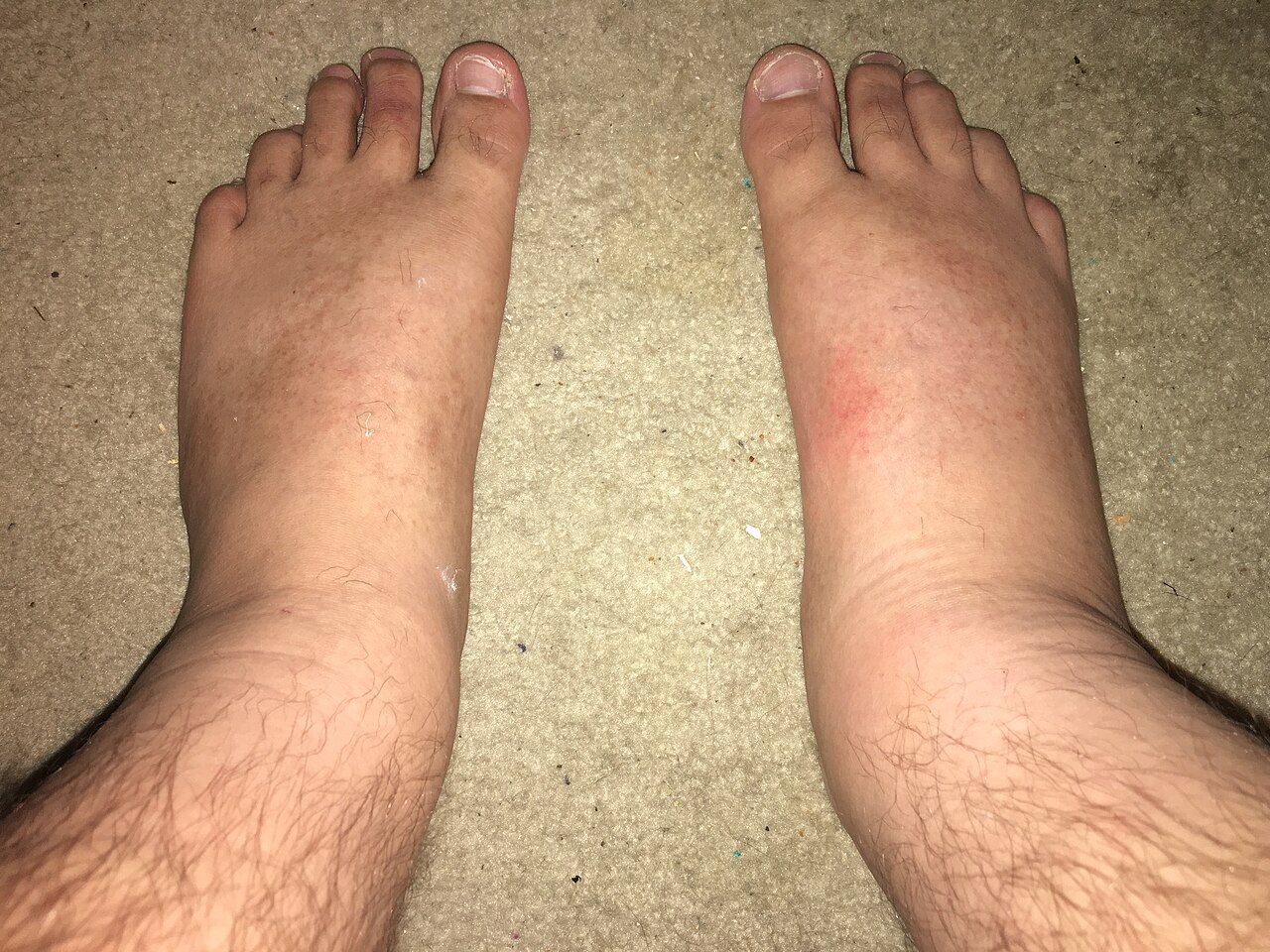
Cellulitis is a bacterial infection of the deep dermis and subcutaneous tissue that enters through a fissure, a bite, a venous ulcer, tinea pedis between the toes or an injection site. The presentation is unilateral, warm, tender, poorly demarcated erythema with oedema, often with lymphangitic streaking and regional lymphadenopathy, and sometimes with fever and leukocytosis. Non-purulent cellulitis is usually beta-haemolytic streptococcal and is treated with cefazolin intravenously or cephalexin orally. Purulent cellulitis with an abscess is usually staphylococcal, frequently MRSA, and needs incision and drainage plus TMP-SMX, doxycycline or clindamycin; without drainage, antibiotics alone commonly fail.
Nursing care is simple and effective. Outline the erythema with a skin marker, write the date and time on the skin, and reassess each shift — expansion beyond the mark within the first 24 to 48 hours of therapy is the earliest objective sign of failure, though a modest initial increase can accompany the inflammatory response to bacterial killing. Elevate the limb above the level of the heart, which does more for the oedema than any drug. Treat the portal of entry: an antifungal for macerated toe webs prevents the next episode. Manage pain, control glucose, and assess for venous insufficiency and lymphoedema, which drive recurrence.
Beware the mimics, because both are common and neither needs antibiotics. Bilateral, symmetric, non-tender erythema with scaling in a patient with venous insufficiency is stasis dermatitis, not cellulitis. Acute unilateral calf swelling with pain but little erythema may be deep vein thrombosis. If the leg is red and the patient is well and afebrile with normal inflammatory markers, argue for a second look before committing to a week of antibiotics.
Necrotising soft-tissue infection: the surgical emergency
Necrotising fasciitis spreads along the fascial plane, thrombosing the perforating vessels that feed the skin. Because the destruction is deep and the skin is initially spared, the outside of the limb underestimates the inside of it. The single most important early finding is pain out of proportion to the physical findings — agony in a limb that looks only mildly red. Then come rapid hourly progression, systemic toxicity with fever, tachycardia, hypotension and confusion, tense woody oedema extending beyond the erythema, and later the ominous signs: dusky or violaceous skin, haemorrhagic bullae, crepitus, and skin anaesthesia where cutaneous nerves have infarcted. Risk factors are diabetes, injection drug use, cirrhosis, immunosuppression, obesity, recent surgery and trauma.
Definitive treatment is emergent surgical debridement, and the strongest predictor of survival is time to the operating room. Laboratory scores and imaging support the diagnosis but must never delay surgery; computed tomography showing fascial gas is confirmatory, its absence is not reassuring. Antimicrobial therapy is broad and includes an agent for toxin suppression: vancomycin or linezolid plus piperacillin-tazobactam or a carbapenem, plus clindamycin, which shuts down streptococcal exotoxin production at the ribosome. Resuscitate aggressively — these patients third-space enormous volumes — keep the patient nil by mouth, obtain consent and surgical clearance, type and cross-match, and expect serial debridements.
Osteomyelitis and the diabetic foot
Bone becomes infected by contiguous spread from an overlying ulcer or open fracture, by direct inoculation at surgery or with hardware, or haematogenously — in adults the haematogenous form most often lands in the vertebral bodies. Staphylococcus aureus is the leading organism. Infection raises intra-osseous pressure, thromboses the nutrient vessels, and produces avascular dead bone called sequestrum; because no antibiotic reaches dead bone, chronic osteomyelitis is a surgical disease with antibiotic support rather than the reverse. Acute presentation is deep, constant, unremitting pain with fever, local tenderness and swelling; vertebral disease presents as unrelenting focal back pain that is worse at night and does not ease with rest, and new weakness, numbness, or bladder or bowel change signals epidural abscess with cord compression — an emergency that needs immediate imaging.
Diagnosis rests on inflammatory markers for trending (ESR and CRP), magnetic resonance imaging as the most sensitive imaging modality, and bone biopsy with culture as the gold standard; swab cultures of an overlying ulcer grow colonisers and mislead. In the diabetic foot, a sterile blunt probe that reaches bone through the ulcer strongly predicts osteomyelitis. Treatment is four to six weeks of targeted therapy, historically all intravenous, though high-bioavailability oral regimens are now accepted in selected patients, alongside debridement and, when infection involves hardware, removal or exchange of the implant.
The nursing load is long-haul. If therapy will run through a peripherally inserted central catheter at home, teach line care, flushing, signs of exit-site infection and what to do if the line dislodges, and confirm home-infusion arrangements before discharge. Reinforce adherence for the full course even after the patient feels well — the commonest cause of relapse is stopping early. Protect the limb, offload the diabetic foot with the prescribed device, optimise glucose and nutrition (adequate protein, vitamin C and zinc support healing), and arrange weekly laboratory monitoring appropriate to the drug.
Infective endocarditis
Endocarditis begins where turbulent flow or a foreign surface damages endothelium; platelets and fibrin form a sterile vegetation, and a transient bacteraemia seeds it. Risk factors are prosthetic valves and intracardiac devices, prior endocarditis, congenital heart disease, degenerative valve disease, haemodialysis and injection drug use, which characteristically produces right-sided tricuspid disease with septic pulmonary emboli. Once established, the vegetation is a mass of organisms shielded from immune cells, which is why therapy is intravenous, bactericidal and prolonged — four to six weeks.
Suspect it in anyone with unexplained fever plus a new or changed murmur. Classic peripheral findings are uncommon but worth knowing: splinter haemorrhages under the nails, painless erythematous macules on palms and soles called Janeway lesions, painful nodules on finger and toe pads called Osler nodes (Osler equals ouch), Roth spots on funduscopy, and petechiae. Draw three sets of blood cultures from separate sites before antibiotics; the bacteraemia is continuous, so timing them apart is less important than sampling adequately. Transthoracic echocardiography is the first study; transoesophageal is far more sensitive for vegetations, abscess and prosthetic valves.
Complications drive the nursing surveillance. Valve destruction causes acute regurgitation and heart failure — watch for new dyspnoea, orthopnoea, crackles, an S3 and rising oxygen requirement. Fragments embolise: to brain (sudden focal deficit or altered consciousness), to spleen and kidney (flank or left upper quadrant pain, haematuria), to limbs (a cold pulseless extremity), and from right-sided disease to lung (pleuritic pain, hypoxia). Perform a neurological check every shift and more often if the vegetation is large. Surgical referral is indicated for heart failure from valve destruction, uncontrolled infection or perivalvular abscess, persistently positive cultures on appropriate therapy, and recurrent emboli or a mobile vegetation larger than 10 mm. Antibiotic prophylaxis before dental procedures that manipulate gingival tissue or perforate oral mucosa — typically amoxicillin 2 g orally 30 to 60 minutes prior — is now reserved for the highest-risk patients only: prosthetic valves or prosthetic material, previous endocarditis, specified congenital heart disease and cardiac transplant valvulopathy. Teach meticulous daily oral hygiene, which prevents more bacteraemia than any single prophylactic dose.
In skin, bone and valve infection the drug is only half the treatment — drainage, debridement, hardware removal and valve surgery are the other half, and pain out of proportion to appearance means the operating room, not another dose.
Section 5Tuberculosis, HIV and Viral Hepatitis
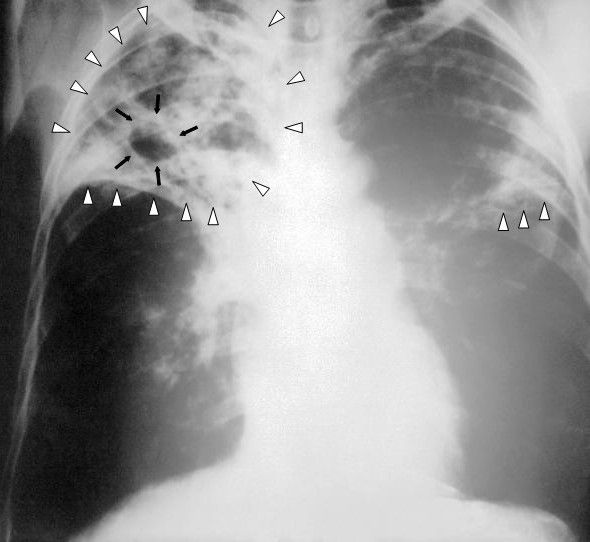
Tuberculosis: airborne, chronic, treated for months
Mycobacterium tuberculosis is inhaled in droplet nuclei small enough to stay suspended for hours and to reach the alveoli. Macrophages ingest the bacilli but cannot kill them; the organism survives intracellularly, and cell-mediated immunity walls it into a granuloma. In roughly 90 percent of immunocompetent people that containment holds for life — latent tuberculosis infection: positive test, no symptoms, normal chest film, not infectious. When immunity falters (HIV, tumour necrosis factor inhibitors, chronic steroids, transplantation, diabetes, malnutrition, dialysis, ageing), the granuloma breaks down and caseous material erodes into a bronchus, producing the cavity, the cough and the transmissibility of active disease.
Active pulmonary tuberculosis presents with a cough lasting more than two to three weeks, haemoptysis, drenching night sweats, low-grade afternoon fever, anorexia, fatigue and weight loss. Screening uses the tuberculin skin test, read at 48 to 72 hours by palpating induration rather than measuring redness, or an interferon-gamma release assay, which is not confounded by prior BCG vaccination. Induration of 5 mm is positive in people with HIV, recent close contacts, fibrotic changes on chest film and organ-transplant or otherwise immunosuppressed patients; 10 mm is positive for recent immigrants from high-prevalence countries, injection drug users, healthcare workers, residents and staff of congregate settings, children under four (and any child or adolescent exposed to an adult in a high-risk category) and people with diabetes, silicosis or chronic kidney disease; 15 mm is required in people with no risk factors. A positive screen means infection, not disease — a chest radiograph and symptom review distinguish latent from active, and active disease is confirmed by sputum acid-fast bacilli smear, nucleic acid amplification and culture, which remains the standard for susceptibility.
Suspected active pulmonary tuberculosis goes immediately into a negative-pressure airborne infection isolation room with at least six air changes per hour (twelve in newer construction), door closed. Staff wear a fit-tested N95 or a PAPR; the patient wears a surgical mask — never an N95 with an exhalation valve — whenever they must leave the room, and transport is minimised. Precautions may be lifted only when the patient has clinically improved on effective therapy and has three consecutive negative sputum smears collected 8 to 24 hours apart with at least one early-morning specimen. Standard treatment for drug-susceptible disease is RIPE: rifampin, isoniazid, pyrazinamide and ethambutol for two months, then rifampin and isoniazid for four more, for six months in total, usually given by directly observed therapy. Latent infection is treated with shorter regimens such as weekly isoniazid plus rifapentine for 12 weeks or daily rifampin for four months.
| Drug | Signature toxicity | Teaching and monitoring |
|---|---|---|
| Rifampin | Hepatotoxicity; potent CYP450 inducer | Orange-red urine, sweat and tears; permanently stains soft contact lenses; makes oral contraceptives, warfarin, methadone and many antiretrovirals fail — use a barrier method |
| Isoniazid | Hepatitis; peripheral neuropathy | Give pyridoxine (vitamin B6) 25–50 mg daily; report numbness or tingling; avoid alcohol; baseline and monthly LFTs in at-risk patients |
| Pyrazinamide | Hepatotoxicity; hyperuricaemia | Report joint pain and swelling; push fluids; monitor uric acid and LFTs |
| Ethambutol | Optic neuritis | Baseline and monthly visual acuity and red-green colour discrimination; report any change in vision immediately |
Three of the four are hepatotoxic, so teach every patient to stop the drugs and call for nausea, vomiting, right upper quadrant pain, dark urine, pale stools or jaundice, and to avoid alcohol and unsupervised acetaminophen. The therapeutic problem is time: patients feel well within weeks but must continue for months, and non-adherence is what breeds multidrug-resistant tuberculosis. Directly observed therapy, incentives, transport support, an interpreter and a named public health contact are not niceties — they are the treatment. Tuberculosis is reportable to public health, and contact investigation is a public health function you support with accurate history rather than perform yourself.
HIV: the labs that define stage and the therapy that ends transmission
HIV is a retrovirus that binds CD4 receptors, transcribes its RNA into DNA with reverse transcriptase, integrates into the host genome with integrase, and destroys CD4 T-lymphocytes — the coordinators of cell-mediated immunity. Untreated, the CD4 count falls over years and opportunistic infections appear in a predictable order. Screening uses a fourth-generation antigen and antibody combination immunoassay that detects p24 antigen and shortens the window period to roughly two to three weeks; a reactive result is confirmed with an HIV-1/HIV-2 antibody differentiation assay, and an indeterminate result is resolved with nucleic acid testing. A normal CD4 count is 500 to 1,600 cells/mm³. AIDS is defined by a CD4 count below 200 cells/mm³ or by an AIDS-defining illness regardless of count.
Antiretroviral therapy is started for everyone with HIV, as soon as possible after diagnosis, regardless of CD4 count. Modern first-line regimens are integrase strand transfer inhibitor based — commonly bictegravir with tenofovir alafenamide and emtricitabine as a single daily tablet, or dolutegravir with a nucleoside backbone. The measure of success is the viral load: suppression to undetectable, generally under 50 copies/mL, within three to six months. Teach the two facts that change lives. First, adherence must be near-perfect, because partially suppressive levels select resistance and burn a whole drug class. Second, undetectable equals untransmittable: a person with sustained viral suppression does not transmit HIV sexually. Prevention for those without HIV includes pre-exposure prophylaxis, either a daily oral tenofovir-emtricitabine tablet or long-acting injectable cabotegravir, and post-exposure prophylaxis started within 72 hours — ideally within 2 hours — and continued for 28 days.
Prophylaxis against opportunistic infection is keyed to the CD4 count: TMP-SMX when the count falls below 200 cells/mm³ to prevent Pneumocystis jirovecii pneumonia, which presents insidiously with dry cough, dyspnoea on exertion and marked hypoxia with a near-normal-sounding chest; TMP-SMX also covers toxoplasmosis below 100 cells/mm³ in patients with positive serology; and azithromycin has traditionally been used below 50 cells/mm³ for Mycobacterium avium complex. Watch for immune reconstitution inflammatory syndrome in the first weeks after starting therapy in a severely immunosuppressed patient — a paradoxical worsening of fever and inflammation as returning immunity attacks an existing occult infection. It is generally not a reason to stop antiretrovirals. Finally, remember that HIV status is protected health information: discuss it in private, disclose only to those involved in care, and do not assume the family in the room knows.
Hepatitis B and C, and occupational exposure
Hepatitis B is transmitted percutaneously, sexually and perinatally, and its serology can be read like a sentence. HBsAg positive means the virus is present now; persisting beyond six months defines chronic infection. Anti-HBs alone means immunity from vaccination. Anti-HBs plus anti-HBc (total) means resolved natural infection. Anti-HBc IgM indicates acute infection. HBeAg and a high HBV DNA level indicate high replication and high infectivity. Chronic hepatitis B is suppressed — not usually cured — with tenofovir or entecavir, and it requires lifelong surveillance for cirrhosis and hepatocellular carcinoma. Hepatitis B is vaccine-preventable, and universal adult vaccination through age 59 is now recommended.
Hepatitis C is predominantly bloodborne, most often through injection drug use. Screening is an anti-HCV antibody test; because antibody persists after cure, current infection must be confirmed with HCV RNA. Direct-acting antiviral regimens taken for 8 to 12 weeks cure over 95 percent of patients, measured as an undetectable viral load 12 weeks after therapy ends. Teach that reinfection is possible — cure is not immunity — and screen for hepatitis B before treatment, because HBV can reactivate. Volume I covered the hepatic failure end of this spectrum; here the nursing emphasis is screening, linkage to care, harm reduction and dismantling the stigma that keeps people from returning for the tablets that would cure them.
For an occupational exposure — a needlestick, a splash to mucous membrane or non-intact skin — act in this order. Wash the site with soap and water immediately, or flush mucous membranes with water or saline for several minutes; do not squeeze the wound and do not apply caustics. Report at once to the supervisor and occupational health, because the clock matters. Source and healthcare-worker testing follow institutional protocol. Approximate per-exposure transmission risk from a percutaneous injury with an infected source is up to 30 percent for hepatitis B in an unvaccinated person, roughly 1.8 percent for hepatitis C and about 0.3 percent for HIV. Hepatitis B is managed with vaccine and, when indicated, hepatitis B immune globulin; there is no post-exposure prophylaxis for hepatitis C, so the response is serial testing and immediate treatment if infection occurs; HIV prophylaxis is a three-drug regimen begun within 72 hours and taken for 28 days.
In chronic communicable disease the pharmacology is the easy part — the nursing work is adherence over months, protection of confidentiality, and the isolation and public-health steps that stop the next case.
The chapter in one breath
Infection is a race between a pathogen and a host, and the findings you assess — fever, tachycardia, tachypnoea, leukocytosis with a left shift, local heat and erythema — are the host’s response made visible. Build the picture before the first dose: two sets of blood cultures from two sites, the right specimen from the right compartment, and markers you can actually interpret against reference values. Then remember that source control beats drug selection — drain the abscess, debride the fascia, pull the line, relieve the obstruction. Choose antimicrobials by mechanism, because the mechanism names the monitoring: creatinine and hearing for aminoglycosides and vancomycin, creatine kinase for daptomycin, a CBC for prolonged linezolid, a stool watch for anything that empties the colon of anaerobes. Practise stewardship at the bedside by getting cultures first, giving doses on time, delabelling false penicillin allergies, prompting the 48-hour time-out, converting to oral and stopping on the intended day. Isolate correctly, wash with soap and water for spores, and honour the device bundles that prevent most of what we treat. Escalate immediately for pain out of proportion, a suddenly quiet distended abdomen in C. difficile, a new neurological deficit in endocarditis and fever in neutropenia. In older adults, expect confusion instead of fever — and never treat a urine culture that has no patient attached to it.
Words worth knowing
| Term | What it means |
|---|---|
| Antimicrobial stewardship | Coordinated practice of giving the right drug, dose, route and duration — or no drug — to preserve effectiveness and limit harm |
| De-escalation | Narrowing broad empiric therapy to a targeted agent once culture and susceptibility results return, usually at 48–72 hours |
| Left shift / bandemia | Increased immature band neutrophils (over roughly 10 percent), signalling that the marrow is releasing cells early under bacterial stress |
| Absolute neutrophil count | WBC times (segs plus bands) divided by 100; under 500/mm³ is severe neutropenia and a medical emergency with fever |
| Procalcitonin | Peptide that rises in bacterial infection; used chiefly to support stopping antibiotics rather than starting them |
| Minimum inhibitory concentration | Lowest drug concentration that prevents visible organism growth; the basis of the susceptible, intermediate and resistant report |
| Time-dependent vs concentration-dependent killing | Time-dependent efficacy depends on time above MIC (beta-lactams) — doses must be on time, sometimes as extended infusions; concentration-dependent efficacy depends on peak-to-MIC ratio (aminoglycosides, fluoroquinolones) — the basis of extended-interval dosing |
| Vancomycin infusion reaction | Histamine-mediated flushing and pruritus from rapid infusion; a rate problem, not an allergy |
| MRSA | Methicillin-resistant Staphylococcus aureus; resistant to all beta-lactams except ceftaroline — treat with vancomycin, daptomycin or linezolid |
| ESBL / CRE | Extended-spectrum beta-lactamase organisms (need a carbapenem) and carbapenem-resistant Enterobacterales (need newer agents) |
| Pseudomembranous colitis | Yellow-white mucosal plaques of C. difficile toxin injury; requires soap-and-water hand hygiene and sporicidal cleaning |
| Toxic megacolon | Colonic dilation with systemic toxicity; suspect when diarrhoea suddenly stops and the abdomen becomes distended and quiet |
| Asymptomatic bacteriuria | Bacteria in urine without urinary symptoms; treated only in pregnancy or before mucosa-traumatising urologic procedures |
| Necrotising fasciitis | Fascial-plane infection with pain out of proportion, crepitus, bullae and skin anaesthesia; treated by emergent surgical debridement |
| Sequestrum | Devascularised dead bone in chronic osteomyelitis that no antibiotic can penetrate; must be removed surgically |
| Osler nodes / Janeway lesions | Painful finger and toe nodules versus painless palm and sole macules in infective endocarditis (Osler equals ouch) |
| Latent tuberculosis infection | Positive skin test or IGRA with no symptoms and a normal chest film; not infectious, but treatable to prevent reactivation |
| Induration | The palpable raised firmness measured when reading a tuberculin skin test at 48–72 hours — redness alone is not measured |
| Undetectable equals untransmittable | Sustained HIV viral suppression on antiretroviral therapy prevents sexual transmission |
| Immune reconstitution inflammatory syndrome | Paradoxical inflammatory worsening after starting antiretroviral therapy as immunity returns and attacks an occult infection |