Blood, the Lymphatic System and Immunity
Blood is the only tissue you can send to a laboratory in a tube, and that fact has generated an enormous, unusually precise vocabulary. Every cell in the blood has a name built from a color, a shape or a nucleus; every deficiency has a -penia; every excess has an -osis; and the whole system is measured, counted and charted daily on patients you will care for. Running alongside blood is the lymphatic system — the drainage network that returns fluid to the circulation and houses the immune cells that patrol it. The two systems share roots, share diseases and share a chapter for good reason. Once you can decode erythr/o, leuk/o, thromb/o, -emia, -poiesis, lymphaden/o and immun/o, a phrase such as “pancytopenia with hepatosplenomegaly and diffuse lymphadenopathy” stops being a wall of letters and becomes a sentence you can read.
By the end of this chapter you can
- Decode terms built on the core blood roots hem/o, hemat/o, plasm/o and ser/o, and use the suffix -emia correctly.
- Distinguish the formed-element roots erythr/o, leuk/o and thromb/o, and attach -cyte, -blast, -osis, -penia and -poiesis to each.
- Explain why myel/o means marrow in a hematology note and spinal cord in a neurology note, and why my/o means neither.
- Interpret red-cell descriptors built from micr/o, macr/o, anis/o, poikil/o, spher/o, chrom/o and sider/o.
- Name the five white-cell types, their combining forms, and the meaning of the suffix -phil.
- Separate thromb/o, embol/o, coagul/o and fibrin/o, and use -stasis, -rrhage and -lysis in bleeding and clotting terms.
- Decode lymphatic terms built on lymph/o, lymphaden/o, lymphangi/o, splen/o, thym/o and tonsill/o.
- Apply the plural rules to thrombus, embolus, petechia, ecchymosis, lymphoma and bacterium.
- Read the abbreviations that fill a real chart, including CBC, H&H, MCV, ANC, PT/INR, aPTT, ESR, DIC, HSCT and CD4.
Section 1The Vocabulary of Blood
Two roots for blood, and one suffix that outranks them
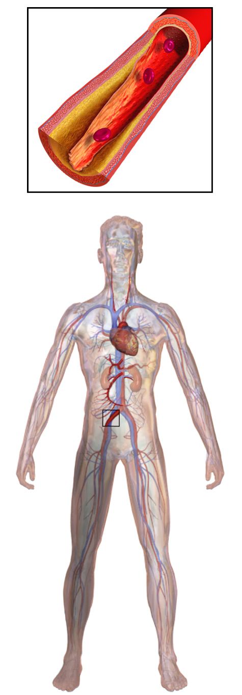
Blood carries two Greek combining forms that mean exactly the same thing: hem/o and hemat/o. Neither is more correct than the other; usage simply settled differently on different words. Hem/o gives us hemorrhage, hemostasis, hemolysis and hemoglobin. Hemat/o gives us hematology, hematoma, hematuria and hematopoiesis. When you are unsure which one a word takes, look it up rather than guess — but when you are decoding, either spelling tells you the same thing: this word is about blood.
The suffix -emia does the heavier lifting. It means “blood condition” or, more usefully, “a substance present in the blood.” Whatever sits in front of -emia is the thing that is present, absent or abnormal in the bloodstream. Hyperkalemia is hyper- + kal/i + -emia = excessive + potassium + in the blood. Bacteremia is bacteria in the blood. Uremia is urea in the blood. Septicemia is a systemic infection in the blood, though modern charts increasingly say sepsis instead. And anemia is an- + -emia = without + blood — a slightly overstated name for a deficiency of red cells or hemoglobin rather than a true absence of blood.
Practice the reassembly out loud, because that is the habit that transfers. Hemat/oma = blood + tumor or mass, so a hematoma is a localized collection of clotted blood, not a neoplasm. Hem/o/lysis (hee-MOL-ih-sis) = blood + breakdown, the destruction of red cells. Hem/o/stasis (hee-moh-STAY-sis) = blood + stopping, the body’s process of arresting bleeding. Note that last one carefully: -stasis means stopping or controlling, so hemostasis is a good thing your body does, while hemorrhage (hem/o + -rrhage, bursting forth) is the opposite.
| Word part | Meaning | Example term | What the example means |
|---|---|---|---|
| hem/o, hemat/o | blood | hematology | the study and specialty of blood |
| -emia | blood condition; substance in the blood | bacteremia | bacteria present in the bloodstream |
| plasm/o | plasma; formation | plasmapheresis | removal of plasma from withdrawn blood |
| ser/o | serum; serous fluid | serology | the study of serum, especially its antibodies |
| cyt/o, -cyte | cell | cytology | the study of cells |
| -poiesis | formation, production | hematopoiesis | the formation of blood cells |
| -poietin | substance that forms | erythropoietin | hormone that stimulates red cell production |
| -blast | immature or developing cell | myeloblast | an immature marrow cell |
| myel/o | bone marrow; also spinal cord | myelofibrosis | fibrous scarring of the bone marrow |
| -globin, -globulin | protein | hemoglobin | the oxygen-carrying protein of red cells |
| -crit | to separate | hematocrit | the percentage of blood volume that is red cells |
| -phil, -philia | attraction for, affinity | eosinophilia | increased eosinophils, cells attracted to rosy dye |
Plasma, serum and the parts you can see in a tube
Spin a tube of blood and it divides into layers, and those layers name themselves. About fifty-five percent of the volume is plasma, the straw-colored liquid that carries proteins, hormones, nutrients and clotting factors. About forty-five percent is packed red cells — that percentage is the hematocrit (hee-MAT-oh-krit), built from hemat/o + -crit = blood + to separate. Between them sits a pale buffy coat of white cells and platelets, normally less than one percent of the volume, which is why a raised white count that looks alarming on paper still occupies almost no space in the tube.
Students confuse plasma and serum constantly, and the distinction is worth ten seconds of your attention. Plasma is the liquid portion of blood with its clotting factors intact; you get it from a tube containing an anticoagulant. Serum is what remains after blood has been allowed to clot — plasma minus fibrinogen and the other consumed clotting factors. That is why the blood bank transfuses fresh frozen plasma to replace clotting factors, while the laboratory runs antibody tests on serum. The root ser/o shows up in serology, seroconversion and serosanguineous (see-roh-sang-GWIN-ee-us) drainage, which is thin fluid tinged with blood.
The suffix -poiesis (poy-EE-sis) means formation or production, and it is the engine of this chapter. Hematopoiesis (hee-mah-toh-poy-EE-sis) is the making of blood cells, which happens in red bone marrow. Erythropoiesis is the making of red cells specifically; leukopoiesis, white cells; thrombopoiesis, platelets. Add -poietin and you get the hormone that drives the process: erythropoietin (eh-rith-roh-POY-eh-tin), or EPO, released by the kidney when oxygen delivery falls. That single word explains why patients with chronic kidney disease become anemic — a failing kidney makes less erythropoietin.
Where blood cells are born: myel/o
All blood cells arise from a single hematopoietic stem cell in the red bone marrow, and the root for marrow is myel/o. From it come myeloid (relating to the marrow or to the cell line that produces granulocytes), myelodysplastic (myel/o + dys- + -plastic = marrow + faulty + formation), myelofibrosis and myeloma. Multiple myeloma (my-eh-LOH-mah) is a malignancy of plasma cells in the marrow, and the term names its home tissue rather than its cell of origin — a reminder that some terms are historical labels rather than perfect descriptions.
The suffix -blast marks an immature or developing cell. An erythroblast is a developing red cell, a myeloblast is a developing granulocyte, a lymphoblast is a developing lymphocyte. When a chart reports “blasts on the peripheral smear,” it means immature cells have escaped the marrow into circulating blood, which is a hallmark of acute leukemia. A megakaryocyte (meg-ah-KAR-ee-oh-site) — megal/o + kary/o + -cyte = large + nucleus + cell — is the giant marrow cell that sheds fragments of its own cytoplasm to make platelets.
Blood terms are built from two interchangeable roots at the front — hem/o and hemat/o — and the suffix -emia at the back, so the first thing to identify in an unfamiliar blood word is which end the blood idea is sitting on.
Section 2Red Cells, Oxygen and the Anemias
Erythr/o and the shape of a red cell
The red blood cell is an erythrocyte (eh-RITH-roh-site): erythr/o + -cyte = red + cell. It is the most numerous cell in the body, it has no nucleus once mature, and it exists to carry hemoglobin (hem/o + -globin = blood + protein), the iron-containing molecule that binds oxygen. Because the cell is essentially a bag of hemoglobin, every red-cell disease is described in two dimensions: how big the cells are, and how much hemoglobin they contain. Medical terminology gives you a root for each.
Size uses micr/o (small), macr/o (large) and norm/o (normal), attached to -cytic. Microcytic (my-kroh-SIT-ik) cells are abnormally small; macrocytic cells are abnormally large; normocytic cells are the right size. Color — which is really hemoglobin content — uses chrom/o. Hypochromic (hy-poh-KROH-mik) cells are pale because they are underfilled with hemoglobin; normochromic cells stain normally. Put the two systems together and you get the phrase that appears in thousands of charts: microcytic hypochromic anemia, small pale cells, the classic picture of iron deficiency.
Two more descriptors appear on smear reports and both are pure word parts. Anisocytosis (an-eye-soh-sye-TOH-sis) is anis/o + cyt/o + -osis = unequal + cell + condition, meaning the red cells vary in size. Poikilocytosis (poy-kih-loh-sye-TOH-sis) is poikil/o + cyt/o + -osis = varied or irregular + cell + condition, meaning they vary in shape. Add spher/o (round) for spherocytosis, in which cells lose their biconcave dish shape, and drepan/o (sickle) for drepanocyte, the formal name for a sickle cell.
| Word part | Meaning | Example term | What the example means |
|---|---|---|---|
| erythr/o | red | erythrocyte | red blood cell |
| micr/o | small | microcytic | pertaining to abnormally small cells |
| macr/o, megal/o | large | macrocytic | pertaining to abnormally large cells |
| norm/o | normal, usual | normochromic | having normal hemoglobin color |
| chrom/o, chromat/o | color | hypochromic | having less color, meaning less hemoglobin |
| anis/o | unequal | anisocytosis | red cells of unequal size |
| poikil/o | varied, irregular | poikilocytosis | red cells of irregular shape |
| spher/o | round, sphere | spherocyte | an abnormally round red cell |
| drepan/o | sickle | drepanocyte | a sickle-shaped red cell |
| sider/o | iron | hemosiderin | an iron-storage pigment left after red cells break down |
| ferr/o, ferrit/o | iron (Latin) | ferritin | the protein that stores iron; measured to assess iron stores |
| reticul/o | net, network | reticulocyte | a young red cell with a netlike remnant inside |
The anemias, decoded rather than memorized
Anemia (ah-NEE-mee-ah) is an- + -emia = without + blood, meaning a deficiency of red cells or of hemoglobin. It is a finding, never a final diagnosis, and the adjective in front of it tells you the mechanism. Iron deficiency anemia is microcytic and hypochromic because there is not enough iron to fill the cells. Pernicious anemia is macrocytic because vitamin B12 deficiency — caused here by loss of intrinsic factor from the stomach — disrupts DNA synthesis, so the cells enlarge without dividing properly. Aplastic anemia uses a- + -plastic = without + formation: the marrow itself has failed, so all three cell lines fall.
Hemolytic anemia is hem/o + -lytic = blood + destroying, meaning red cells are being broken down faster than they can be replaced. When hemoglobin is released and metabolized, bilirubin rises, which is why jaundice so often accompanies hemolysis. Sickle cell disease is an inherited hemoglobinopathy (hee-moh-gloh-bih-NOP-ah-thee) — hem/o + globin + -pathy = blood + protein + disease — in which abnormal hemoglobin polymerizes and deforms the cell, producing both hemolysis and painful vaso-occlusive crises. Thalassemia (thal-ah-SEE-mee-ah) is another inherited defect, this one in the rate at which globin chains are produced.
The opposite of anemia takes an -osis or a -cytosis. Polycythemia (pol-ee-sye-THEE-mee-ah) is poly- + cyt/o + -hemia = many + cell + blood condition, an excess of red cells that thickens the blood and raises clot risk. Polycythemia vera is the primary marrow disorder; secondary polycythemia is the body’s appropriate response to chronic hypoxia, which is why you may meet it in a patient with long-standing COPD or in someone living at high altitude. A rising reticulocyte count in any anemia is good news: the marrow is responding by shipping out young cells.
| Term | Word parts | What it means clinically |
|---|---|---|
| Anemia | an- (without) + -emia (blood) | Deficiency of red cells or hemoglobin; a finding, not a diagnosis |
| Hemolysis | hem/o (blood) + -lysis (breakdown) | Destruction of red cells, releasing hemoglobin and raising bilirubin |
| Hemoglobinopathy | hem/o + globin + -pathy (disease) | Inherited disease of the hemoglobin molecule, such as sickle cell disease |
| Polycythemia | poly- (many) + cyt/o (cell) + -hemia | Excess red cells; raises blood viscosity and clot risk |
| Pancytopenia | pan- (all) + cyt/o + -penia (deficiency) | Deficiency of all three cell lines — red cells, white cells and platelets |
| Erythropoiesis | erythr/o (red) + -poiesis (formation) | Production of red cells in the bone marrow |
| Hemochromatosis | hem/o + chromat/o (color) + -osis (condition) | Iron overload with pigment deposition in liver, heart and pancreas |
| Erythema | erythr/o (red) | Redness of the skin — the same root working in a different tissue |
Red-cell terms describe size, color and shape with a small set of interchangeable roots — micr/o, macr/o, chrom/o, anis/o, poikil/o — so the adjective in front of the word anemia names the mechanism, not merely the severity.
Section 3White Cells, Platelets and the Language of Clotting
Leuk/o and the differential
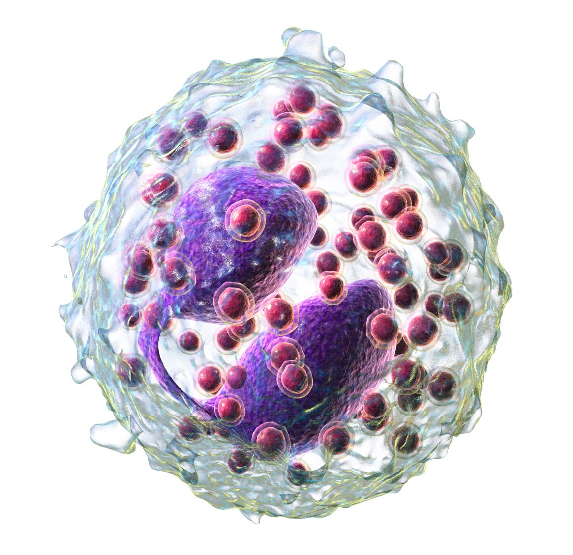
A white blood cell is a leukocyte (LOO-koh-site): leuk/o + -cyte = white + cell. There are five kinds, and the laboratory report that names their proportions is called the differential, usually shortened on a chart to “diff.” Three of the five carry visible granules and are collectively granulocytes; two do not and are agranulocytes. The names of the granulocytes come from nineteenth-century staining, and the suffix that unlocks all three is -phil, meaning attraction or affinity for.
A neutrophil (NOO-troh-fil) is neutr/o + -phil = neutral + attracted to: its granules take up neither the acidic nor the basic dye. Neutrophils are the first responders to bacterial infection, and because a mature one has a segmented nucleus you will see them charted as segs, or as polys and PMNs for polymorphonuclear leukocyte — poly- + morph/o + nucle/o = many + shape + nucleus. Immature neutrophils are bands, and a rise in bands during acute infection is the classic “left shift.” An eosinophil (ee-oh-SIN-oh-fil) is attracted to eosin, a rosy acidic dye, and rises in allergy and parasitic infection. A basophil takes up basic dye and releases histamine and heparin.
The two agranulocytes are the lymphocyte — the cell of specific immunity, discussed in Section 5 — and the monocyte, from mon/o + -cyte = one + cell, named for its single unlobed nucleus. When a monocyte leaves the bloodstream and enters tissue it becomes a macrophage (MAK-roh-fayj): macr/o + -phage = large + eater. That suffix belongs to the root phag/o, to eat or swallow, and it will follow you through microbiology and immunology. Phagocytosis is the process by which such a cell engulfs and digests a pathogen.
| Word part | Meaning | Example term | What the example means |
|---|---|---|---|
| leuk/o | white | leukocyte | white blood cell |
| granul/o | granule, small grain | agranulocytosis | a severe deficiency of granulocytes |
| neutr/o | neutral | neutropenia | deficiency of neutrophils |
| eosin/o | rosy, dawn-colored | eosinophilia | increased eosinophils in the blood |
| bas/o | base, basic (opposite of acidic) | basophil | white cell whose granules take up basic dye |
| mon/o | one, single | monocyte | white cell with a single unlobed nucleus |
| phag/o, -phage | eat, swallow | macrophage | large tissue cell that engulfs debris and microbes |
| nucle/o, kary/o | nucleus | polymorphonuclear | having a many-shaped nucleus, as a neutrophil does |
| morph/o | shape, form | morphology | the study of shape, as of cells on a smear |
| -phil, -philia | attraction for, affinity | neutrophilia | an increase in neutrophils |
Counts move in two directions and take the suffixes you already know. Leukocytosis (loo-koh-sye-TOH-sis) is an increase in white cells, usually a response to infection, inflammation or stress. Leukopenia is a decrease, and its most dangerous form is neutropenia (noo-troh-PEE-nee-ah), a deficiency of the very cells that fight bacteria. Chemotherapy causes neutropenia predictably, which is why you will chart the absolute neutrophil count (ANC) and place a patient on neutropenic precautions when it falls. Agranulocytosis (ay-gran-yoo-loh-sye-TOH-sis) — a- + granul/o + cyt/o + -osis = without + granule + cell + condition — is a near-total loss of granulocytes and is a medical emergency, however innocent the -osis ending may look.
Leukemia (loo-KEE-mee-ah) is leuk/o + -emia = white + blood condition: a malignancy of the blood-forming tissue in which abnormal white cells crowd out normal marrow. Its four common forms are named by two axes, and both axes are word parts you can decode. Acute means immature blast cells and rapid onset; chronic means more mature cells and a slower course. Lymphocytic names the lymphoid line; myelogenous (my-eh-LOJ-eh-nus), from myel/o + -genous = marrow + produced by, names the granulocyte line. Cross them and you get ALL, AML, CLL and CML — four abbreviations that describe themselves once you know the four parts.
Thromb/o, coagul/o and the vocabulary of bleeding
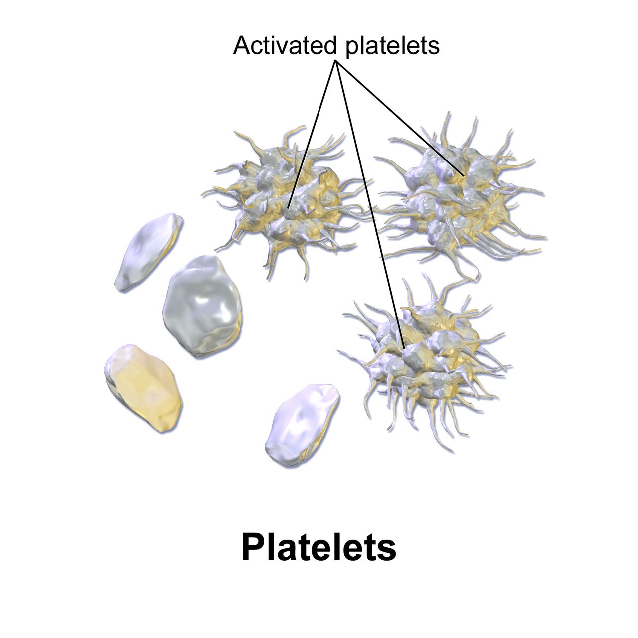
A platelet is a thrombocyte (THROM-boh-site): thromb/o + -cyte = clot + cell. It is not a whole cell at all but a fragment shed from a megakaryocyte, and it begins the clotting sequence by plugging an injured vessel. Thrombocytopenia (throm-boh-sye-toh-PEE-nee-ah) is a deficiency of platelets and produces bleeding; thrombocytosis is an excess and can produce clotting. Hemostasis is the whole coordinated process of stopping blood loss: vessel spasm, platelet plug, then a fibrin mesh built by the coagulation cascade, from coagul/o, to clot.
The distinction between two nouns matters enormously in practice. A thrombus (THROM-bus) is a clot that forms and stays where it formed. An embolus (EM-boh-lus), from embol/o meaning plug or stopper, is anything that travels through the bloodstream and lodges elsewhere — a broken-off clot, but also fat, air or amniotic fluid. So a deep vein thrombosis becomes a pulmonary embolism when the clot breaks loose and travels to the lung. Both nouns take the Latin plural: one thrombus, two thrombi; one embolus, two emboli.
Bleeding into skin has its own precise ladder, and clinicians expect you to use the right rung. Petechiae (peh-TEE-kee-ee) are pinpoint hemorrhages, singular petechia. Purpura (PUR-pyoo-rah) are larger purple patches. An ecchymosis (ek-ih-MOH-sis) is a bruise, plural ecchymoses. A hematoma is a pocket of clotted blood with mass and volume. The suffix family that names bleeding itself deserves a moment: -rrhage and -rrhagia mean bursting forth of blood (hemorrhage, menorrhagia), -rrhea means flow or discharge (rhinorrhea), -rrhexis means rupture, and -rrhaphy means suturing. Four suffixes, all with the doubled r, all describing entirely different events.
| Word part | Meaning | Example term | What the example means |
|---|---|---|---|
| thromb/o | clot | thrombosis | abnormal condition of clot formation in a vessel |
| embol/o | plug, stopper | embolism | obstruction by material carried in the bloodstream |
| coagul/o | clotting | anticoagulant | an agent that works against clot formation |
| fibrin/o | fibrin, fibers | fibrinolysis | breakdown of the fibrin mesh that holds a clot together |
| -stasis | stopping, controlling | hemostasis | the arrest of bleeding |
| -rrhage, -rrhagia | bursting forth of blood | hemorrhage | rapid, excessive loss of blood |
| -lysis, -lytic | breakdown, destruction | thrombolytic | a drug that dissolves an existing clot |
| -penia | deficiency | thrombocytopenia | deficiency of platelets, producing bleeding risk |
| hem/o + -philia | blood + attraction, tendency | hemophilia | inherited deficiency of a clotting factor |
| purpur/o | purple | purpura | purple discoloration from bleeding under the skin |
White cells are named for the dye their granules take or the shape of their nucleus, platelets are named for the clot they start, and the suffixes -osis, -penia, -emia and -rrhage convert those names into countable, chartable findings.
Section 4The Lymphatic System: Vessels, Nodes and Organs
What lymph is and where it goes
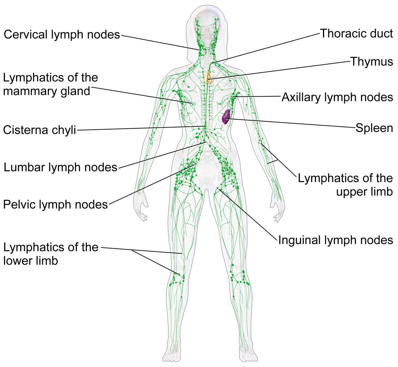
Every minute, plasma leaks out of capillaries into the tissue spaces. Most of it returns to the venous side, but roughly three liters a day do not, and if that fluid stayed where it was you would swell and eventually die. The lymphatic system collects it. Once the fluid enters a lymphatic capillary it is called lymph, from the Latin for clear water, and the combining form is lymph/o. Lymph travels through progressively larger lymphatic vessels — combining form lymphangi/o, from angi/o meaning vessel — passes through filtering lymph nodes (lymphaden/o), and finally empties back into the bloodstream through the thoracic duct and the right lymphatic duct at the base of the neck.
The system has no pump. Lymph moves by skeletal muscle contraction, breathing and one-way valves, which is why immobility produces swelling and why elevation and compression are the standard nursing interventions for it. In the small intestine, specialized lymphatic capillaries called lacteals absorb dietary fat, turning the lymph there milky white; that fluid is chyle (KILE). The term chylothorax — chyle in the pleural space — tells you the thoracic duct has been injured, and it is a good example of a word whose parts fully explain the clinical problem.
Three organs join the vessels and nodes. The spleen (splen/o) filters blood rather than lymph, removes worn-out red cells and stores platelets. The thymus (thym/o) sits behind the sternum, is large in childhood and shrinks in adulthood, and is where T lymphocytes mature — the T stands for thymus. The tonsils (tonsill/o) guard the entrance to the throat. Note the spelling trap: the organ has one l, but the surgery tonsillectomy has two, because the Latin root is tonsill/o.
| Word part | Meaning | Example term | What the example means |
|---|---|---|---|
| lymph/o | lymph, lymphatic tissue | lymphocyte | white cell of the lymphatic and immune system |
| lymphaden/o | lymph node | lymphadenopathy | disease or enlargement of the lymph nodes |
| lymphangi/o | lymphatic vessel | lymphangitis | inflammation of a lymphatic vessel |
| aden/o | gland | adenopathy | disease of a gland (loosely used for node enlargement) |
| adenoid/o | adenoids | adenoidectomy | surgical removal of the adenoids |
| splen/o | spleen | splenomegaly | enlargement of the spleen |
| thym/o | thymus gland | thymectomy | surgical removal of the thymus |
| tonsill/o | tonsil | tonsillitis | inflammation of the tonsils |
| lymphat/o | lymphatic system | lymphatic | pertaining to lymph and its vessels |
| chyl/o | chyle, fatty lymph | chylothorax | chyle leaking into the pleural cavity |
| -edema | swelling from fluid | lymphedema | swelling caused by obstructed lymph drainage |
| -megaly | enlargement | hepatosplenomegaly | enlargement of both liver and spleen |
Lymphatic pathology, from swollen nodes to lymphoma
Start with the word you will hear on rounds most often. Lymphadenopathy (lim-fad-eh-NOP-ah-thee) is lymphaden/o + -pathy = lymph node + disease: enlarged or abnormal lymph nodes. It is a sign, not a diagnosis, and the note will always qualify it — cervical, axillary, inguinal, localized or generalized. Lymphadenitis adds -itis and specifies inflammation, usually from infection draining into that node. Lymphangitis (lim-fan-JYE-tis) is inflammation of the vessel itself, and it produces the red streak tracking up a limb from an infected wound that should send you to a provider quickly.
Lymphedema (lim-feh-DEE-mah) is swelling caused by obstructed lymph drainage. Its most common cause in developed countries is treatment for cancer: removing axillary nodes during breast cancer surgery interrupts the drainage of the arm, which is why that arm is protected from blood draws, blood pressure cuffs and injections for life. Worldwide, the leading cause is filariasis, a parasitic infection whose severe chronic form is called elephantiasis. Elevation, compression and manual lymphatic drainage are the mainstays of care because there is no pump to restart.
Lymphoma (lim-FOH-mah) is lymph/o + -oma = lymph tissue + tumor, a malignancy of lymphocytes that lives in the nodes rather than circulating like leukemia. It is divided by an eponym that has survived because the distinction still matters: Hodgkin lymphoma (HOJ-kin), identified by the presence of the giant Reed-Sternberg cell on biopsy, and non-Hodgkin lymphoma, a large family of everything else. Modern style has dropped the possessive — write Hodgkin lymphoma, not Hodgkin’s disease — and the same convention now applies to Down syndrome and Crohn disease. The plural of a Greek -oma is technically -omata (lymphomata), but English plurals are standard in charting: lymphomas, hematomas, sarcomas.
The spleen supplies its own vocabulary. Splenomegaly (splee-noh-MEG-ah-lee) is enlargement, common in mononucleosis, portal hypertension and hematologic malignancy; an enlarged spleen is fragile, which is why contact sports are restricted after infectious mononucleosis — mon/o + nucle/o + -osis, named for the abundant single-nucleus cells the Epstein-Barr virus provokes. Hypersplenism is an overactive spleen destroying too many cells; splenectomy (splee-NEK-toh-mee) is its removal, after which the patient needs vaccination against encapsulated bacteria for the rest of their life. A thymoma is a tumor of the thymus, notable for its association with myasthenia gravis.
| Term | Word parts | What it means clinically |
|---|---|---|
| Lymphadenopathy | lymphaden/o (node) + -pathy (disease) | Enlarged or abnormal lymph nodes; a sign requiring a cause |
| Lymphadenitis | lymphaden/o + -itis (inflammation) | Inflamed, tender nodes, usually draining an infection |
| Lymphangiography | lymphangi/o (vessel) + -graphy (process of recording) | Imaging of lymphatic vessels after contrast injection |
| Lymphadenectomy | lymphaden/o + -ectomy (removal) | Surgical removal of lymph nodes, often for cancer staging |
| Lymphedema | lymph/o + -edema (swelling) | Chronic swelling from blocked or removed lymphatic drainage |
| Splenomegaly | splen/o (spleen) + -megaly (enlargement) | Enlarged spleen, palpable below the left costal margin |
| Splenorrhaphy | splen/o + -rrhaphy (suturing) | Surgical repair of a lacerated spleen instead of removing it |
| Thymectomy | thym/o (thymus) + -ectomy | Removal of the thymus, sometimes done in myasthenia gravis |
| Tonsillectomy | tonsill/o (tonsil) + -ectomy | Removal of the palatine tonsils |
| Lymphoma | lymph/o + -oma (tumor) | Malignancy of lymphocytes arising in lymphoid tissue |
Lymphatic terms name one of three things — the fluid, the vessel or the node — and the roots lymph/o, lymphangi/o and lymphaden/o keep those three separate no matter what suffix is attached.
Section 5Immunity, Diagnostics and the Abbreviations on the Chart
Immun/o and the two arms of defense
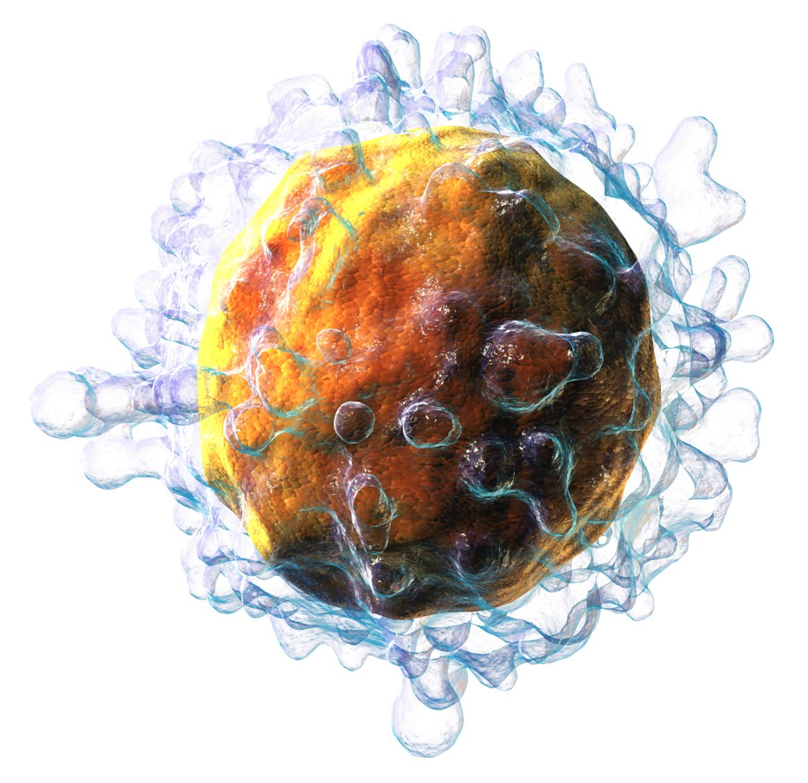
The root immun/o comes from the Latin for exempt or safe, and it heads nearly every term in this field. Defense arrives in two layers. Innate immunity is present from birth, is nonspecific, and includes skin, mucous membranes, stomach acid, fever, inflammation and phagocytes. Adaptive (or acquired) immunity is specific, learns from exposure and remembers — and it is built from lymphocytes. B lymphocytes mature in the bone marrow and produce antibodies; T lymphocytes mature in the thymus and either kill infected cells directly or direct the rest of the response.
Two nouns anchor everything else, and students reverse them constantly. An antigen is any substance the body recognizes as foreign — the term is anti- + -gen = against + producing, meaning it produces a response against itself. An antibody, also called an immunoglobulin (im-yoo-noh-GLOB-yoo-lin), is the protein the body makes in response. The suffix -globulin simply means protein, and the five classes are abbreviated IgG, IgA, IgM, IgE and IgD. IgE drives allergy; IgM is the first antibody made in a new infection; IgG is the long-term memory antibody and the only one that crosses the placenta.
How that immunity is obtained produces four labels worth decoding once. Active immunity means your own body made the antibodies; passive means they were given to you ready-made. Natural means it happened through ordinary life; artificial means medicine arranged it. Cross the two and you get: natural active (you had the infection), artificial active (you were vaccinated), natural passive (maternal antibodies crossing the placenta or arriving in breast milk), and artificial passive (an injection of immune globulin, which protects immediately but only for weeks).
| Word part | Meaning | Example term | What the example means |
|---|---|---|---|
| immun/o | protection, safe, immune | immunosuppression | deliberate or disease-related weakening of immune response |
| -gen, -genic | producing, forming | pathogenic | capable of producing disease |
| path/o, -pathy | disease | pathogen | an organism that produces disease |
| tox/o, toxic/o | poison | antitoxin | an antibody that works against a specific poison |
| anti- | against | antibody | protein produced against a specific antigen |
| auto- | self | autoimmune | immune attack directed against one’s own tissue |
| allo- | other, different | allogeneic | from a genetically different donor of the same species |
| phylax/o, -phylaxis | protection | anaphylaxis | a severe systemic allergic reaction |
| ser/o | serum | seroconversion | the point at which antibody first becomes detectable |
| -globulin | protein | immunoglobulin | an antibody protein |
| vaccin/o | vaccine | vaccination | giving an antigen to stimulate active immunity |
| -osis | abnormal condition | sarcoidosis | inflammatory disease forming granulomas in many organs |
When immunity misfires
Too much immune response is hypersensitivity, and its everyday name is allergy. A substance that triggers it is an allergen — again -gen, producing. The most dangerous form is anaphylaxis (an-ah-fih-LAK-sis), from ana- + -phylaxis = again or excessive + protection: a whole-body IgE-mediated reaction with airway swelling, bronchospasm and circulatory collapse, treated with epinephrine without delay. Milder terms you will chart include urticaria (er-tih-KAIR-ee-ah) for hives and angioedema for deep swelling of the lips, tongue and face.
Immunity aimed at the wrong target is autoimmune disease, from auto- meaning self. Systemic lupus erythematosus, rheumatoid arthritis, Hashimoto thyroiditis, type 1 diabetes and myasthenia gravis all belong here, and the treatment is usually immunosuppression — which then creates the opposite problem. Too little immune response is immunodeficiency. It may be congenital, or acquired, as in HIV, the human immunodeficiency virus, which destroys CD4 T lymphocytes; when the CD4 count falls below 200 or an opportunistic infection appears, the diagnosis becomes AIDS, acquired immunodeficiency syndrome. A patient who is immunocompromised for any reason — chemotherapy, steroids, transplant drugs, advanced age — needs protection from organisms that would not trouble anyone else.
Transplantation adds two more terms built on donor relationships. Autologous tissue comes from the patient’s own body; allogeneic tissue comes from another person. Rejection is the recipient’s immune system attacking the graft; graft-versus-host disease is the reverse, donor immune cells attacking the recipient, and it is a distinctive risk of allogeneic stem cell transplant. Blood transfusion runs on the same logic in miniature: the ABO and Rh antigens on red cells must be matched, because mismatched antigen meets preformed antibody and the cells clump — agglutination — and hemolyze. Type O negative blood, carrying none of those antigens, is the universal donor; type AB positive is the universal recipient.
Reading the chart: tests, procedures and abbreviations
Most hematology begins with the complete blood count, which reports red cells, white cells, platelets and the red cell indices. The differential breaks the white count into its five types. Clotting is assessed with the prothrombin time and its standardized INR, which follow warfarin, and the activated partial thromboplastin time, which follows heparin. The erythrocyte sedimentation rate and C-reactive protein are nonspecific markers of inflammation. A D-dimer detects fragments of broken-down fibrin and is used to help rule out clot. The Coombs test, an eponym still in daily use alongside its newer name “direct antiglobulin test,” detects antibodies stuck to red cells in hemolytic anemia.
Procedures follow the suffixes you already own. Phlebotomy (fleh-BOT-oh-mee) is phleb/o + -tomy = vein + incision, the drawing of blood; venipuncture is its Latin equivalent. Bone marrow aspiration withdraws liquid marrow through a needle, usually from the posterior iliac crest, while a bone marrow biopsy takes a core of bone as well. Apheresis (af-eh-REE-sis), from the Greek for taking away, withdraws blood, removes one component and returns the rest: plasmapheresis removes plasma, leukapheresis removes white cells. Hematopoietic stem cell transplantation, still often charted as bone marrow transplant, replaces a diseased marrow with healthy stem cells. And splenectomy, lymphadenectomy and thymectomy mean exactly what their -ectomy promises.
| Abbreviation | What it stands for |
|---|---|
| CBC | Complete blood count |
| H&H | Hemoglobin and hematocrit |
| WBC, RBC, PLT | White blood cell, red blood cell and platelet counts |
| MCV, MCH, MCHC | Mean corpuscular volume, hemoglobin and hemoglobin concentration |
| RDW | Red cell distribution width, a measure of size variation |
| diff | Differential — the percentages of each white cell type |
| ANC | Absolute neutrophil count |
| ESR | Erythrocyte sedimentation rate (also called sed rate) |
| CRP | C-reactive protein |
| PT/INR | Prothrombin time and international normalized ratio |
| aPTT | Activated partial thromboplastin time |
| T&C | Type and crossmatch |
| PRBC, FFP | Packed red blood cells; fresh frozen plasma |
| DIC | Disseminated intravascular coagulation |
| DVT, PE | Deep vein thrombosis; pulmonary embolism |
| ITP | Immune thrombocytopenic purpura |
| SCD | Sickle cell disease |
| ALL, AML, CLL, CML | Acute or chronic lymphocytic or myelogenous leukemia |
| HL, NHL | Hodgkin lymphoma; non-Hodgkin lymphoma |
| HSCT, BMT | Hematopoietic stem cell transplant; bone marrow transplant |
| Ig (IgG, IgA, IgM, IgE, IgD) | Immunoglobulin and its five classes |
| HIV, AIDS | Human immunodeficiency virus; acquired immunodeficiency syndrome |
| CD4 | The T lymphocyte subset counted to stage HIV infection |
| SLE | Systemic lupus erythematosus |
Immunology vocabulary turns on knowing which side of the reaction a term names — antigen provokes, antibody defends, auto- attacks self, and immuno- prefixed words describe how much of that defense the patient still has.
The chapter in one breath
Blood carries two equal roots, hem/o and hemat/o, at the front of a term and the suffix -emia at the back, so the first question in any blood word is which end the blood idea occupies. The formed elements name themselves: erythr/o for the red oxygen carrier, leuk/o for the white defender, thromb/o for the clot-starting platelet, each taking -cyte, -blast, -poiesis, -osis and -penia in turn. Red cells are described by size, color and shape — microcytic, hypochromic, anisocytosis, poikilocytosis — so the adjectives in front of the word anemia state the mechanism. White cells are named for the dye their granules take or the shape of their nucleus, and their counts rise with -osis and fall with -penia. Clotting language separates the stationary thrombus from the traveling embolus, and the doubled-r suffixes -rrhage, -rrhea, -rrhexis and -rrhaphy keep bleeding, flow, rupture and repair apart. The lymphatic system distinguishes fluid (lymph/o), vessel (lymphangi/o) and node (lymphaden/o), and adds splen/o, thym/o and tonsill/o. Immunity turns on one relationship: the antigen provokes, the antibody answers, and every immun/o term describes how much of that answer the patient can still mount.
Words worth knowing
| Term | What it means |
|---|---|
| Anemia (ah-NEE-mee-ah) | Deficiency of red blood cells or hemoglobin; a finding whose modifier names the cause. |
| Anisocytosis (an-eye-soh-sye-TOH-sis) | Red blood cells of unequal size on a peripheral smear. |
| Antigen (AN-tih-jen) | Any substance recognized as foreign that provokes an immune response. |
| Antibody (AN-tih-bod-ee) | Immunoglobulin protein produced against a specific antigen. |
| Anaphylaxis (an-ah-fih-LAK-sis) | Severe systemic IgE-mediated allergic reaction; a medical emergency. |
| Ecchymosis (ek-ih-MOH-sis), plural ecchymoses | A bruise; bleeding under the skin larger than petechiae or purpura. |
| Erythropoietin (eh-rith-roh-POY-eh-tin) | Kidney hormone that stimulates red cell production in the marrow. |
| Hematocrit (hee-MAT-oh-krit) | Percentage of total blood volume made up of red cells. |
| Hematopoiesis (hee-mah-toh-poy-EE-sis) | Formation of blood cells, occurring in red bone marrow. |
| Hemolysis (hee-MOL-ih-sis) | Destruction of red blood cells, releasing hemoglobin into plasma. |
| Hemostasis (hee-moh-STAY-sis) | The body’s process of stopping bleeding: spasm, platelet plug, fibrin clot. |
| Leukocytosis (loo-koh-sye-TOH-sis) | Increase in circulating white blood cells, usually a response to infection. |
| Lymphadenopathy (lim-fad-eh-NOP-ah-thee) | Enlarged or otherwise abnormal lymph nodes. |
| Lymphedema (lim-feh-DEE-mah) | Chronic swelling caused by obstructed or removed lymphatic drainage. |
| Neutropenia (noo-troh-PEE-nee-ah) | Deficiency of neutrophils, leaving the patient vulnerable to bacterial infection. |
| Petechiae (peh-TEE-kee-ee), singular petechia | Pinpoint hemorrhages in the skin, classically seen with low platelets. |
| Polycythemia (pol-ee-sye-THEE-mee-ah) | Excess of red blood cells, raising blood viscosity and clot risk. |
| Splenomegaly (splee-noh-MEG-ah-lee) | Enlargement of the spleen, palpable below the left costal margin. |
| Thrombocytopenia (throm-boh-sye-toh-PEE-nee-ah) | Deficiency of platelets, producing easy bruising and bleeding. |
| Thrombus (THROM-bus), plural thrombi | A blood clot that forms and remains in the vessel where it developed. |