Microbiology
The Microbial World & Why It Matters to Nursing
You are outnumbered. For every human cell you carry, there is roughly one bacterial cell living on or inside you — and the vast majority are harmless, even helpful. Microbiology is the study of that invisible world: the bacteria, viruses, fungi, and parasites that share your body and, occasionally, make your patients sick. This chapter introduces the cast of characters, explains the difference between a microbe that lives with you and one that harms you, tells the story of how we figured out that germs cause disease, and shows you how those germs are named. Everything else in this course — and much of what you will do at the bedside to keep patients safe — grows out of these first ideas.
By the end of this chapter you can
- Define microbiology and identify the major groups of microorganisms.
- Distinguish cellular from acellular microbes, and prokaryotic from eukaryotic cells.
- Explain the human microbiome and contrast normal flora, opportunistic pathogens, and true pathogens.
- Distinguish colonization, infection, and disease, and explain why infection prevention is central to nursing.
- Summarize the germ theory of disease and the contributions of Semmelweis, Pasteur, and Koch.
- State Koch's postulates and what they are for.
- Correctly write and read a microbe's binomial name and place organisms in the three-domain system.
Section 1Meet the microbes
What counts as a microbe
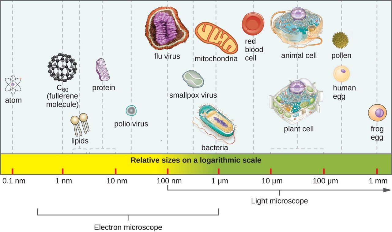
A microorganism (or microbe) is simply a living thing too small to see with the naked eye — you need a microscope. That single size rule sweeps together a wildly diverse group of organisms that are otherwise unrelated. Learning microbiology begins with sorting that group into a handful of families, because the family a microbe belongs to tells you almost everything that matters clinically: how it is built, how it grows, what disease it causes, and — crucially — which drugs will kill it.
The main groups
Microbes split first into cellular organisms (made of one or more true cells) and acellular agents (not cells at all). Among the cellular microbes, the deepest divide is between prokaryotes — small, simple cells with no nucleus — and eukaryotes, larger cells that keep their DNA in a membrane-bound nucleus, just like your own cells do.
| Group | Cell type | Key features | Example disease |
|---|---|---|---|
| Bacteria | Prokaryote | Single-celled, no nucleus; enormous variety; many are our normal flora | Strep throat, UTIs, TB |
| Archaea | Prokaryote | Prokaryotes of extreme environments; none are known to cause human disease | — |
| Fungi | Eukaryote | Yeasts (single-celled) and molds (filamentous) | Thrush (Candida), ringworm |
| Protozoa | Eukaryote | Single-celled eukaryotic microorganisms (protists); often motile; many are parasites | Malaria, giardiasis |
| Helminths | Eukaryote (animal) | Parasitic worms — not micro, but their eggs/larvae are, and they're studied here | Pinworm, tapeworm |
| Viruses | Acellular | Genetic material in a protein coat; must hijack a host cell to replicate | Influenza, COVID-19, HIV |
Keep the prokaryote–eukaryote line firmly in mind, because it is the reason many drugs are safe to give: an antibiotic that attacks a structure found only in bacterial (prokaryotic) cells — a cell wall, a bacterial ribosome — can wipe out the infection while leaving your patient's eukaryotic cells untouched. That principle, called selective toxicity, is the whole reason antimicrobial therapy is possible.
Tap through the six groups of microbes below.
- A microbe is any organism too small to see unaided.
- Cellular microbes are bacteria, archaea, fungi, protozoa (plus helminths by tradition); viruses are acellular.
- Prokaryotes (bacteria, archaea) have no nucleus; eukaryotes (fungi, protozoa) do.
- Selective toxicity — hitting a microbial-only target — is why antimicrobial drugs can cure without poisoning the patient.
Section 2Microbes on and in us
The microbiome: your resident microbes
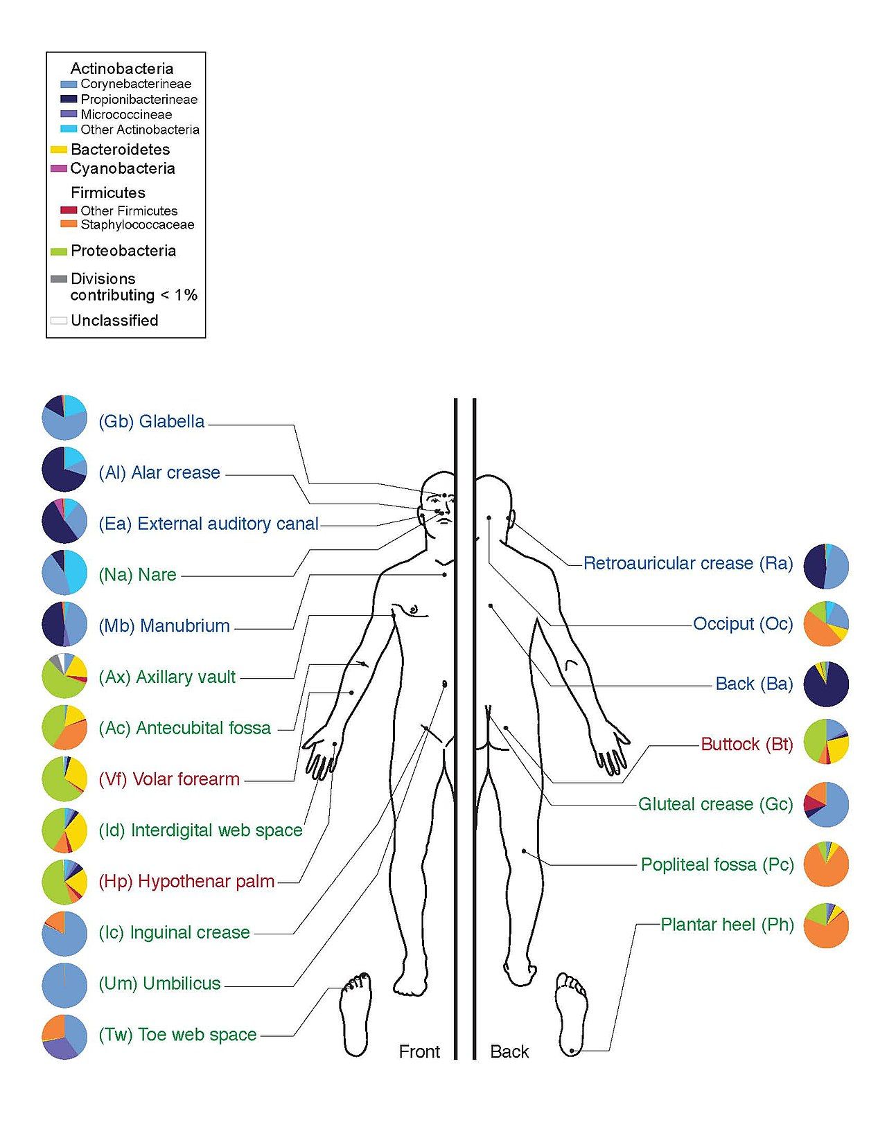
You are not sterile, and you were never meant to be. Trillions of microbes — collectively the microbiota, and together with their genes the microbiome — colonize your skin, mouth, gut, and other surfaces from the moment you are born. These normal flora are not freeloaders. They earn their keep: gut bacteria synthesize vitamin K and some B vitamins, they crowd out invaders by occupying space and consuming nutrients (competitive exclusion), and they help train the developing immune system to tell friend from foe.
Flora come in two flavors. Resident flora live with you long-term and re-establish themselves if disturbed. Transient flora are just passing through — picked up from a doorknob or a patient's bedrail — and this is precisely the population that hand hygiene is designed to remove before it moves from one patient to the next.
When a good microbe goes bad
Many of the organisms that cause healthcare infections are not exotic invaders at all — they are the patient's own normal flora, turned dangerous by opportunity. An opportunistic pathogen causes no trouble in its usual home but causes disease when it is moved somewhere new, when the normal flora are wiped out, or when the host's defenses fall. Escherichia coli is harmless in the colon but causes a urinary tract infection if it reaches the bladder; Candida overgrows into thrush when antibiotics clear its bacterial competitors; and Clostridioides difficile blooms into severe colitis after antibiotics flatten the gut's normal community.
| What normal flora do | Why it matters |
|---|---|
| Make vitamin K & some B vitamins | Nutrition; K is needed for clotting |
| Competitive exclusion of pathogens | They physically and chemically crowd out invaders |
| Train the immune system | Early exposure calibrates immune responses |
| Maintain low pH / hostile niches | e.g., vaginal lactobacilli keep pH low, blocking pathogens |
Explore where your normal flora live.
- The microbiome is the community of microbes that normally live on and in the body.
- Normal flora are mostly beneficial — nutrients, competitive exclusion, immune training.
- Opportunistic pathogens are normal flora that cause disease when displaced, unopposed, or the host is weakened.
- Removing transient flora is the entire point of hand hygiene.
Section 3When microbes cause disease
Colonization, infection, disease
These three words are often used loosely in conversation, but in the clinical world they mean different things, and confusing them leads to over-treatment. Colonization means a microbe is simply present and multiplying on a body surface without causing harm — the patient has no symptoms. Infection means a microbe has invaded and the body is now responding to it. Disease is the step further, when that infection produces recognizable signs and symptoms and impairs function.
The distinction is intensely practical. A wound swab growing bacteria in a patient with no redness, warmth, or fever usually reflects colonization, not infection — treating it with antibiotics does harm without benefit. A pathogen, meanwhile, is any microbe capable of causing disease, and its virulence is the degree of harm it can do (we unpack the machinery of virulence in Chapter 10).
| State | Microbe present? | Host responding? | Symptoms? |
|---|---|---|---|
| Colonization | Yes (multiplying) | No | No |
| Infection | Yes (invading) | Yes | Not necessarily |
| Disease | Yes | Yes | Yes — signs & symptoms |
Why this is a nursing problem
Infections are among the most common and most preventable threats a hospitalized patient faces. Healthcare-associated infections (HAIs) — catheter-associated UTIs, central-line bloodstream infections, surgical-site infections, ventilator-associated pneumonia — affect roughly one in every thirty-one hospital patients on any given day. The nurse is the person at the bedside most often, which makes the nurse the single most important line of defense: every hand wash, every sterile field, every catheter removed a day earlier prevents an infection that would otherwise have happened.
Step through the three clinical states below.
- Colonization (present, no harm) ≠ infection (invading, host responding) ≠ disease (symptoms & impairment).
- A pathogen can cause disease; virulence is how much harm it does.
- HAIs are common and largely preventable, and the nurse is the front line of prevention.
Section 4How we found out microbes cause disease
From bad air to germ theory
For most of history, disease was blamed on miasma — bad air — or on spontaneous generation, the idea that life simply arose from non-living matter. The germ theory of disease, the recognition that specific microbes cause specific diseases, is only about 150 years old, and it is the intellectual foundation of everything nurses do to prevent infection.
Three people worth knowing
Ignaz Semmelweis, a Viennese obstetrician in the 1840s, noticed that mothers died of childbed fever far more often on the ward staffed by doctors (who came straight from autopsies) than on the ward staffed by midwives. He ordered handwashing with a chlorine solution, and the death rate plummeted. He could not explain why it worked — germ theory did not yet exist — and his colleagues ridiculed and rejected him. He is now remembered as the father of hand hygiene.
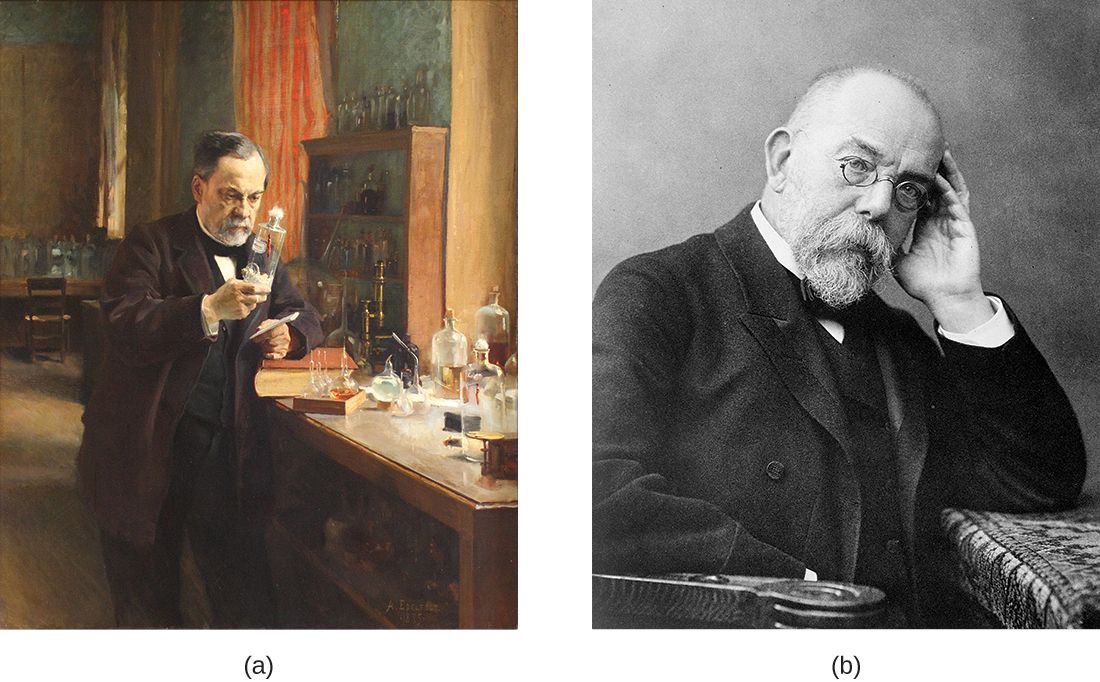
Louis Pasteur disproved spontaneous generation with his elegant swan-neck flask experiments, showing that broth stayed sterile until microbes from the air reached it. He gave us pasteurization and helped establish that microbes drive fermentation and spoilage. Robert Koch then supplied the rigor: a systematic way to prove that one particular microbe causes one particular disease, now called Koch's postulates.
| Koch's postulate | In plain English |
|---|---|
| 1 | The microbe is found in every case of the disease, and absent in healthy hosts |
| 2 | It can be isolated from a sick host and grown in pure culture |
| 3 | The cultured microbe causes the same disease when introduced into a healthy host |
| 4 | The same microbe can be re-isolated from that newly diseased host |
Walk through Koch's four-step proof below.
- Germ theory — specific microbes cause specific diseases — is the basis of all infection control.
- Semmelweis = hand hygiene; Pasteur = disproved spontaneous generation, pasteurization; Koch = postulates linking microbe to disease.
- Koch's postulates are the logical test for proving a given microbe causes a given disease.
Section 5Naming and classifying microbes
The three domains
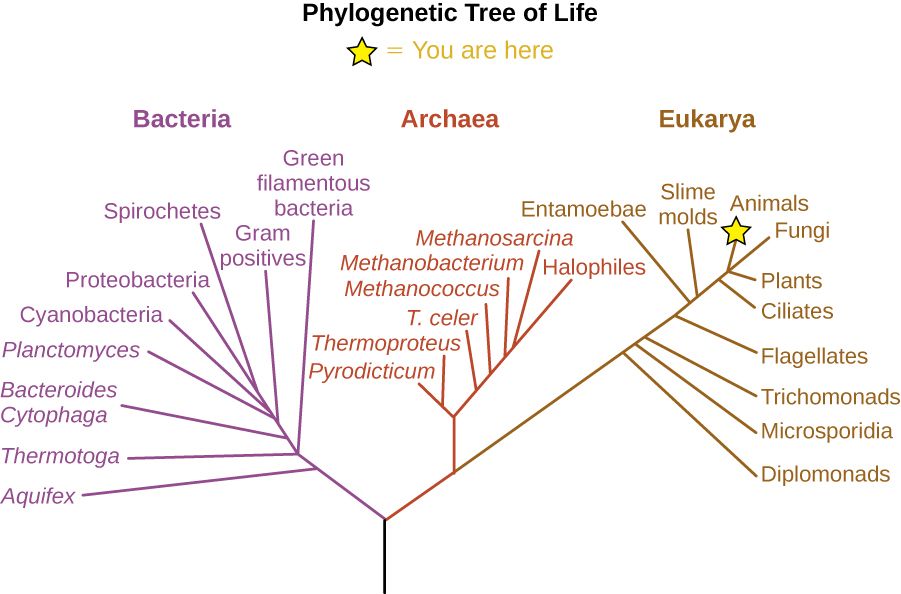
Every living organism belongs to one of three great domains, a scheme worked out by Carl Woese from differences in ribosomal RNA. Two domains, Bacteria and Archaea, are prokaryotes. The third, Eukarya, holds everything with a nucleus — protozoa, fungi, plants, animals, and you. Notice where the microbes land: bacteria are their own domain, archaea are a second, and the microbial eukaryotes (fungi and protozoa) sit alongside us in the third. Viruses, being acellular, are not placed on this tree of life at all.
Binomial names, and how to read them
Every organism gets a two-part Latin name — binomial nomenclature, a system from Carl Linnaeus. The first word is the genus (capitalized), the second is the species (lowercase), and the whole name is italicized: Staphylococcus aureus, Escherichia coli, Clostridioides difficile. After the full name is written once, it is abbreviated to the genus initial: S. aureus, E. coli, C. difficile (or the familiar "C. diff"). Getting this right matters at the bedside, because a chart, a culture result, and a handoff report all assume you can read these names fluently.
| Domain | Cell type | Members | Human disease? |
|---|---|---|---|
| Bacteria | Prokaryote | True bacteria | Yes — most bacterial pathogens |
| Archaea | Prokaryote | Extremophiles & others | No known pathogens |
| Eukarya | Eukaryote | Protozoa, fungi, plants, animals | Yes — fungi, protozoa, helminths |
Decode a few real microbe names below.
- All life falls into three domains: Bacteria, Archaea (both prokaryotic), and Eukarya.
- Viruses are acellular and are not on the tree of life.
- Binomial names are Genus species, italicized, abbreviated to G. species after first use.
The chapter in one breath
- Microbes are organisms too small to see: bacteria, archaea, fungi, protozoa, helminths, and (acellular) viruses.
- The prokaryote/eukaryote divide underlies selective toxicity — why antimicrobials can cure without harming the patient — and antibiotics do nothing to viruses.
- The microbiome of normal flora is mostly beneficial; opportunistic pathogens are normal flora that harm when displaced or unopposed.
- Colonization ≠ infection ≠ disease; a pathogen causes disease and virulence is how much harm it does.
- HAIs are common and preventable, and the nurse's hand hygiene is the most powerful infection-control tool there is.
- Germ theory (Semmelweis, Pasteur, Koch) grounds all infection control; Koch's postulates prove a microbe causes a disease.
- Life sorts into three domains; microbes are named with italic binomial names abbreviated after first use.
ReferenceWords worth knowing
A quick reference for the terms in this chapter — skim it now, or circle back whenever a word trips you up.
- Acellular
- Not composed of cells; describes viruses, viroids, and prions.
- Archaea
- A domain of prokaryotes distinct from bacteria; none are known to cause human disease.
- Bacteria
- A domain of single-celled prokaryotes; the source of most bacterial infections and much of our normal flora.
- Binomial nomenclature
- The two-part naming system (italic Genus species) used for every organism.
- Colonization
- The presence and multiplication of a microbe on a body surface without harm or symptoms.
- Domain
- The highest level of classification: Bacteria, Archaea, or Eukarya.
- Disease
- An infection that produces recognizable signs and symptoms and impairs function.
- Eukaryote
- A cell (or organism) whose DNA is enclosed in a membrane-bound nucleus; includes fungi and protozoa.
- Germ theory of disease
- The principle that specific microorganisms cause specific diseases.
- Healthcare-associated infection (HAI)
- An infection a patient acquires while receiving care, e.g., CAUTI, CLABSI, or surgical-site infection.
- Infection
- Invasion of body tissues by a microbe and the host's response to it.
- Koch's postulates
- Four criteria used to establish that a particular microbe causes a particular disease.
- Microbiome
- The community of microbes normally living on and in the body, together with their genes.
- Microorganism (microbe)
- Any living organism too small to see without a microscope.
- Normal flora (microbiota)
- The resident microbes that normally inhabit a healthy body, mostly beneficial.
- Opportunistic pathogen
- A normally harmless microbe that causes disease when displaced, unopposed, or the host is weakened.
- Pathogen
- A microbe capable of causing disease.
- Prokaryote
- A cell (or organism) with no membrane-bound nucleus; bacteria and archaea.
- Selective toxicity
- Harming a microbe while sparing the host by targeting a structure the microbe has and the host lacks.
- Virulence
- The degree of harm (pathogenicity) a microbe is capable of causing.
Check yourself
Microscopy, Staining & the Microbiology Lab
The organisms you spent Chapter 1 meeting share one inconvenient trait: not one of them can be seen with the naked eye. Every practical thing microbiology does — identifying the bug in a wound, choosing the right antibiotic, confirming that a sterilizer worked — begins with a way to make the invisible visible. This chapter is about that toolkit: the units we use to measure things a thousand times smaller than a grain of salt, the microscope that magnifies them, the dyes that give them color and reveal their secrets, and the lab workflow that turns a swab from your patient into a culture-and-sensitivity report you can act on. Master this chapter and a lab result stops being a mystery and starts being a set of instructions.
By the end of this chapter you can
- State the metric units used to measure microbes and place bacteria, viruses, and human cells on the size scale.
- Explain why the unaided eye cannot resolve a bacterium.
- Name the major parts of a compound light microscope and describe what each does.
- Distinguish magnification from resolution, and explain the role of the oil-immersion lens.
- Explain why cells must be stained, and contrast simple, differential, and negative stains.
- Walk through the four steps of the Gram stain and explain what makes a cell Gram-positive or Gram-negative.
- Trace a specimen from collection through culture, colony, identification, and sensitivity testing.
- Describe the nurse's role in specimen collection and in reading a culture-and-sensitivity report.
Section 1Why microbes are invisible
The metric ladder
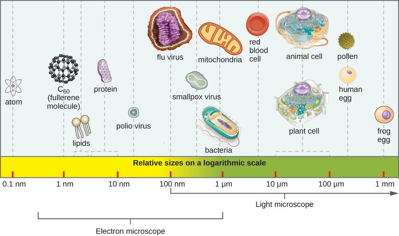
Microbiology runs on the metric system, and the whole subject makes more sense once the units are second nature. Start with the meter and divide by a thousand at each step: a millimeter (mm) is a thousandth of a meter, a micrometer (µm, sometimes called a micron) is a thousandth of a millimeter, and a nanometer (nm) is a thousandth of a micrometer. Each rung down is 1000 times smaller than the one above it. You already live at the millimeter scale — the smallest lines on a ruler are about a millimeter apart. Microbes live one or two rungs below that, in a world your eye was never built to enter.
How big is a microbe, really?
A useful benchmark is the red blood cell, roughly 7 µm across. Most bacteria are smaller still — a typical rod is about 0.5 µm wide and 2–3 µm long, and a round coccus is around 1 µm. Yeasts and protozoa, being eukaryotes, run larger, from several to tens of micrometers. Viruses are in a different league altogether, measured in nanometers: most fall between 20 and 300 nm, so dozens of them — and up to roughly a hundred of the smallest — could line up across a single bacterium. This is why viruses were invisible even to early microscopists and why we needed the electron microscope to see them at all.
| Unit | Size | What lives here |
|---|---|---|
| Millimeter (mm) | 1/1000 of a meter | The smallest thing the naked eye resolves (about 0.1 mm) |
| Micrometer (µm) | 1/1000 of a mm | Human cells (~10–100 µm), red blood cells (~7 µm), bacteria (~1–3 µm) |
| Nanometer (nm) | 1/1000 of a µm | Viruses (~20–300 nm), large molecules, ribosomes |
Why your eye can't see them
The human eye can separate two points only if they are roughly 0.1 mm (100 µm) apart — anything closer blurs into a single dot. A bacterium at 1 µm is about a hundred times below that limit, so it simply cannot register, no matter how good your eyesight or how bright the light. This is not a matter of things being faint or far away; it is a hard physical limit called resolution, and defeating it is the entire job of the microscope you will meet in the next section.
- Metric ladder: 1 mm = 1000 µm, and 1 µm = 1000 nm — each step is 1000× smaller.
- Most bacteria are ~1–3 µm; viruses are ~20–300 nm; a red blood cell (~7 µm) is a handy benchmark.
- The naked eye resolves only to about 0.1 mm — roughly 100× too coarse to see a bacterium.
- Because microbes are invisible individually, the lab grows them into visible colonies before it can work with them.
Section 2The light microscope
The parts and what they do
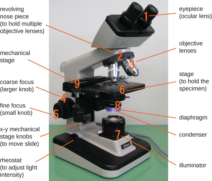
The workhorse of every clinical lab is the compound light microscope — "compound" because it uses two lens systems in series to build up its magnifying power. Light starts at the illuminator in the base and travels upward. The condenser gathers and focuses that light onto the specimen, and the iris diaphragm adjusts how much passes through, which controls contrast. The specimen sits on the stage. Above it, the objective lenses — mounted on a rotating nosepiece — do the first stage of magnifying, and the ocular (eyepiece) you look through does the second. The coarse and fine focus knobs raise and lower the stage until the image is sharp.
Magnification: stacking two lenses
Total magnification is simply the ocular multiplied by the objective. Oculars are almost always 10×. Objectives come in a standard set: scanning (4×), low power (10×), high-dry (40×), and oil-immersion (100×). So with the 10× ocular, the oil-immersion objective gives 10 × 100 = 1000× total magnification — enough to see bacterial shape and arrangement clearly. You almost always find your target on a low-power objective first, center it, then rotate up to higher power.
| Objective | Objective power | With 10× ocular | Typical use |
|---|---|---|---|
| Scanning | 4× | 40× | Find the specimen, survey the slide |
| Low power | 10× | 100× | Larger cells, general orientation |
| High-dry | 40× | 400× | Fungi, cells, parasites |
| Oil immersion | 100× | 1000× | Bacteria — Gram stains, cell arrangement |
Resolution is what really matters
Beginners chase magnification, but the property that decides whether an image is useful is resolution — the smallest distance between two points that still lets you see them as separate. Magnifying a blurry image just gives you a bigger blur (lab staff call this empty magnification). The resolution of a light microscope is capped near 0.2 µm by the wavelength of visible light itself — you cannot resolve two things closer than about half a wavelength apart. That 0.2 µm limit is good enough to see bacteria but not viruses, which is exactly why viruses need an electron microscope. To squeeze the best resolution out of the 100× lens, we add a drop of immersion oil between the slide and the objective. Oil has the same light-bending (refractive) index as glass, so it keeps light rays from scattering away as they leave the slide, funneling more of them into the lens and sharpening the image.
- The compound light microscope uses two lenses; light travels illuminator → condenser → specimen → objective → ocular.
- Total magnification = ocular × objective; the 10× ocular with the 100× oil lens gives 1000×.
- Resolution (~0.2 µm for light) — not magnification — determines how much real detail you see.
- Immersion oil matches the refractive index of glass, capturing more light rays and improving resolution at 100×.
Section 3Why we stain
The contrast problem
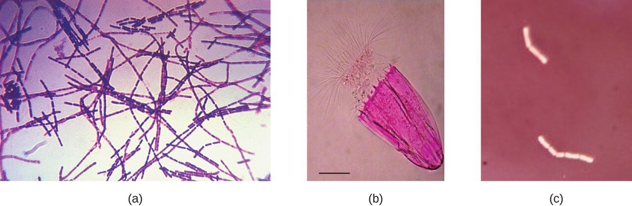
Living cells are mostly water and nearly transparent, so under an ordinary light microscope they are almost invisible against a bright background — there just isn't enough contrast to make out shape or structure. Staining solves this by coating the cell in colored dye. Before you can stain a bacterial slide, you make a smear: spread a thin film of the sample on a slide, let it air-dry, then pass it through a flame or warm it to heat-fix it. Fixing does three jobs — it kills most of the organisms, glues them to the glass so they don't rinse off, and preserves their shape. Note that heat-fixing is not sterilization: it kills most vegetative bacteria, but mycobacteria and bacterial endospores can survive it. Most bacterial dyes are basic (positively charged) dyes — methylene blue, crystal violet, safranin — because the bacterial cell surface carries a negative charge that attracts them.
Simple stains
A simple stain uses a single dye to add color and contrast. It tells you a cell's size, shape, and arrangement — whether the organism is a round coccus or a rod-shaped bacillus, and whether it forms clusters, chains, or pairs — but nothing more. It's fast and it's the right tool when all you need is to confirm that organisms are present and see their basic morphology.
Differential and special stains
A differential stain uses more than one reagent to divide bacteria into groups based on how they react. The two you must know are the Gram stain (the entire next section) and the acid-fast stain, which singles out the waxy-walled organisms like Mycobacterium tuberculosis that a Gram stain misses. Special stains go after one specific structure — capsule stains, endospore stains, flagella stains. Many of these use negative staining, in which an acidic dye like India ink colors the background and leaves the cell unstained and glowing against it. Negative staining skips heat-fixing, so it doesn't distort delicate cells or shrink a capsule, which is why it's the go-to for capsules and for fragile organisms.
| Stain type | Reagents | What it tells you | Example |
|---|---|---|---|
| Simple | One dye | Size, shape, arrangement | Methylene blue |
| Differential | Two or more reagents | Sorts cells into groups | Gram stain, acid-fast stain |
| Special | Structure-specific | Reveals one feature | Capsule, endospore, flagella |
| Negative | Acidic dye (stains background) | Shape without distortion; capsules | India ink, nigrosin |
- Cells are nearly transparent; staining adds the contrast needed to see shape and structure.
- A smear is air-dried and heat-fixed — killing most vegetative organisms, fixing them to the slide, and preserving shape; it does not sterilize.
- Simple stains (one dye) show morphology; differential stains (multiple reagents) sort cells into groups.
- Negative staining colors the background and avoids heat, so it's ideal for capsules and fragile cells.
Section 4The Gram stain
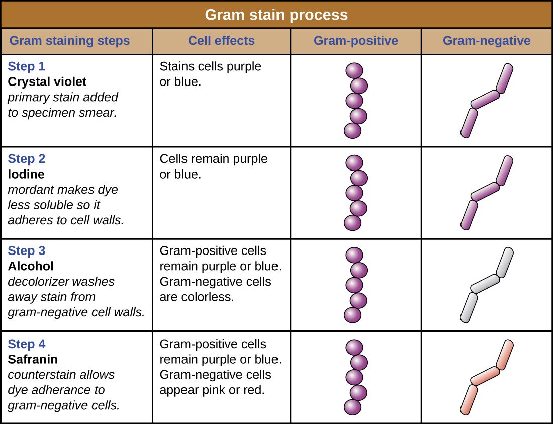
What makes a cell Gram-positive or Gram-negative
The Gram stain, devised by Hans Christian Gram in 1884, is the single most important stain in clinical microbiology, and it works because of a real difference in the bacterial cell wall. Gram-positive bacteria have a thick outer layer of peptidoglycan, a mesh-like polymer that acts like a sponge. Gram-negative bacteria have only a thin peptidoglycan layer, wrapped by an extra outer membrane. That structural difference is what the four reagents exploit: the thick wall traps the first dye, and the thin one lets it wash out.
The four steps
The sequence is always the same, and every step has a job. First, the primary stain crystal violet floods every cell purple. Second, Gram's iodine acts as a mordant — it binds the crystal violet into a large complex too big to slip out of a thick wall easily. Third comes the decolorizer, alcohol or an alcohol-acetone mix, and this is the step that actually separates the two groups: it dehydrates and tightens the thick Gram-positive wall so the dye complex stays locked in, but it dissolves the Gram-negative outer membrane and washes the dye straight out, leaving those cells colorless. Fourth, the pink counterstain safranin re-colors the now-colorless Gram-negative cells; the Gram-positive cells are already saturated with purple and don't change. The end result: Gram-positive = purple, Gram-negative = pink/red.
| Step | Reagent | Role | Gram-positive | Gram-negative |
|---|---|---|---|---|
| 1 | Crystal violet | Primary stain | Purple | Purple |
| 2 | Gram's iodine | Mordant (traps dye) | Purple | Purple |
| 3 | Alcohol / acetone | Decolorizer (the key step) | Stays purple | Becomes colorless |
| 4 | Safranin | Counterstain | Stays purple | Turns pink/red |
Reading the result at the bedside
A Gram stain can be read in under an hour, long before a culture matures, so it is the first real clue to guide empiric antibiotics while the lab waits 24–48 hours for growth. The report pairs the Gram reaction with the shape and arrangement, and each combination narrows the field: Gram-positive cocci in clusters point to Staphylococcus; Gram-positive cocci in chains point to Streptococcus; Gram-negative rods suggest gut organisms like E. coli; Gram-negative diplococci raise Neisseria. That structural difference also has treatment consequences — the Gram-negative outer membrane blocks some antibiotics and carries endotoxin (Chapter 10), which is why Gram-negative bloodstream infections can be so dangerous.
- The Gram stain sorts bacteria by cell wall: thick peptidoglycan (Gram-positive) vs. thin peptidoglycan plus an outer membrane (Gram-negative).
- Four steps: crystal violet → iodine mordant → alcohol decolorizer → safranin counterstain.
- Decolorization is the make-or-break step that distinguishes the two groups.
- Result: Gram-positive = purple, Gram-negative = pink; read in under an hour, it guides early antibiotic choice.
Section 5From specimen to answer
Collecting and culturing the specimen
Every lab result is only as good as the specimen it started from, and specimen collection is squarely a nursing responsibility. The rules are simple but unforgiving: sample the right site (the infection, not the surrounding skin), collect before antibiotics whenever possible, use the correct container and transport medium, and label it accurately at the bedside. Back in the lab, the sample is spread onto a culture medium — usually a nutrient agar plate (a solid jelly) or a liquid broth. Some media are chosen deliberately: selective media suppress unwanted organisms, and differential media (like blood agar or MacConkey) make different species look visibly different, giving the lab an early hint about what's growing.
The colony: an invisible bug made visible
To isolate a single organism, the lab uses a streak plate, dragging the sample thinner and thinner across the agar until individual cells are separated. Each isolated cell, left overnight in a warm incubator, divides again and again into a visible mound of millions of identical clones — a colony. Because every cell in a colony descends from one parent, a colony is a pure sample to work from. Its morphology — size, shape, color, texture, and whether it destroys the blood in blood agar (hemolysis) — is itself a first clue to the organism's identity. From there, biochemical panels, automated analyzers, or rapid MALDI-TOF mass spectrometry nail down the species.
Sensitivity testing and the antibiogram
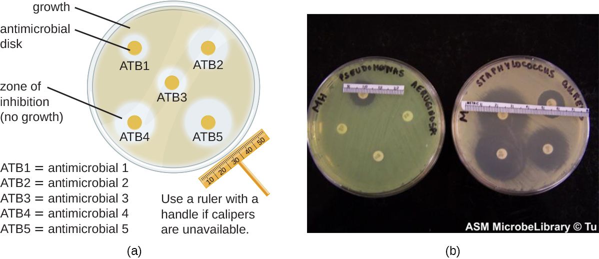
Knowing the organism's name is only half the answer; the clinician also needs to know which antibiotics will kill it. That is sensitivity (or susceptibility) testing. In the classic Kirby-Bauer disk-diffusion method, small paper disks soaked with different antibiotics are placed on a lawn of the organism; where a drug works, it diffuses out and clears a zone of inhibition, and a larger zone generally means the organism is more susceptible to that drug (each zone is measured against standardized breakpoints, not sized up against the neighboring disk). Other methods measure the minimum inhibitory concentration (MIC) — the lowest drug concentration that stops growth — using broth microdilution or a gradient Etest strip. Results come back reported as S, I, or R (susceptible, intermediate, resistant). This is the report that lets the team de-escalate: start broad while waiting, then narrow to the most targeted, effective drug once the sensitivities return.
| Stage | Typical timing | What you learn |
|---|---|---|
| Gram stain | < 1 hour | Gram reaction, shape, arrangement — guides empiric therapy |
| Colony growth | 18–48 hours | Preliminary identity from colony morphology |
| Identification & sensitivity | 48–72 hours | Species and the S/I/R susceptibility report |
- Good results start with good collection: right site, before antibiotics, correct container, accurate label.
- A streak plate isolates single cells; each grows into a pure colony whose morphology hints at identity.
- Sensitivity testing (Kirby-Bauer zones, or an MIC) reports each drug as S/I/R.
- The culture-and-sensitivity report lets the team de-escalate from broad empiric therapy to a targeted drug.
The chapter in one breath
- Microbes are measured in micrometers (bacteria, ~1–3 µm) and nanometers (viruses, ~20–300 nm); the naked eye stops at ~0.1 mm, so microbes are invisible individually.
- The compound light microscope stacks two lenses; total magnification is ocular × objective, reaching 1000× with the oil lens.
- Resolution, not magnification, sets useful detail; light microscopy bottoms out near 0.2 µm, and immersion oil sharpens the 100× image.
- Cells are transparent, so we make a heat-fixed smear and stain it; simple stains show shape, differential stains sort cells into groups, and negative stains color the background.
- The Gram stain's four steps (crystal violet, iodine, alcohol, safranin) exploit the cell wall: Gram-positive = purple, Gram-negative = pink, with decolorization as the key step.
- A specimen becomes an answer through culture, a pure colony, species identification, and sensitivity testing reported as S/I/R.
- Nurses own the front end (proper collection, cultures before antibiotics) and use the culture-and-sensitivity report to advocate for the right drug.
ReferenceWords worth knowing
A quick reference for the terms in this chapter — skim it now, or circle back whenever a word trips you up.
- Acid-fast stain
- A differential stain that identifies organisms with waxy cell walls, such as Mycobacterium tuberculosis, which a Gram stain does not detect well.
- Agar
- A solid gel derived from seaweed, used as the base of culture plates on which microbes are grown.
- Antibiogram
- The report of an organism's antibiotic susceptibilities, listing each drug as susceptible, intermediate, or resistant.
- Colony
- A visible mound of millions of genetically identical cells descended from a single original cell.
- Compound light microscope
- A microscope that uses two lens systems (ocular and objective) in series to magnify a specimen.
- Counterstain
- The second-color dye in a differential stain (safranin in the Gram stain) that recolors the cells the decolorizer has left colorless.
- Culture
- The growth of microorganisms on or in a nutrient medium in the lab.
- Decolorizer
- The alcohol/acetone step of the Gram stain that removes dye from Gram-negative cells while Gram-positive cells retain it.
- Differential stain
- A staining procedure using two or more reagents to distinguish between groups of bacteria (e.g., Gram stain).
- Fixation
- Heat- or chemical-treating a smear to kill the organisms, adhere them to the slide, and preserve their shape before staining.
- Gram stain
- The key differential stain that sorts bacteria into Gram-positive (purple) and Gram-negative (pink) based on cell-wall structure.
- Magnification
- How much larger an image appears than the real object; ocular power multiplied by objective power.
- Micrometer (µm)
- One thousandth of a millimeter; the unit used to measure most bacteria and eukaryotic cells.
- Minimum inhibitory concentration (MIC)
- The lowest concentration of an antibiotic that prevents visible growth of an organism.
- Mordant
- A substance (Gram's iodine) that binds a dye more firmly to the cell, trapping it in the wall.
- Negative staining
- Staining the background with an acidic dye so cells appear clear; avoids heat and preserves capsules and fragile cells.
- Objective lens
- The lens nearest the specimen (4×, 10×, 40×, 100×) that performs the first stage of magnification.
- Oil immersion
- Placing immersion oil between the slide and the 100× lens; the oil's refractive index matches glass, improving resolution.
- Peptidoglycan
- The mesh-like polymer of the bacterial cell wall; thick in Gram-positive cells, thin in Gram-negative cells.
- Resolution
- The smallest distance between two points at which they can still be seen as separate; ~0.2 µm for a light microscope.
- Simple stain
- A stain using a single dye to reveal a cell's size, shape, and arrangement.
- Zone of inhibition
- The clear ring around an antibiotic disk in disk-diffusion testing where the drug has stopped bacterial growth.
Check yourself
Prokaryotic vs Eukaryotic Cells
Almost every microbe that concerns a nurse is a single cell — and that cell is built one of two ways. It is either a prokaryote, a small cell with its DNA loose in the cytoplasm, or a eukaryote, a larger cell that locks its DNA inside a nucleus, exactly like your own cells do. Bacteria are prokaryotes; fungi and protozoa are eukaryotes; your patient is made of eukaryotic cells. That single architectural split is not just trivia — it is the reason an antibiotic can flood a patient's bloodstream and destroy the bacteria while leaving the patient unharmed. This chapter takes a bacterial cell apart piece by piece, from the wall that gives it shape to the spore that lets it survive a decade on a bedrail, and shows you why each structure matters at the bedside.
By the end of this chapter you can
- Contrast prokaryotic and eukaryotic cells by size, nucleus, organelles, and ribosomes.
- Explain how the prokaryote–eukaryote divide creates selective toxicity for antimicrobial drugs.
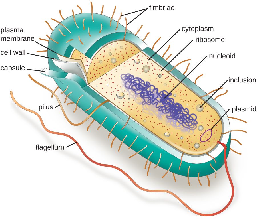
- Describe the interior of a bacterial cell — plasma membrane, cytoplasm with ribosomes, nucleoid, and plasmids — and what each part does.
- Explain the structure of peptidoglycan and contrast Gram-positive and Gram-negative cell walls.
- Connect the cell wall and endotoxin (LPS) to how antibiotics work and to Gram-negative sepsis.
- Describe the roles of the capsule, flagella, pili, and fimbriae in disease and gene transfer.
- Explain how endospores form, why they resist heat and disinfectants, and what that means for infection control.
Section 1Two kinds of cell
The single deepest divide in biology
Of all the ways you could sort living cells, one line runs deeper than any other: does the cell keep its DNA inside a membrane-bound nucleus, or not? Cells that do not are prokaryotes (from Greek roots meaning "before the nut," the "nut" being the nucleus). Cells that do are eukaryotes ("true nut"). This is not a small stylistic difference — it comes with a whole package of other features, and knowing which side of the line a microbe sits on tells you how it is built, how fast it grows, and which drugs can touch it.
Prokaryotes are the bacteria and the archaea. They are small — typically 0.2 to 2 µm across — and simple, with their single circular chromosome floating loose in the cytoplasm. Eukaryotes — the fungi, protozoa, plants, animals, and every cell in your patient's body — are usually ten to a hundred times larger across and far more elaborate inside. When you say a patient has a "bacterial" infection versus a "fungal" one, you are really naming which kind of cell you are fighting.
What prokaryotes lack — and what they have
The headline is what prokaryotes are missing. They have no nucleus, and no other membrane-bound organelles either — no mitochondria, no endoplasmic reticulum, no Golgi apparatus. All of that chemistry happens either in the open cytoplasm or against the inner face of the plasma membrane. But "simple" does not mean "defenseless." Prokaryotes make up for the lack of internal compartments with tough external gear: most wear a rigid cell wall, many add a slimy capsule, and many carry flagella to swim and pili to stick — the structures the rest of this chapter is about.
One difference is easy to overlook but medically enormous: the ribosome. Both cell types build proteins on ribosomes, but the machines are not the same size. Prokaryotes use a 70S ribosome; eukaryotes use a larger 80S ribosome. That size gap is a gift to medicine, because a drug can be designed to jam the 70S machine while ignoring the 80S one.
Why the difference is a drug target
Here is the payoff. Because bacterial cells have structures your cells simply do not have — a peptidoglycan wall, a 70S ribosome, unique enzymes — a drug can attack those targets and destroy the bacterium while leaving the patient's eukaryotic cells alone. This principle is called selective toxicity, and it is the entire reason antibacterial therapy is possible. Penicillin blocks cell-wall building, which human cells never do. Many antibiotics latch onto the 70S ribosome, which human cells do not have. The wider the gap between microbe and host, the safer the drug.
| Feature | Prokaryote (bacteria) | Eukaryote (fungi, protozoa, human) |
|---|---|---|
| Nucleus | Absent — DNA free in nucleoid | Present — DNA in membrane-bound nucleus |
| Typical size | 0.2–2 µm | 10–100 µm |
| Membrane-bound organelles | None | Mitochondria, ER, Golgi, etc. |
| DNA shape | Usually one circular chromosome | Multiple linear chromosomes |
| Ribosomes | 70S | 80S (cytoplasmic) |
| Cell wall | Usually peptidoglycan | Fungi: chitin; protozoa: none; human: none |
| Reproduction | Binary fission | Mitosis / meiosis |
- The deepest split among cells is prokaryote (no nucleus) vs eukaryote (nucleus + organelles).
- Bacteria and archaea are prokaryotes; fungi, protozoa, and human cells are eukaryotes.
- Prokaryotes are small, lack organelles, and use a 70S ribosome; eukaryotes use an 80S ribosome.
- Prokaryote-only structures give antibiotics their selective toxicity — the reason they cure without poisoning the patient.
Section 2Inside the bacterial cell
The plasma membrane: the real boundary
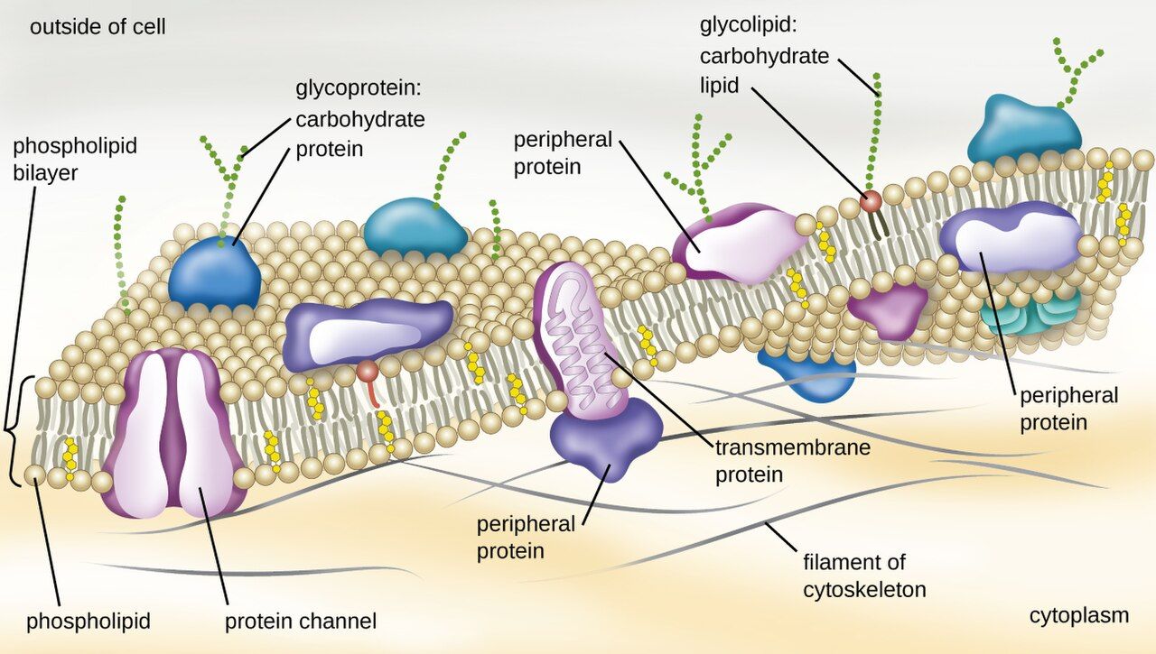
The layers that enclose a bacterium — the plasma membrane, the cell wall, and, in Gram-negatives, an outer membrane — are together called the cell envelope; everything the envelope encloses (cytoplasm, ribosomes, nucleoid, plasmids) is cell contents. Working from the outside in, the wall gives a bacterium its shape, but the plasma membrane is the layer that actually keeps the cell alive. It is a phospholipid bilayer — two sheets of fatty molecules with their water-loving heads facing out and their water-fearing tails tucked inside — peppered with proteins. It is selectively permeable, meaning it decides what crosses: nutrients in, wastes out, ions carefully balanced. Because prokaryotes have no mitochondria, the membrane also does a job your cells hand off to organelles: it runs the electron transport chain and generates the cell's energy right there in the membrane.
That makes the membrane a tempting drug target, but a risky one — bacterial and human membranes are chemically similar, so drugs that punch holes in membranes tend to be toxic. The polymyxins (colistin) do exactly this and are held in reserve as last-resort agents against highly resistant Gram-negative bacteria precisely because they can also injure the patient's kidneys.
Cytoplasm, ribosomes, and inclusions
Fill the membrane and you have the cytoplasm — a crowded, water-based gel where nearly all of the cell's chemistry happens. Suspended in it are tens of thousands of 70S ribosomes, the protein factories introduced in Section 1 and the target of whole families of antibiotics (aminoglycosides, tetracyclines, macrolides). Many bacteria also stockpile spare nutrients in inclusions — little granules of stored carbon, phosphate, or sulfur they can draw on when times are lean.
The nucleoid and plasmids
A prokaryote's DNA is not gone — it is just not gift-wrapped in a nucleus. The single, circular chromosome coils into a dense region called the nucleoid, which has no surrounding membrane, so the DNA sits in direct contact with the cytoplasm. This is one reason bacteria can grow so fast: transcription and translation happen in the same space, at the same time.
Many bacteria carry extra, much smaller loops of DNA called plasmids, separate from the chromosome. Plasmids are not usually needed for basic survival, but they often carry the genes that matter most to nurses: antibiotic-resistance genes, toxin genes, and the machinery to pass those genes to other bacteria. A plasmid can hop from one bacterium to another, which is how resistance spreads through a ward faster than any single organism could reproduce.
| Structure | What it is | Why it matters clinically |
|---|---|---|
| Plasma membrane | Phospholipid bilayer with proteins | Controls transport; makes energy; polymyxin target |
| Cytoplasm | Water-based gel filling the cell | Site of metabolism and growth |
| Ribosomes (70S) | Protein-building machines | Target of many antibiotics (selective toxicity) |
| Nucleoid | Region holding the circular chromosome | No membrane → fast, coupled gene expression |
| Plasmid | Small extra loop of DNA | Carries and spreads resistance & toxin genes |
- The plasma membrane is a selectively permeable phospholipid bilayer that also generates the cell's energy.
- The cytoplasm holds 70S ribosomes — the target of many antibiotics.
- The circular chromosome coils into a membrane-free nucleoid.
- Plasmids are small DNA loops that carry resistance and toxin genes and can move between bacteria.
Section 3The cell wall
Peptidoglycan: the mesh that holds shape
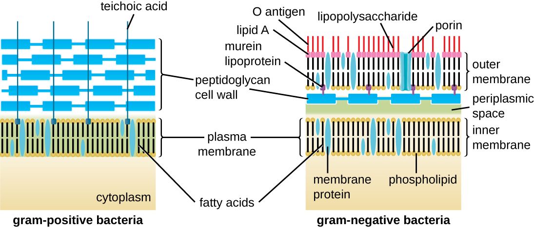
Outside the membrane, most bacteria wear a rigid cell wall made of peptidoglycan (also called murein) — a material found nowhere in the human body. Picture a chain-link fence wrapped completely around the cell: long sugar chains running in parallel, cross-linked to each other by short chains of amino acids. That cross-linking turns a floppy bag into a rigid mesh strong enough to resist the cell's internal osmotic pressure, which would otherwise make it swell and burst. The wall is what gives a bacterium its shape — rod, sphere, or spiral — and its very existence, absent from human cells, is why it is such a clean drug target.
This is exactly where the beta-lactam antibiotics work. Penicillins, cephalosporins, and carbapenems block the enzymes that stitch the peptidoglycan cross-links. The bacterium keeps trying to grow, the wall can no longer hold, and the cell ruptures. Because human cells have no wall to protect, these drugs are among the safest antibiotics we have.
Gram-positive vs Gram-negative walls
Not all walls are built alike, and the difference is the single most useful early clue in a microbiology lab. The Gram stain sorts bacteria into two great camps by how their walls hold a purple dye:
Gram-positive bacteria have a thick peptidoglycan layer on the outside. It traps the crystal-violet dye and the cell stays purple. Examples: Staphylococcus, Streptococcus, Enterococcus, Clostridioides.
Gram-negative bacteria have only a thin peptidoglycan layer, but they add a second, outer membrane on top of it. That outer membrane lets the purple dye wash out during decolorizing, so the cell picks up the pink counterstain and appears red/pink. Examples: Escherichia coli, Klebsiella, Pseudomonas, Neisseria.
Why the Gram stain still runs the show
The Gram result is not just a color — it predicts behavior. That extra outer membrane makes Gram-negative bacteria harder to kill, because it blocks many drugs from ever reaching the wall. And the outer membrane carries a molecule that matters enormously to nurses: lipopolysaccharide (LPS), better known as endotoxin. When Gram-negative bacteria die and break apart, LPS is released into the blood and triggers a violent immune response — fever, plummeting blood pressure, and, at the extreme, septic shock. This is why a Gram-negative bloodstream infection can spiral so fast.
| Feature | Gram-positive | Gram-negative |
|---|---|---|
| Peptidoglycan | Thick, on the surface | Thin, between two membranes |
| Outer membrane | No | Yes — carries LPS |
| Gram stain color | Purple (retains crystal violet) | Pink/red (takes counterstain) |
| Endotoxin (LPS) | No | Yes — drives septic shock |
| Examples | Staph, Strep, Enterococcus, C. diff | E. coli, Klebsiella, Pseudomonas |
- The wall is made of peptidoglycan, a human-free target — where beta-lactam antibiotics act.
- Gram-positive = thick peptidoglycan, stains purple; Gram-negative = thin wall + outer membrane, stains pink.
- The Gram-negative outer membrane blocks many drugs and carries LPS (endotoxin).
- Released endotoxin can drive fever, hypotension, and septic shock.
Section 4External structures
The capsule: a slippery shield
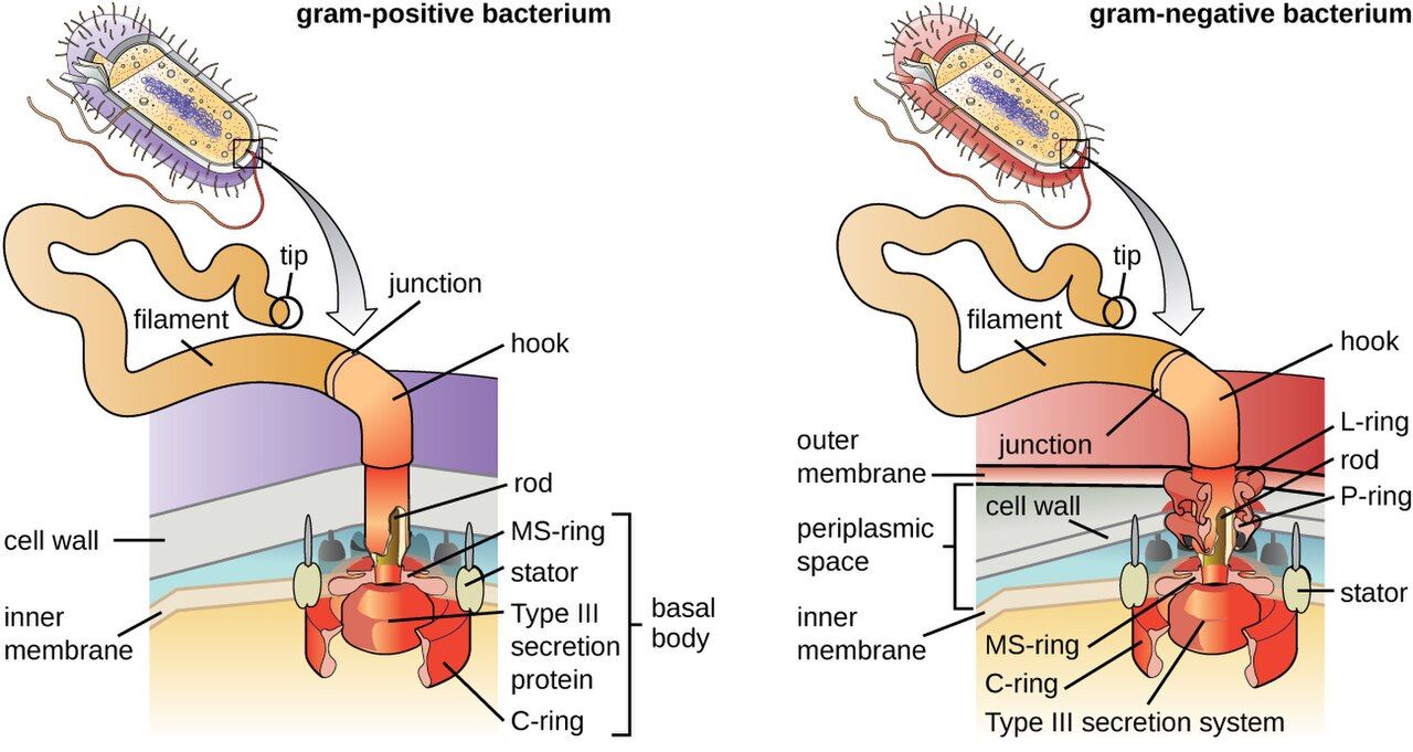
Some bacteria wrap themselves in a thick, sticky outer layer called a capsule (a looser version is called a slime layer). It is not needed to live, but where it matters, it matters a great deal. A capsule is a major virulence factor: it is slippery enough that the patient's immune cells struggle to grab and swallow the bacterium, a trick called resisting phagocytosis. This is why the most dangerous strains of Streptococcus pneumoniae, Haemophilus influenzae, and Neisseria meningitidis are the encapsulated ones. Reassuringly, the capsule is also what several key vaccines are aimed at — teach the immune system to recognize the capsule, and it can clear the organism before it causes meningitis or pneumonia.
Flagella: swimming and chemotaxis
Flagella are long, whip-like tails that rotate like a propeller to push a bacterium through liquid. They are not random wanderers — bacteria steer toward nutrients and away from toxins in a directed movement called chemotaxis. Motility can be a virulence trait: it helps organisms like E. coli and Pseudomonas climb a urinary catheter or reach and colonize new tissue. The number and position of flagella (one end, both ends, or all over) is also a useful clue to a bacterium's identity.
Pili and fimbriae: sticking and swapping genes
Two shorter appendages round out the outside of the cell, and the naming trips students up, so hold them apart:
Fimbriae are numerous, short, hair-like fibers that a bacterium uses to attach to surfaces and to host cells. Attachment is the essential first step of nearly every infection — a microbe that cannot stick gets flushed away. Fimbriae are exactly how E. coli grips the lining of the bladder to start a urinary tract infection, and how bacteria anchor into the slimy communities called biofilms on catheters and implants.
Pili (specifically the sex pilus, or conjugation pilus) are usually longer and far fewer — often just one or two. Their job is different: a pilus builds a bridge to another bacterium and pulls it close so the two can swap DNA in a process called conjugation. This is one of the main ways antibiotic-resistance plasmids move from cell to cell.
| Structure | Main job | Clinical relevance |
|---|---|---|
| Capsule | Sticky outer coat | Resists phagocytosis; vaccine target |
| Flagella | Swimming (chemotaxis) | Motility helps reach and colonize tissue |
| Fimbriae | Attachment (many, short) | Adhesion to host cells; biofilms; UTIs |
| Pili (sex pilus) | DNA transfer (few, long) | Conjugation spreads resistance genes |
- The capsule resists phagocytosis (a virulence factor) and is a target of several vaccines.
- Flagella provide directed movement (chemotaxis) that can help bacteria colonize tissue.
- Fimbriae (many, short) mediate attachment — the first step of infection and of biofilms.
- Pili (few, long) transfer DNA by conjugation, spreading resistance genes.
Section 5Endospores
What an endospore is
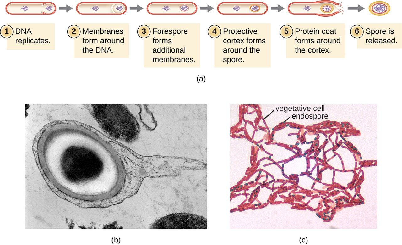
A handful of Gram-positive bacteria — most importantly the genera Bacillus and Clostridioides/Clostridium — can do something no other microbes can: when conditions turn hostile, they seal a copy of their DNA inside an armored survival capsule called an endospore. The spore is not a way to reproduce — one cell makes one spore — it is a way to wait out disaster. Inside its tough coat, the spore is metabolically dormant: no growth, no chemistry, no vulnerability. It can persist for years, even decades, in soil, on surfaces, or on hospital equipment.
How a spore forms and wakes
The normal, actively growing bacterium is called the vegetative cell. When it senses starvation or other stress, it begins sporulation: it copies its chromosome, walls off a small portion of cytoplasm around that DNA, and wraps it in layer after protective layer — a dense core stripped of most of its water, a thick cortex, and a tough protein coat. The rest of the original cell then disintegrates, releasing the finished endospore.
The spore stays dormant until conditions improve. When nutrients return, it germinates — the coat cracks, water rushes back in, metabolism restarts, and it grows back into an ordinary, dividing vegetative cell. Crucially, the vegetative cell is the form that causes active disease and that ordinary antibiotics and disinfectants can kill; the spore is the form that survives to start the cycle again.
Why spores defeat routine disinfection
The endospore's low water content and layered armor make it extraordinarily resistant — to heat, drying, radiation, and most chemical disinfectants, including the alcohol-based hand rubs that work so well against almost everything else. This is not an academic point; it changes bedside practice. Two organisms make it real:
Clostridioides difficile spreads by spores that survive on bedrails, commodes, and hands. Alcohol gel does not kill them — so for a patient with C. diff, the rule is soap-and-water handwashing (which physically washes spores away) plus a sporicidal cleaner such as diluted bleach for surfaces. Clostridium tetani and Clostridium botulinum spores lurk in soil and improperly canned food. And because spores can shrug off boiling, true sterilization of instruments requires an autoclave — pressurized steam at 121°C — not just hot water.
| Organism | Disease | Why the spore matters |
|---|---|---|
| Clostridioides difficile | Antibiotic-associated colitis | Spores survive alcohol gel → soap & water + bleach |
| Clostridium tetani | Tetanus | Soil spores enter wounds |
| Clostridium botulinum | Botulism | Spores survive in improperly canned food |
| Bacillus anthracis | Anthrax | Durable spores; bioterror concern |
- An endospore is a dormant, armored survival form — not a method of reproduction.
- Made mainly by Gram-positive Bacillus and Clostridioides/Clostridium; the growing form is the vegetative cell.
- Sporulation packages DNA in protective layers under stress; germination revives it when conditions improve.
- Spores resist heat, drying, and disinfectants — hence soap-and-water for C. diff and autoclaving for sterilization.
The chapter in one breath
- Cells are prokaryotic (no nucleus — bacteria) or eukaryotic (nucleus + organelles — fungi, protozoa, human).
- Prokaryote-only structures — peptidoglycan wall, 70S ribosome — give antibiotics their selective toxicity.
- The cell envelope is the plasma membrane (transport + energy) plus the cell wall and, in Gram-negatives, an outer membrane; inside it sit the cytoplasm with 70S ribosomes and the DNA-holding nucleoid, while plasmids carry and spread resistance.
- The wall is peptidoglycan, the target of beta-lactams; Gram-positive = thick wall/purple, Gram-negative = thin wall + outer membrane/pink.
- The Gram-negative outer membrane carries LPS (endotoxin), a driver of septic shock.
- External structures — capsule (resists phagocytosis), flagella (motility), fimbriae (attachment), pili (gene transfer) — are key virulence and resistance-spreading tools.
- Endospores let Bacillus and Clostridioides survive heat and disinfectants — the reason C. diff needs soap-and-water and instruments need autoclaving.
ReferenceWords worth knowing
A quick reference for the terms in this chapter — skim it now, or circle back whenever a word trips you up.
- Capsule
- A thick, sticky outer layer around some bacteria that resists phagocytosis; a virulence factor and vaccine target.
- Cell envelope
- The layers enclosing a bacterium — the plasma membrane, the cell wall, and any outer membrane.
- Cell wall
- The rigid peptidoglycan layer outside the membrane that gives a bacterium its shape and resists osmotic bursting.
- Chemotaxis
- Directed movement by flagella toward nutrients or away from harmful chemicals.
- Conjugation
- Transfer of DNA (often a plasmid) between bacteria through a sex pilus.
- Endospore
- A dormant, highly resistant survival form made by some Gram-positive bacteria; not a means of reproduction.
- Endotoxin (LPS)
- Lipopolysaccharide in the Gram-negative outer membrane that triggers fever, hypotension, and septic shock when released.
- Eukaryote
- A cell whose DNA is enclosed in a membrane-bound nucleus and that contains organelles; fungi, protozoa, and human cells.
- Fimbriae
- Numerous short hair-like appendages used to attach to surfaces and host cells; enable biofilms and adhesion.
- Flagella
- Long whip-like appendages that rotate to propel a bacterium through liquid.
- Gram-negative
- Bacteria with a thin peptidoglycan wall plus an outer membrane; stain pink and carry endotoxin.
- Gram-positive
- Bacteria with a thick peptidoglycan wall and no outer membrane; retain crystal violet and stain purple.
- Nucleoid
- The membrane-free region of a prokaryote where the circular chromosome is coiled.
- Peptidoglycan
- The mesh of cross-linked sugars and amino acids that makes up the bacterial cell wall; the target of beta-lactam antibiotics.
- Pili (sex pilus)
- Long, few appendages that bridge two bacteria to transfer DNA by conjugation.
- Plasma membrane
- The selectively permeable phospholipid bilayer that controls transport and generates energy in prokaryotes.
- Plasmid
- A small circular loop of DNA separate from the chromosome that often carries resistance or toxin genes.
- Prokaryote
- A cell with no membrane-bound nucleus or organelles; bacteria and archaea.
- Ribosome (70S / 80S)
- The protein-building machine; prokaryotes use the smaller 70S, eukaryotes the larger 80S — a key antibiotic target.
- Selective toxicity
- Harming a microbe while sparing the host by targeting a structure the microbe has and the host lacks.
- Sporulation
- The process by which a stressed vegetative cell forms an endospore; reversed by germination.
- Vegetative cell
- The active, growing, dividing form of a bacterium — the form that causes disease and is killed by drugs and disinfectants.
Check yourself
Bacterial Diversity & Classification
There are more kinds of bacteria than there are kinds of everything else alive, and only a small fraction ever make a person sick — but that fraction is the fraction you will meet at the bedside. The trick to keeping the clinically important organisms straight is not memorizing a list; it is learning the handful of features microbiologists use to sort bacteria into groups. Shape, staining, oxygen needs, and a few special quirks together place almost any pathogen into a recognizable box, and the box tells you how it spreads, what illness it causes, and which drugs will work. This chapter walks through those sorting features one at a time and hangs the most important pathogens on them, so that a culture result or a Gram-stain report starts to read like a sentence you already understand.
By the end of this chapter you can
- Name the three basic bacterial shapes and the common arrangements, and read the clues hidden in a genus name.
- Explain what the Gram stain reveals and why gram-positive and gram-negative cell walls respond differently.
- Identify the major gram-positive pathogens — Staphylococcus, Streptococcus, and Clostridium/Clostridioides — and the diseases they cause.
- Identify the major gram-negative pathogens — E. coli, Salmonella, and Pseudomonas — and explain why endotoxin makes them dangerous.
- Describe the special bacteria — mycobacteria, spirochetes, and mycoplasmas — that break the usual staining and cell-wall rules.
- Classify bacteria by their oxygen requirements and explain why this changes how specimens are collected and where infections form.
- List the other axes — temperature, pH, salt, intracellular lifestyle — used to describe and identify bacteria.
- Connect each classification feature to a concrete nursing action at the bedside.
Section 1Shapes & arrangements
Three basic shapes
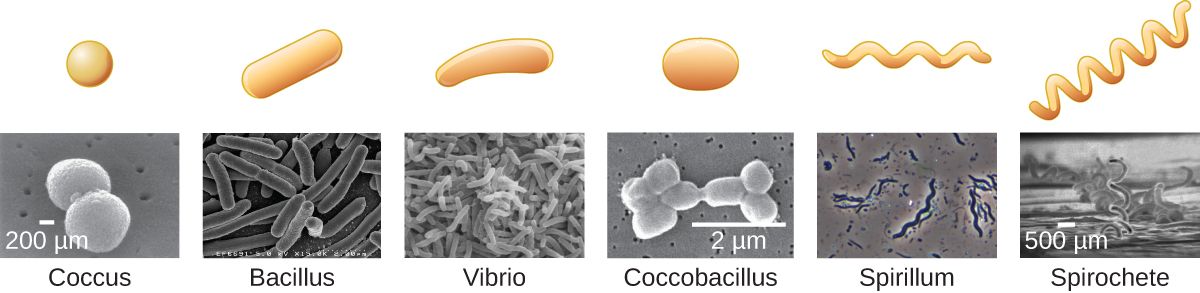
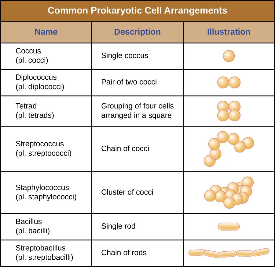
Under the microscope, the overwhelming majority of bacteria take one of three shapes, and the shape is the very first thing a lab reports because it narrows the field so quickly. A coccus (plural cocci) is round or spherical, like a tiny ball. A bacillus (plural bacilli) is a rod, shaped like a stick or a grain of rice. A spirillum (plural spirilla) is spiral or curved, coiled like a corkscrew or bent like a comma. Between these three there are in-between forms worth knowing: a coccobacillus is a short, plump rod that can be mistaken for a coccus, and a vibrio is a short comma-shaped curve — the shape that gives Vibrio cholerae its name.
Shape is not just description. It is built into the name of nearly every organism you will chart, so the moment you learn to hear it, the vocabulary of microbiology starts teaching itself. Staphylococcus and Streptococcus are spheres; Bacillus and Lactobacillus are rods; Spirochetes are coils. Reading the shape out of a genus name is a habit worth building on day one.
Arrangements: how the cells hang together
When a bacterium divides, the two daughter cells may drift apart or may stay stuck together, and the pattern they form is a second identifying clue — especially for cocci. The prefixes are logical once you learn them. Diplo- means the cells sit in pairs (diplococci, as in Neisseria). Strepto- means they form chains, like a string of beads (streptococci). Staphylo- means they clump in irregular grape-like clusters (staphylococci). Cocci can also form flat squares of four (tetrads) or cubical packets of eight (sarcinae). Rods more often line up end-to-end in short chains or side-by-side like a fence (a palisade), the arrangement typical of Corynebacterium.
| What you see | Name | Classic organism |
|---|---|---|
| Round, in grape-like clusters | Staphylococci | Staphylococcus aureus |
| Round, in chains | Streptococci | Streptococcus pyogenes |
| Round, in pairs | Diplococci | Neisseria, S. pneumoniae |
| Rod-shaped | Bacillus | E. coli, Clostridium |
| Comma / curved rod | Vibrio | Vibrio cholerae |
| Spiral / corkscrew | Spirochete | Treponema pallidum |
- The three basic shapes are coccus (sphere), bacillus (rod), and spirillum (spiral); vibrios and coccobacilli are in-between forms.
- Arrangement prefixes — diplo- (pairs), strepto- (chains), staphylo- (clusters) — add a second layer of identification, especially for cocci.
- Shape and arrangement are usually built into the genus name, so the name itself carries clues.
- The preliminary Gram-stain report (shape + arrangement + color) guides empiric therapy before the final identification arrives.
Section 2Gram-positive pathogens
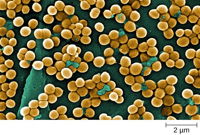
What "gram-positive" means
The Gram stain, devised by Hans Christian Gram in 1884, sorts almost all bacteria into two great camps based on how their cell wall holds a purple dye. Gram-positive bacteria have a thick outer layer of peptidoglycan that traps the crystal-violet stain and turns the cells deep purple. Because that wall is exposed on the surface, gram-positive organisms are the classic targets of the penicillin family, which sabotages peptidoglycan as it is built. The three genera below account for a huge share of the gram-positive disease a nurse will ever see.
Staphylococcus — the cluster
Staphylococcus are gram-positive cocci in grape-like clusters. The species that matters most, Staphylococcus aureus, is a spectacularly versatile pathogen: it lives harmlessly in the nose of roughly a third of healthy people, yet given a break in the skin it causes boils, abscesses, cellulitis, wound infections, bloodstream infections, and endocarditis, and its toxins cause a form of food poisoning and toxic shock syndrome. MRSA — methicillin-resistant S. aureus — is the resistant form that drives contact precautions on so many units. A related group, the coagulase-negative staphylococci such as S. epidermidis, are low-virulence skin residents that nonetheless cause trouble by colonizing catheters, prosthetic joints, and heart valves.
Streptococcus — the chain
Streptococcus are gram-positive cocci in chains, and they are sorted clinically by the way they break down blood on an agar plate and by surface antigens. Group A strep (S. pyogenes) causes strep throat, impetigo, cellulitis, scarlet fever, the flesh-destroying necrotizing fasciitis, and the delayed immune complications of rheumatic fever and post-streptococcal glomerulonephritis. Group B strep (S. agalactiae) colonizes the vagina in some pregnant patients and is a leading cause of newborn sepsis and meningitis — the reason for routine screening late in pregnancy. Streptococcus pneumoniae (a diplococcus) is a top cause of community-acquired pneumonia, otitis media, and meningitis, and is the target of the pneumococcal vaccines.
Clostridium — the spore-former
Clostridium and its relative Clostridioides are gram-positive rods with two dangerous properties: they are obligate anaerobes (Section 5), and they form endospores — dormant, armored survival capsules that shrug off heat, drying, and alcohol. The roll call is memorable: Clostridioides difficile (antibiotic-associated colitis), Clostridium tetani (tetanus), Clostridium botulinum (botulism), and Clostridium perfringens (gas gangrene and food poisoning). Because their spores resist alcohol, an alcohol hand rub does not remove C. difficile — hands must be washed with soap and water, and surfaces cleaned with a sporicidal (bleach) agent.
| Genus | Look | Key species & disease | Nursing flag |
|---|---|---|---|
| Staphylococcus | Cocci in clusters | S. aureus — skin/wound/blood; MRSA | Contact precautions for MRSA |
| Streptococcus | Cocci in chains | Group A (throat, necrotizing); Group B (newborn sepsis); pneumoniae (pneumonia) | Screen GBS in pregnancy; vaccinate for pneumococcus |
| Clostridium / Clostridioides | Rods, spore-forming, anaerobic | C. difficile, tetanus, botulism, gas gangrene | Soap & water, not alcohol, for C. diff |
- Gram-positive bacteria have thick peptidoglycan, stain purple, and are the classic targets of penicillins.
- Staphylococcus = cocci in clusters; S. aureus and MRSA cause skin, wound, and bloodstream infections.
- Streptococcus = cocci in chains; Group A (throat, necrotizing fasciitis, rheumatic fever), Group B (newborn sepsis), pneumoniae (pneumonia/meningitis).
- Clostridium/Clostridioides = anaerobic, spore-forming rods; C. diff spores resist alcohol, so wash with soap and water.
Section 3Gram-negative pathogens
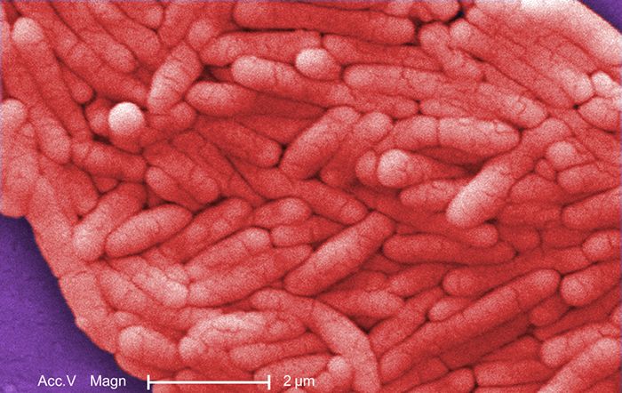
What "gram-negative" means — and why endotoxin matters
Gram-negative bacteria have only a thin layer of peptidoglycan, so they lose the purple crystal violet during decolorizing and take up the pink counterstain instead. What makes them clinically distinct, though, is a second membrane wrapped outside the peptidoglycan — the outer membrane — whose outer face is studded with lipopolysaccharide (LPS). The lipid A portion of LPS is a potent endotoxin: when gram-negative bacteria die and break apart in the bloodstream, released endotoxin triggers a runaway inflammatory cascade — fever, falling blood pressure, and, at the extreme, septic shock. This is why a gram-negative bloodstream infection is a medical emergency, and why the sepsis alerts nurses respond to so often trace back to these organisms.
Escherichia coli — the everyday enteric
Escherichia coli is a gram-negative rod and a normal resident of the colon, where it is harmless. Out of place, it is the single most common cause of urinary tract infections, and it is a frequent cause of gram-negative sepsis and neonatal meningitis. Certain strains are outright intestinal pathogens: enterohemorrhagic E. coli (such as O157:H7) produces Shiga toxin and can cause bloody diarrhea and hemolytic uremic syndrome, a cause of acute kidney failure in children. Because E. coli is a facultative anaerobe, it thrives both in the oxygen-poor gut and in the bladder.
Salmonella — the food-borne invader
Salmonella are gram-negative rods spread by contaminated food and water and by contact with reptiles and poultry. Most infections are a self-limited gastroenteritis — the classic food poisoning from undercooked eggs or chicken — but Salmonella Typhi causes typhoid fever, a systemic illness with sustained high fever. Salmonella is a reminder that hydration and hand hygiene, not routine antibiotics, are the mainstay for uncomplicated infectious diarrhea in an otherwise healthy adult.
Pseudomonas aeruginosa — the opportunist of wet places
Pseudomonas aeruginosa is a gram-negative rod, an obligate aerobe, and one of the great opportunists of the hospital. It lives in moist reservoirs — sink drains, respiratory equipment, standing water, even flower vases — and it attacks patients whose defenses are down: it causes ventilator-associated pneumonia, infections of burns and chronic wounds, catheter-associated UTIs, and the chronic lung colonization seen in cystic fibrosis. It is famous for producing a blue-green pigment and a grape-like or "corn tortilla" odor, and for being intrinsically resistant to many antibiotics, which makes it hard to treat and a frequent driver of multidrug-resistant outbreaks.
| Organism | Oxygen | Signature disease | Where it lurks |
|---|---|---|---|
| Escherichia coli | Facultative anaerobe | UTI, gram-negative sepsis; O157:H7 → HUS | Colon (normal flora) |
| Salmonella | Facultative anaerobe | Gastroenteritis; typhoid fever | Eggs, poultry, reptiles |
| Pseudomonas aeruginosa | Obligate aerobe | VAP, burn/wound infection, CF lung | Water, moist equipment |
- Gram-negative bacteria stain pink and carry an outer membrane with LPS/endotoxin that can drive septic shock.
- E. coli is the top cause of UTIs and a common cause of gram-negative sepsis; some strains (O157:H7) cause bloody diarrhea and HUS.
- Salmonella is food-borne; most infections are self-limited gastroenteritis, but S. Typhi causes typhoid fever.
- Pseudomonas aeruginosa is a water-loving, drug-resistant opportunist behind ventilator pneumonia, burn/wound, and CF infections.
Section 4Special bacteria
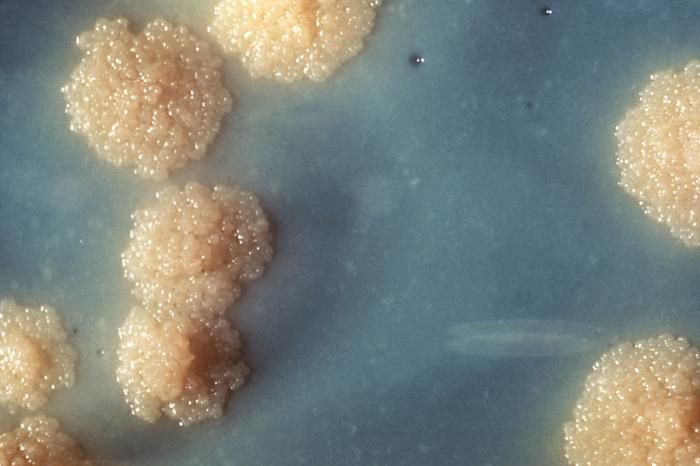
Mycobacteria — the waxy, acid-fast rods
A few important bacteria refuse to fit the Gram-stain scheme, and the most important of these are the mycobacteria. Their cell wall is loaded with a waxy lipid called mycolic acid, which repels ordinary dyes; to see them you need a special acid-fast stain that forces dye into the wall and then resists decolorizing with acid-alcohol. Mycobacterium tuberculosis causes tuberculosis, and its features drive real nursing practice: the waxy wall makes it slow-growing (cultures take weeks) and hardy in the environment, it spreads through the air on tiny droplet nuclei, and it therefore requires airborne isolation — a negative-pressure room and a fitted N95 respirator, not a simple surgical mask. Its cousin Mycobacterium leprae causes leprosy (Hansen disease).
Spirochetes — the corkscrews
Spirochetes are long, thin, tightly coiled bacteria that move with a distinctive corkscrew motion driven by internal axial filaments (endoflagella). They are so slender that many cannot be seen on a routine Gram stain or grown on ordinary media, so they are usually diagnosed by blood tests rather than culture. The three to know are Treponema pallidum (syphilis), Borrelia burgdorferi (Lyme disease, spread by tick bite), and Leptospira (leptospirosis, from water contaminated with animal urine). Their stealthy, hard-to-detect nature is exactly why serologic testing and a careful history matter so much for these infections.
Mycoplasma — the wall-less minimalists
Mycoplasma are the smallest free-living bacteria and are unique in having no cell wall at all. Without a rigid wall they have no fixed shape (they are pleomorphic) and, crucially, no peptidoglycan for a penicillin to attack — so beta-lactam antibiotics are useless against them. Mycoplasma pneumoniae causes "walking pneumonia," a mild, drawn-out atypical pneumonia common in younger people, and is treated with a macrolide or tetracycline instead of a penicillin. The wall-less lesson is a clean illustration of selective toxicity from the other direction: no wall means no target.
| Group | What breaks the rules | Key organism / disease | Practical consequence |
|---|---|---|---|
| Mycobacteria | Waxy mycolic-acid wall; acid-fast | M. tuberculosis — TB | Airborne isolation; slow culture |
| Spirochetes | Thin coils; hard to stain/culture | T. pallidum (syphilis), B. burgdorferi (Lyme) | Diagnosed by serology, not culture |
| Mycoplasma | No cell wall at all | M. pneumoniae — walking pneumonia | Penicillins fail; use macrolide/tetracycline |
- Mycobacteria have a waxy mycolic-acid wall, are acid-fast, grow slowly, and (as M. tuberculosis) require airborne isolation.
- Spirochetes are thin corkscrews (Treponema, Borrelia, Leptospira) usually diagnosed by serology because they resist staining and culture.
- Mycoplasma have no cell wall, so beta-lactam antibiotics do not work; M. pneumoniae causes walking pneumonia.
- Each "special" group breaks a rule — a reminder to match staining, isolation, and drug choice to the organism's structure.
Section 5Oxygen & other groupings
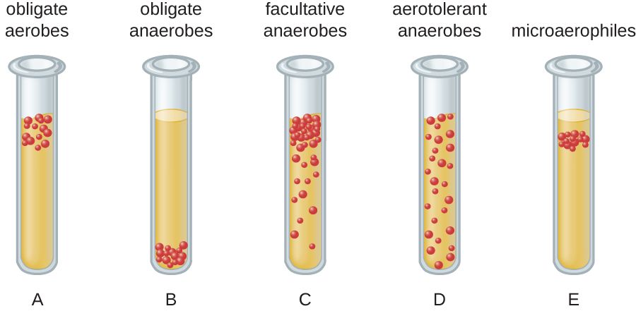
Sorting bacteria by their relationship to oxygen
Oxygen is life-giving to your cells, but to many bacteria it is either essential, irrelevant, or lethal — and how an organism relates to oxygen is a core classification axis with direct bedside consequences. An obligate aerobe must have oxygen to grow (Pseudomonas, Mycobacterium tuberculosis). An obligate anaerobe is poisoned and killed by oxygen and grows only where it is absent (Clostridium, Bacteroides). A facultative anaerobe is the flexible middle: it prefers oxygen but can switch to fermentation without it (E. coli, Staphylococcus, most of the organisms we culture easily). A microaerophile needs oxygen but only at low levels (Campylobacter, Helicobacter pylori), and an aerotolerant anaerobe does not use oxygen at all but survives being exposed to it.
Why oxygen grouping changes nursing care
This is not an academic distinction. Anaerobes flourish in exactly the places oxygen cannot reach: the depths of a puncture wound, the center of an abscess, dead and crushed tissue, the gut, and the pockets of the mouth — which is why deep, dirty, or bite wounds and gas gangrene are anaerobic problems. It also shapes specimen handling: because oxygen kills obligate anaerobes on contact, a specimen collected to look for them must be protected from air and transported to the lab quickly in special anaerobic conditions, or the organism dies before it can be grown and the culture falsely comes back negative.
| Group | Relationship to oxygen | Example |
|---|---|---|
| Obligate aerobe | Requires oxygen | Pseudomonas, M. tuberculosis |
| Obligate anaerobe | Killed by oxygen | Clostridium, Bacteroides |
| Facultative anaerobe | Uses oxygen if present, ferments if not | E. coli, Staphylococcus |
| Microaerophile | Needs low oxygen | Campylobacter, H. pylori |
| Aerotolerant anaerobe | Ignores oxygen but tolerates it | Some lactobacilli |
The other axes of classification
Oxygen is only one of several dials microbiologists turn. Bacteria are also grouped by temperature preference (most human pathogens are mesophiles that thrive near body temperature, 37°C), by pH and salt tolerance (a few extremophiles love acid or high salt), and by whether they can survive only inside host cells. Two clinically important groups are obligate intracellular bacteria — Chlamydia and Rickettsia — which cannot reproduce outside a host cell at all, so they too cannot be grown on standard plates and are diagnosed by other means. Put together, these axes — shape, Gram reaction, oxygen need, growth requirements, and lifestyle — are the coordinates that let a lab pin down almost any organism.
- Bacteria sort by oxygen into obligate aerobes, obligate anaerobes, facultative anaerobes, microaerophiles, and aerotolerant anaerobes.
- Anaerobes cause deep-wound, abscess, and dead-tissue infections, and their specimens must be protected from air during transport.
- Other axes include temperature, pH, salt, and an intracellular lifestyle (e.g., Chlamydia, Rickettsia).
- Combining all the features — shape, stain, oxygen, growth needs — is how the lab identifies an organism.
The chapter in one breath
- Bacteria are sorted first by shape (coccus, bacillus, spirillum) and arrangement (pairs, chains, clusters), clues usually built into the genus name.
- The Gram stain splits bacteria into gram-positive (thick peptidoglycan, purple) and gram-negative (thin wall + outer membrane, pink).
- Key gram-positive pathogens: Staphylococcus (clusters), Streptococcus (chains), and spore-forming anaerobic Clostridium/Clostridioides.
- Key gram-negative pathogens: E. coli, Salmonella, and Pseudomonas — their endotoxin (LPS) can drive septic shock.
- Special bacteria break the rules: mycobacteria are acid-fast, spirochetes are hard-to-stain coils, and mycoplasmas have no cell wall (so penicillins fail).
- Oxygen requirements (aerobe, anaerobe, facultative, microaerophile) shape where infections form and how specimens must be collected.
- Additional axes — temperature, pH, salt, and an intracellular lifestyle — complete the coordinate system labs use to identify any organism.
ReferenceWords worth knowing
A quick reference for the terms in this chapter — skim it now, or circle back whenever a word trips you up.
- Acid-fast
- A staining property of bacteria whose waxy, mycolic-acid wall resists decolorizing by acid-alcohol; the way mycobacteria are identified.
- Aerobe (obligate)
- A bacterium that requires oxygen to grow, such as Pseudomonas or M. tuberculosis.
- Anaerobe (obligate)
- A bacterium that is killed by oxygen and grows only where oxygen is absent, such as Clostridium.
- Arrangement
- The pattern bacterial cells form after dividing — pairs (diplo-), chains (strepto-), or clusters (staphylo-).
- Bacillus
- A rod-shaped bacterium (plural bacilli).
- Coccus
- A spherical or round bacterium (plural cocci).
- Endospore
- A dormant, highly resistant structure formed by Clostridium and Bacillus that survives heat, drying, and alcohol.
- Endotoxin
- The lipid A part of the lipopolysaccharide in a gram-negative outer membrane; released when cells die, it can trigger fever and septic shock.
- Facultative anaerobe
- A bacterium that uses oxygen when available but can grow without it, such as E. coli.
- Gram stain
- The staining procedure that divides bacteria into gram-positive (purple) and gram-negative (pink) by cell-wall structure.
- Gram-negative
- Bacteria with a thin peptidoglycan wall and an outer membrane bearing LPS; they stain pink.
- Gram-positive
- Bacteria with a thick peptidoglycan wall that traps crystal violet and stains purple.
- Lipopolysaccharide (LPS)
- The molecule on the gram-negative outer membrane whose lipid A component is endotoxin.
- Microaerophile
- A bacterium that needs oxygen but only at low levels, such as Campylobacter or H. pylori.
- MRSA
- Methicillin-resistant Staphylococcus aureus; a resistant strain that drives contact precautions.
- Mycolic acid
- The waxy lipid in the mycobacterial cell wall responsible for acid-fastness and environmental hardiness.
- Peptidoglycan
- The mesh-like polymer that forms the bacterial cell wall and is the target of penicillin-family drugs.
- Pleomorphic
- Having no fixed shape; describes wall-less bacteria such as Mycoplasma.
- Spirillum
- A spiral or corkscrew-shaped bacterium (plural spirilla).
- Spirochete
- A thin, tightly coiled bacterium that moves by internal axial filaments, such as Treponema pallidum.
- Vibrio
- A short, comma-shaped curved rod, as in Vibrio cholerae.
Check yourself
Viruses, Fungi & Parasites
Not every microbe is a bacterium. Once you step outside the bacterial world you meet a stranger cast of characters: viruses, which are barely alive and cannot reproduce without hijacking one of your cells; fungi, which are built more like us than like bacteria and are therefore harder to poison selectively; and the parasites — single-celled protozoa and multicellular worms — that cause some of the most widespread diseases on earth. This chapter walks through how each of these agents is built, how it makes copies of itself, and how it makes your patients sick. The through-line for the nurse is simple: what a pathogen is made of decides how you kill it, how it spreads, and how you protect the next patient from it.
By the end of this chapter you can
- Describe the parts of a virus — capsid, genome, and (sometimes) envelope — and explain why an envelope changes how a virus is disinfected.
- Walk through the viral replication cycle and explain how latency lets a virus hide and return.
- Distinguish yeasts from molds, name the common mycoses, and explain why antifungal drugs are harder on the patient than antibiotics.
- Explain how protozoa cause disease, using malaria and giardiasis as anchor examples.
- Separate the parasitic roundworms from the flatworms and recognize the clues (like eosinophilia) that suggest a worm.
- Connect each pathogen's structure to its route of transmission and the nursing precautions that interrupt it.
- Recognize why so many of these infections cluster in immunocompromised patients.
Section 1How a virus is built
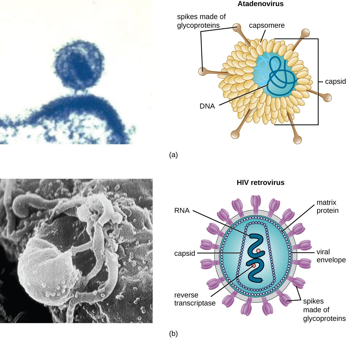
Alive, or not quite?
A virus sits on the fence between living and non-living, and that in-between status is the single most important thing to understand about it. A virus is acellular — it is not a cell. It has no cytoplasm, no ribosomes, no way to make energy, and no machinery to copy itself. Left on a countertop, a virus is inert, more like a complicated chemical than an organism. It only comes to life when it enters a host cell and borrows that cell's machinery. Because it absolutely cannot reproduce on its own, a virus is called an obligate intracellular parasite — "obligate" meaning it has no choice. This is exactly why antibiotics do nothing to a virus: antibiotics attack bacterial cell structures, and a virus has none of them.
The capsid and the genome
Strip a virus down and you find two guaranteed parts. At the core is the genome — the virus's genetic instructions. Unlike your cells, which always use double-stranded DNA, viruses are wildly varied: a virus may carry DNA or RNA, and that nucleic acid may be single-stranded or double-stranded. This matters clinically because RNA viruses (influenza, HIV, SARS-CoV-2, hepatitis C) copy their genomes sloppily, mutate quickly, and are the reason we need a new flu shot every year. Wrapped around the genome is the capsid, a protective shell built from repeating protein subunits called capsomeres. The capsid protects the fragile genome and carries the surface proteins the virus uses to latch onto a host cell. Capsids come in a few classic shapes — helical (rod-like), icosahedral (a roughly spherical 20-sided cage), and complex — and together the capsid plus genome are called the nucleocapsid.
The envelope, and why hand hygiene cares
Some viruses add a third layer: an envelope, a lipid membrane the virus steals from its host cell on the way out. Studded in that envelope are viral spike proteins (glycoproteins) that act as the keys for the virus's next break-in — the SARS-CoV-2 spike and the influenza hemagglutinin are the famous examples. Whether a virus has an envelope or is "naked" turns out to be enormously practical at the bedside. An enveloped virus depends on its lipid coat to infect, and lipids are easy to destroy — soap, alcohol-based sanitizer, and ordinary drying pull the envelope apart, so enveloped viruses like flu, COVID, HIV, and herpes are relatively easy to kill. There is one exception you must not generalize away: hepatitis B is enveloped, yet it stays infectious in dried blood on a surface for a week or more. Blood and body-fluid spills are decontaminated with an EPA-registered hospital disinfectant with a tuberculocidal or HBV claim, or with 1:10 household bleach — not with an alcohol wipe. A non-enveloped (naked) virus, protected only by its tough protein capsid, shrugs off alcohol and survives for days on surfaces; norovirus and the poliovirus are classic hardy naked viruses that demand bleach and scrupulous surface cleaning.
| Feature | Enveloped virus | Non-enveloped (naked) virus |
|---|---|---|
| Outer layer | Lipid envelope with spike proteins | Protein capsid only |
| Environmental survival | Usually fragile — dries out, short-lived on surfaces (exception: hepatitis B survives 7+ days in dried blood) | Hardy — survives for days |
| Alcohol / soap | Usually destroyed — the lipid coat dissolves; blood spills (HBV) still need bleach or a hospital disinfectant | Often resistant |
| Best disinfectant | Alcohol sanitizer, soap and water | Bleach (hypochlorite) and friction |
| Examples | Influenza, SARS-CoV-2, HIV, herpes, hepatitis B | Norovirus, rotavirus, poliovirus, hepatitis A |
- A virus is acellular and an obligate intracellular parasite — it cannot reproduce without a host cell.
- Every virus has a genome (DNA or RNA, single- or double-stranded) inside a protein capsid; RNA viruses mutate fast.
- Some viruses add a stolen lipid envelope with spike proteins that serve as attachment keys.
- Enveloped viruses usually die to soap and alcohol (hepatitis B in dried blood is the exception); naked viruses resist alcohol and need bleach — a direct hand-hygiene decision.
Section 2How viruses replicate
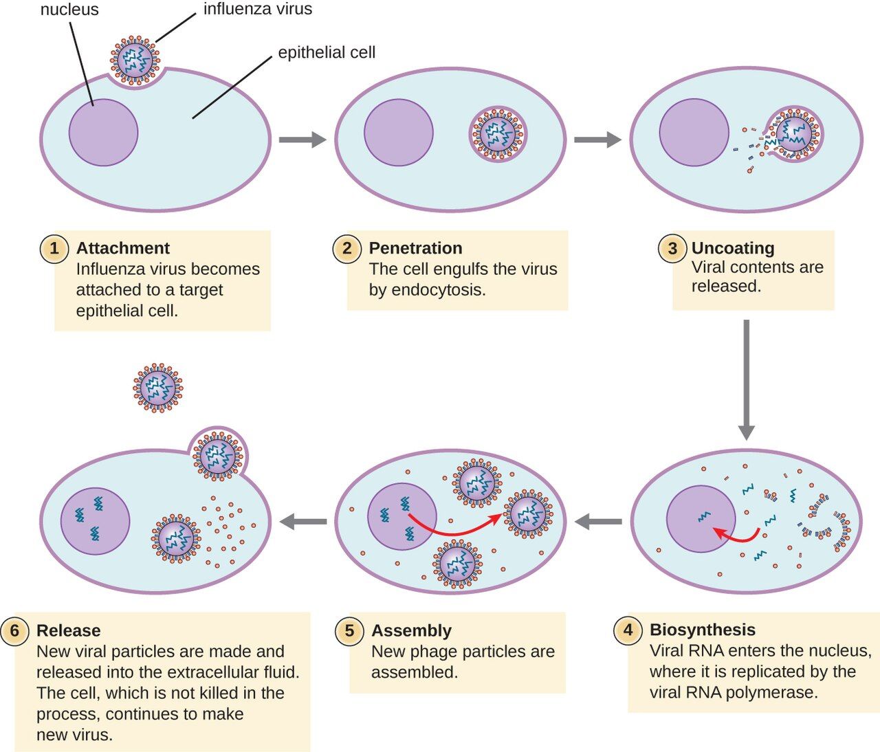
The five-step cycle
Because a virus has no factory of its own, replication is really a story of hijacking. Across almost all viruses the sequence is the same five steps. First is attachment: a surface protein on the virus fits a specific receptor on the host cell, like a key in a lock — and that lock-and-key specificity is why a given virus infects only certain cells (HIV targets CD4 T-cells; rhinovirus targets airway lining). Second is entry (penetration), as the whole virus or just its genome slips inside. Third, and the heart of it, is biosynthesis: the viral genome commandeers the cell's ribosomes, enzymes, and building blocks to mass-produce viral proteins and copies of the viral genome. Fourth is assembly (maturation), where those fresh parts snap together into new virus particles. Fifth is release, when the new viruses escape to infect neighboring cells.
Budding versus bursting
That last step comes in two flavors, and the difference maps onto the envelope from Section 1. Naked viruses usually escape by lysis — the host cell is packed so full of new virus that it bursts and dies, releasing hundreds of particles at once. Enveloped viruses more often leave by budding, pushing through the host membrane and wrapping themselves in a piece of it (that is where the envelope comes from). Budding can be gentler and slower, which is part of how some enveloped viruses set up long, smoldering, chronic infections rather than a quick blaze. Either way, the destruction of host cells — whether by bursting or by wearing the cell out — is a major reason viral infections make people sick.
Latency: the virus that waits
Some viruses do not replicate and leave — they hide. In latency, the virus enters a cell, quiets down, and parks its genome inside the host, producing few or no new particles and causing no symptoms, sometimes for decades. Then, often when the immune system is stressed or weakened, it reactivates and starts replicating again. The herpesvirus family are the masters of this: varicella-zoster causes chickenpox, retreats into nerve roots, and can reawaken years later as painful shingles; herpes simplex hides in nerves between cold-sore or genital outbreaks. HIV does something similar at the DNA level, splicing a DNA copy of itself (a provirus) into the host chromosome, where it can sit silently — which is exactly why HIV cannot yet be cured, only suppressed.
| Step | What happens | Where drugs can strike |
|---|---|---|
| 1. Attachment | Viral protein binds a specific host receptor | Entry blockers (e.g., some HIV drugs) |
| 2. Entry | Virus or its genome enters the cell | Fusion inhibitors |
| 3. Biosynthesis | Host machinery mass-produces viral genome & proteins | Most antivirals (e.g., acyclovir, HIV reverse-transcriptase inhibitors) |
| 4. Assembly | New parts snap into complete particles | Protease inhibitors |
| 5. Release | New virus buds off or bursts the cell | Neuraminidase inhibitors (e.g., oseltamivir for flu) |
- Viral replication has five steps: attachment → entry → biosynthesis → assembly → release.
- Attachment is receptor-specific, which decides exactly which cells and hosts a virus can infect.
- Naked viruses tend to exit by bursting (lysis); enveloped viruses often exit by budding.
- Latency lets herpesviruses and HIV hide and reactivate; antivirals hit active replication, not the dormant genome.
Section 3Fungi
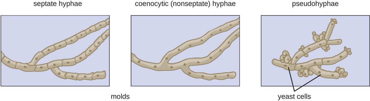
Yeasts and molds
Fungi are eukaryotes — their cells, like ours, keep DNA in a nucleus and are far more complex than bacteria. That family resemblance is exactly what makes fungal infections tricky to treat: a drug that harms a fungal cell can easily harm the patient's cells too. Fungi come in two body plans. A yeast is a single, oval cell that reproduces by budding off a small copy of itself; Candida is the classic yeast. A mold grows as long branching filaments called hyphae, which mat together into a visible fuzzy colony (a mycelium) — the green fur on old bread. Some fungi are dimorphic, living as a mold in the cool environment but switching to a yeast at body temperature; several dangerous lung fungi work this way. Two structural facts drive fungal drug therapy: the fungal cell wall is made of chitin (not the peptidoglycan bacteria use), and the fungal membrane is built on ergosterol where ours uses cholesterol. Ergosterol is the chink in the armor most antifungals aim for.
How fungal infections are grouped
Clinicians sort fungal diseases — the mycoses — largely by how deep they go. Superficial and cutaneous mycoses stay in the skin, hair, and nails; these are the dermatophyte infections nurses see constantly, the "tineas" — ringworm, athlete's foot, jock itch. Subcutaneous mycoses are driven deeper by trauma, like a thorn puncture. Systemic mycoses spread through the body and are the dangerous ones, frequently landing in the lungs. A crucial nursing theme runs through all of this: many serious fungal infections are opportunistic, meaning they take hold only when defenses are down — in patients on chemotherapy, on long courses of antibiotics or steroids, with poorly controlled diabetes, or with HIV/AIDS.
The mycoses nurses actually see
A handful of fungi account for most bedside encounters. Candida albicans is everywhere: it causes oral thrush (white plaques you can scrape off), vaginal yeast infections, diaper rash, and — when it reaches the bloodstream in a critically ill patient — life-threatening candidemia. Dermatophytes cause the itchy, ring-shaped tinea rashes. Aspergillus, a mold breathed in as spores, can carve out serious lung disease in the immunocompromised. Cryptococcus, picked up from bird droppings, causes a dangerous meningitis in AIDS patients. And Pneumocystis jirovecii causes the pneumonia (PJP/PCP) that is a hallmark of advanced HIV. Notice the pattern: the sicker and more immunosuppressed the patient, the more the fungi matter.
| Fungus | Type | Classic infection | Who's at risk |
|---|---|---|---|
| Candida albicans | Yeast | Thrush, vaginal yeast infection, candidemia | Antibiotic/steroid use, diabetes, ICU patients |
| Dermatophytes (Trichophyton & others) | Mold | Ringworm, athlete's foot, jock itch (tinea) | Anyone; warm, moist skin |
| Aspergillus | Mold | Invasive lung aspergillosis | Neutropenic, transplant patients |
| Cryptococcus neoformans | Yeast | Cryptococcal meningitis | Advanced HIV/AIDS |
| Pneumocystis jirovecii | Atypical fungus | PJP pneumonia | Advanced HIV/AIDS, immunosuppressed |
- Fungi are eukaryotes; yeasts are single cells that bud, molds grow as filamentous hyphae, and dimorphic fungi switch between the two.
- The fungal wall is chitin and the membrane uses ergosterol — the target of most antifungal drugs.
- Common mycoses range from skin tinea and Candida thrush to lethal systemic disease in the immunocompromised.
- Because fungi resemble our own cells, antifungals have narrower selective toxicity and more side effects.
Section 4Protozoa
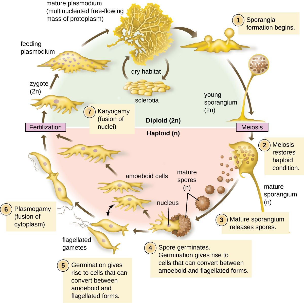
Single-celled animals
Protozoa are single-celled eukaryotes that behave like tiny animals — many hunt, most move, and a great number are parasites of humans. Many protozoa live a two-stage life. The trophozoite is the active, feeding, dividing, disease-causing form. The cyst is a tough, dormant, walled-off form that survives outside the body — in water, on hands, in food — and is the form that transmits the infection to the next person. Understanding that split is genuinely useful: the cyst is what you are trying to block with clean water and hand hygiene, and the cyst's toughness is why some of these parasites survive ordinary chlorination.
Malaria: the world's great killer
Malaria is caused by the protozoan Plasmodium and spread by the bite of the female Anopheles mosquito — a classic vector-borne disease. After a bite, the parasite first multiplies quietly in the liver, then pours into the bloodstream and invades red blood cells, multiplying inside them until they burst in synchronized waves. Those bursts produce malaria's signature: cyclical fever, chills, and drenching sweats that recur every couple of days. The most dangerous species, Plasmodium falciparum, can clog small vessels in the brain and cause fatal cerebral malaria. The single most important nursing takeaway is a habit of mind: fever in a patient who has recently traveled to a malaria region is an emergency until proven otherwise, because falciparum malaria can kill within days.
Giardia and the other clinical protozoa
Giardia is the protozoan a nursing student is most likely to meet at home: its hardy cysts contaminate lakes and streams, so it causes "backpacker's" or "camper's" diarrhea — foul-smelling, greasy, floating stools with gas and cramps, spread fecal–oral through contaminated water. Because the cysts resist normal chlorine levels, prevention means boiling or filtering water, not just treating it. Beyond Giardia, a few other protozoa recur on the wards and in exams: Entamoeba histolytica causes bloody amebic dysentery and liver abscess; Trichomonas vaginalis is a common sexually transmitted infection; Toxoplasma gondii (from cat feces and undercooked meat) is dangerous in pregnancy and in AIDS; and Cryptosporidium causes waterborne diarrhea that becomes severe and prolonged in the immunocompromised.
| Protozoan | Disease | How it spreads | Hallmark |
|---|---|---|---|
| Plasmodium | Malaria | Anopheles mosquito bite (vector) | Cyclic fever/chills; invades red cells |
| Giardia lamblia | Giardiasis | Fecal–oral; contaminated water (cysts) | Greasy, foul, floating diarrhea |
| Entamoeba histolytica | Amebic dysentery | Fecal–oral | Bloody diarrhea; liver abscess |
| Trichomonas vaginalis | Trichomoniasis | Sexual contact | Frothy discharge; itching |
| Toxoplasma gondii | Toxoplasmosis | Cat feces; undercooked meat | Danger in pregnancy & AIDS |
- Protozoa are single-celled eukaryotes; many alternate between an active trophozoite and a tough, transmissible cyst.
- Malaria (Plasmodium, via Anopheles mosquito) invades red blood cells and causes cyclic fevers; falciparum can be rapidly fatal.
- Giardia spreads fecal–oral through cyst-contaminated water and causes greasy, foul diarrhea; cysts resist chlorine.
- Recent travel and pregnancy reshape the protozoal risk list — both are questions worth asking.
Section 5Helminths: the parasitic worms
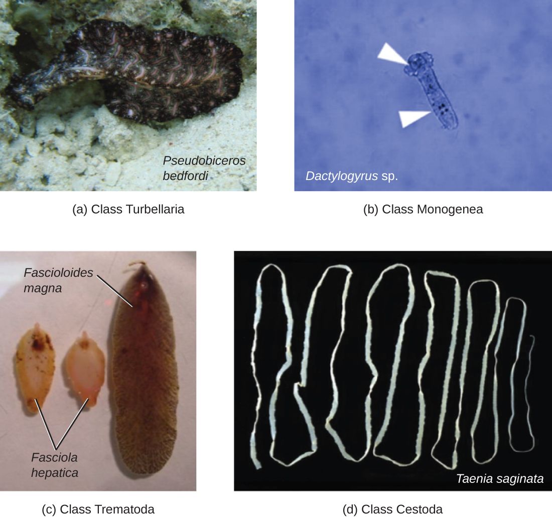
Worms as pathogens
The helminths are parasitic worms — and strictly speaking they are not microbes at all, since an adult tapeworm can be meters long. They earn a place in microbiology because they are diagnosed the way microbes are: by finding their microscopic eggs and larvae in stool, blood, or tissue. Worms make people sick in slow, chronic ways — stealing nutrients, causing anemia and malnutrition, blocking the bowel or bile ducts, and triggering allergic-type immune responses. That last point gives the nurse a valuable clue: worm infections classically drive up the eosinophils, a white-blood-cell type that fights parasites, so an unexplained eosinophilia on a blood count should raise the question, "could this be a worm?"
Roundworms (nematodes)
Nematodes are the roundworms — smooth, cylindrical, tapered at both ends. The one you are guaranteed to meet is the pinworm (Enterobius), the most common worm infection in the United States, especially in children: the female crawls out to lay eggs around the anus at night, causing intense itching, and the eggs spread easily on hands and bedding through whole households. Other important roundworms include Ascaris, a large intestinal worm that can grow the length of a pencil and, in heavy loads, obstruct the bowel; hookworms, which latch onto the gut wall and suck blood, causing iron-deficiency anemia; and Trichinella, caught from undercooked pork.
Flatworms: tapeworms and flukes
The platyhelminths (flatworms) split into two clinical groups. The cestodes are the tapeworms — long, flat, ribbon-like worms with a head that hooks onto the gut wall and a body of repeating segments; people catch them from undercooked beef, pork, or fish. The trematodes are the flukes, leaf-shaped worms with complex life cycles that usually pass through a snail. Globally the most important is Schistosoma (blood flukes), a leading cause of chronic illness in parts of Africa, Asia, and South America, where its eggs damage the bladder, liver, and intestines over years. For a nurse in most settings, tapeworms and pinworm come up far more often than flukes — but the flukes are a reminder of how a travel or immigration history reshapes the differential.
| Worm | Group | Classic scenario | Main harm |
|---|---|---|---|
| Pinworm (Enterobius) | Roundworm | Itchy bottom in a child at night | Perianal itching; spreads in households |
| Ascaris | Roundworm | Large worm in stool; heavy load | Bowel obstruction, malnutrition |
| Hookworm | Roundworm | Walking barefoot on contaminated soil | Iron-deficiency anemia (blood loss) |
| Tapeworm (Taenia) | Flatworm (cestode) | Undercooked beef or pork | Nutrient theft; segments in stool |
| Schistosoma | Flatworm (trematode) | Freshwater exposure abroad | Chronic bladder/liver/gut damage |
- Helminths are parasitic worms, diagnosed microscopically by their eggs and larvae; they cause slow, chronic harm.
- An unexplained eosinophilia is a classic clue that a worm may be present.
- Roundworms (nematodes) include pinworm, Ascaris, and blood-sucking hookworms.
- Flatworms (platyhelminths) are the tapeworms (cestodes) and flukes (trematodes), including Schistosoma.
The chapter in one breath
- A virus is acellular — a genome in a protein capsid, sometimes wrapped in a lipid envelope — and must hijack a host cell to reproduce.
- Enveloped viruses usually die to soap and alcohol — but hepatitis B survives in dried blood for a week; naked viruses resist alcohol and need bleach, a real hand-hygiene decision.
- Viruses replicate in five steps (attach, enter, biosynthesize, assemble, release); latency lets herpesviruses and HIV hide and reactivate.
- Fungi are eukaryotes — yeasts bud, molds form hyphae — and their ergosterol membrane is the antifungal target; many mycoses are opportunistic.
- Protozoa are single-celled parasites; malaria invades red cells and can kill fast, and Giardia spreads through cyst-contaminated water.
- Helminths are worms — roundworms (pinworm, Ascaris, hookworm) and flatworms (tapeworms, flukes) — found by their eggs and hinted at by eosinophilia.
- Across all four groups, the sickest and most immunocompromised patients carry the greatest risk, and pathogen structure dictates prevention.
ReferenceWords worth knowing
A quick reference for the terms in this chapter — skim it now, or circle back whenever a word trips you up.
- Acellular
- Not made of cells; describes viruses, which have no cytoplasm, ribosomes, or metabolism of their own.
- Budding
- Two meanings here: how a yeast reproduces (pinching off a daughter cell), and how an enveloped virus exits a host cell by wrapping itself in host membrane.
- Capsid
- The protein shell, built of repeating capsomeres, that surrounds and protects a virus's genome.
- Cestode
- A tapeworm; a flatworm with a hooking head and a ribbon of repeating segments.
- Cyst
- The tough, dormant, environmentally resistant form of many protozoa, responsible for transmission.
- Dermatophyte
- A mold that infects skin, hair, and nails, causing the ringworm/tinea group of infections.
- Dimorphic fungus
- A fungus that grows as a mold in the environment but converts to a yeast at body temperature.
- Envelope
- A lipid membrane, stolen from the host cell, that coats some viruses and usually makes them easy to kill with soap or alcohol — hepatitis B is the notable exception, surviving in dried blood for a week or more.
- Eosinophilia
- An elevated eosinophil count on a blood test; a classic clue to a parasitic worm infection or allergy.
- Ergosterol
- The sterol in fungal cell membranes (where humans use cholesterol); the main target of antifungal drugs.
- Helminth
- A parasitic worm — a roundworm (nematode) or flatworm (platyhelminth).
- Hyphae
- The branching filaments that make up the body of a mold; a tangled mass of them is a mycelium.
- Latency
- A dormant state in which a virus parks its genome in a host cell, causing no symptoms until it reactivates.
- Mycosis
- Any fungal infection; classified as superficial, cutaneous, subcutaneous, or systemic by how deep it goes.
- Nematode
- A roundworm — smooth and cylindrical — such as pinworm, Ascaris, or hookworm.
- Obligate intracellular parasite
- An agent that cannot reproduce outside a host cell; all viruses qualify.
- Protozoan
- A single-celled, often motile eukaryote; many are human parasites (e.g., Plasmodium, Giardia).
- Provirus
- A DNA copy of a virus's genome integrated into the host chromosome, as HIV does during latency.
- Spike protein
- A glycoprotein projecting from a viral envelope that the virus uses to attach to host-cell receptors.
- Trematode
- A fluke; a leaf-shaped flatworm with a snail stage in its life cycle, such as Schistosoma.
- Trophozoite
- The active, feeding, dividing, disease-causing form of a protozoan.
- Yeast
- A single-celled fungus that reproduces by budding, such as Candida.
Check yourself
Microbial Growth & Metabolism
A single bacterium you cannot see can become a billion in a day — if it is fed, warmed, and left alone. Everything a microbe does to your patient, from spoiling a wound to filling a blood culture bottle, depends on whether its surroundings let it grow. This chapter is about those surroundings: what microbes eat, whether they can tolerate oxygen, the temperature and acidity they demand, the predictable arc a population follows from a few cells to a teeming mass, and how we coax them onto a plate in the lab to name them. Learn what makes microbes grow and you already understand why we refrigerate specimens, why we draw cultures before the first antibiotic dose, and why some infections hide where oxygen cannot reach.
By the end of this chapter you can
- Identify the carbon and energy sources microbes use and place a microbe in a nutritional category.
- List the nutrients microbes require and explain what makes an organism fastidious.
- Classify microbes by their oxygen requirement — obligate aerobe, obligate anaerobe, facultative anaerobe, microaerophile, aerotolerant anaerobe.
- Explain why oxygen is toxic to some microbes and the role of catalase and superoxide dismutase.
- Describe how temperature, pH, and osmotic pressure shape microbial growth, and where human pathogens fall on each scale.
- Diagram the four phases of the bacterial growth curve and define generation time.
- Explain why actively dividing cells matter for antimicrobial therapy and specimen timing.
- Distinguish the major types of culture media and describe how a pure culture is obtained from an isolated colony.
Section 1What microbes need
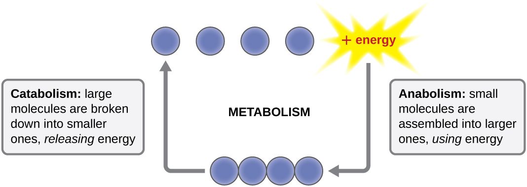
The raw materials of a cell
To grow, a microbe must build more of itself, and to build it needs raw materials. The bulk of any cell is made from just a handful of elements — carbon, hydrogen, oxygen, nitrogen, phosphorus, and sulfur — the so-called macronutrients, needed in large amounts to make proteins, nucleic acids, membranes, and sugars. Alongside these, cells need small amounts of micronutrients (trace elements) such as iron, zinc, and copper, which slot into enzymes as helpers. Some microbes can synthesize every complex molecule they need from these simple ingredients; others cannot, and must be handed finished vitamins or amino acids — these ready-made extras are called growth factors.
An organism that cannot make one or more of its own growth factors, and therefore has demanding, picky nutritional needs, is described as fastidious. This is not a trivia point — it is the reason some pathogens need special enriched plates to grow at all, and the reason a lab that uses the wrong medium reports "no growth" on a patient who is, in fact, infected.
Where the carbon comes from
Because carbon forms the backbone of every biological molecule, one of the first questions we ask about any microbe is where it gets its carbon. There are only two answers. An autotroph ("self-feeder") builds its own organic molecules from inorganic carbon dioxide (CO₂) — think of the environmental bacteria that, like plants, fix carbon from the air. A heterotroph ("other-feeder") cannot do this; it must take in carbon that some other organism already assembled, feeding on organic compounds such as sugars, proteins, and fats. Every microbe that infects humans is a heterotroph — it lives off of you, your tissues, or the nutrients on a hospital surface.
Where the energy comes from
The second question is where the energy comes from. A phototroph captures energy from light; a chemotroph extracts energy from chemical bonds by breaking down compounds. Combine the carbon source with the energy source and you get a microbe's nutritional type. The category that matters most to nursing is the last row of the table: the chemoheterotroph, which gets both its carbon and its energy from organic compounds. Bacteria, fungi, protozoa, and helminths that cause human disease are all chemoheterotrophs — and so are you.
| Nutritional type | Carbon source | Energy source | Who |
|---|---|---|---|
| Photoautotroph | CO₂ | Light | Cyanobacteria, algae, plants |
| Photoheterotroph | Organic compounds | Light | Some purple & green bacteria |
| Chemoautotroph | CO₂ | Inorganic chemicals | Certain soil & deep-sea bacteria |
| Chemoheterotroph | Organic compounds | Organic compounds | All human pathogens, fungi, animals |
- Microbes need macronutrients (C, H, O, N, P, S), micronutrients (trace metals), and sometimes ready-made growth factors.
- Carbon source splits microbes into autotrophs (from CO₂) and heterotrophs (from organic compounds).
- Energy source splits them into phototrophs (light) and chemotrophs (chemicals).
- All human pathogens are chemoheterotrophs; a fastidious organism needs enriched media to grow.
Section 2Oxygen requirements
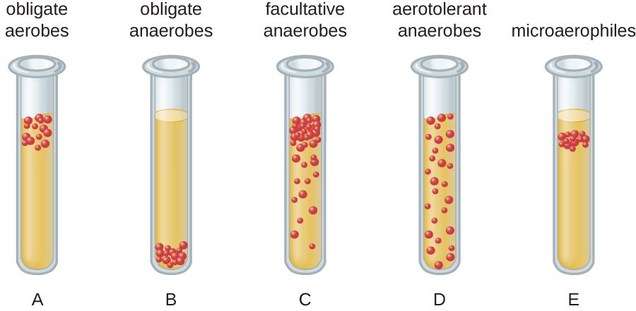
Oxygen: fuel for some, poison for others
We tend to think of oxygen as universally life-giving, but for microbes it is a double-edged sword. Using oxygen to burn fuel yields far more energy than any other option, which is why organisms that can use it grow fast. But oxygen chemistry also produces reactive oxygen species — superoxide, hydrogen peroxide (H₂O₂), and others — that shred proteins, membranes, and DNA. To live with oxygen safely, a microbe must carry detoxifying enzymes: superoxide dismutase (SOD), which neutralizes superoxide, and catalase, which splits hydrogen peroxide into water and O₂. Microbes that lack these enzymes are killed by the very air we breathe.
The five oxygen classes
Microbes sort into five groups by their relationship with oxygen, and the classic way to see it is a thioglycolate tube: a broth that is oxygen-rich at the top and oxygen-free at the bottom, so each organism grows only in the band where it can survive. An obligate aerobe must have oxygen and grows only at the top. An obligate anaerobe is poisoned by oxygen and grows only at the bottom. A facultative anaerobe prefers oxygen but can switch to fermentation without it, so it grows throughout, thickest at the top. A microaerophile needs oxygen but only a little, growing in a thin band just below the surface. An aerotolerant anaerobe ignores oxygen entirely — it never uses it but is not harmed by it — and grows evenly through the tube.
| Class | Uses O₂? | Harmed by O₂? | Clinical example |
|---|---|---|---|
| Obligate aerobe | Yes — required | No | Pseudomonas aeruginosa, M. tuberculosis |
| Obligate anaerobe | No | Yes — killed | Clostridioides difficile, Bacteroides |
| Facultative anaerobe | Yes if present; ferments if not | No | E. coli, Staphylococcus, Klebsiella |
| Microaerophile | Yes — low levels only | Yes at normal levels | Helicobacter pylori, Campylobacter |
| Aerotolerant anaerobe | No | No (tolerates it) | Streptococcus, Cutibacterium, some lactobacilli |
A related group worth knowing is the capnophiles, which grow best in an atmosphere with elevated carbon dioxide — organisms like Neisseria and Haemophilus that the lab incubates in a CO₂-enriched jar.
- Oxygen yields the most energy but generates toxic reactive oxygen species; survival requires catalase and superoxide dismutase.
- Obligate aerobes require O₂; obligate anaerobes are killed by it; facultative anaerobes do fine either way.
- Microaerophiles need low O₂; aerotolerant anaerobes never use it but tolerate it.
- Anaerobic infections live in low-oxygen tissue and demand special specimen handling.
Section 3Temperature, pH & conditions
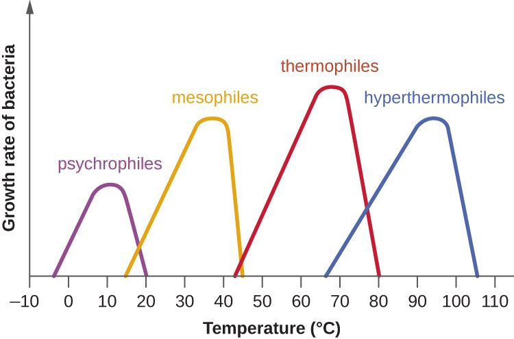
Temperature: everyone has a sweet spot
Every microbe has three cardinal temperatures — a minimum below which it stops growing, an optimum where it grows fastest, and a maximum above which its proteins cook and it dies. Microbes are grouped by where that optimum falls. Psychrophiles love the cold and grow best in near-freezing conditions; mesophiles prefer moderate temperatures around body heat; thermophiles thrive in hot springs and compost; and hyperthermophiles flourish in near-boiling water. The single most clinically important fact here is that human pathogens are mesophiles, with an optimum right around 37°C — body temperature — which is exactly why the human body is such a hospitable home for them and why lab incubators are set to 35–37°C.
| Group | Optimum range | Where you find them |
|---|---|---|
| Psychrophile | ~0–15°C | Arctic waters, deep ocean |
| Psychrotroph | ~4–25°C | Refrigerated food — Listeria, spoilage bacteria |
| Mesophile | ~20–40°C | Human body — nearly all pathogens |
| Thermophile | ~50–60°C | Hot springs, compost heaps |
| Hyperthermophile | ~80°C and above | Volcanic vents, geysers |
pH: living in acid, base, or neutral
Microbes also have a preferred acidity. Most human pathogens are neutrophiles, growing best near neutral pH (about 6.5–7.5), which matches the pH of blood and most tissues. Acidophiles prefer acidic conditions — the lactobacilli that keep the vagina at a protective low pH, or the food-preserving bacteria of yogurt and sauerkraut. Alkaliphiles favor basic environments. The stomach, at a pH near 2, is a chemical barrier that kills most swallowed microbes — which makes Helicobacter pylori, which neutralizes the acid around itself to colonize the stomach lining, all the more remarkable.
Water, salt, and the rest
Microbes need liquid water, and anything that ties up water slows them down. High concentrations of salt or sugar pull water out of a cell by osmosis, which is exactly how salting meat and sugaring jam preserve food — the environment becomes too dry for microbes to grow. A few organisms defy this: halophiles actually require high salt, and osmotolerant organisms simply put up with it. This is why Staphylococcus aureus, which tolerates salt well, can grow on skin and in salty foods that stop other bacteria — a trait the lab exploits with high-salt selective media.
- Every microbe has cardinal temperatures; human pathogens are mesophiles optimized near 37°C.
- Most pathogens are neutrophiles; stomach acid is a defense that H. pylori uniquely defeats.
- Salt and sugar preserve food by pulling water out of microbes via osmosis.
- Listeria grows in the refrigerator, so chilling is not the same as sterilizing.
Section 4The bacterial growth curve
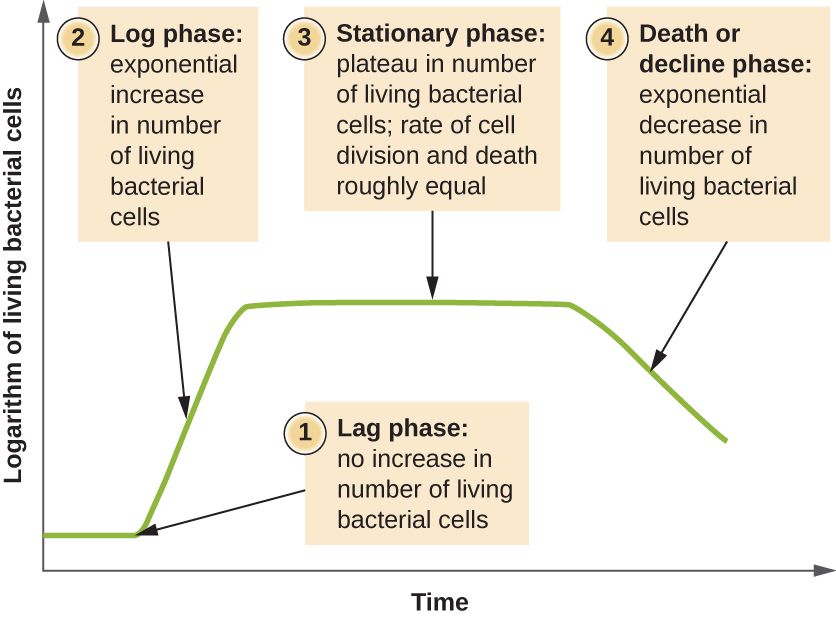
Doubling: how bacteria multiply
Most bacteria reproduce by binary fission: one cell copies its DNA, grows, and splits into two identical daughters. The time it takes one cell to become two is the generation time (or doubling time), and it can be startlingly short — Escherichia coli can double every 20 minutes under ideal conditions. Because each generation doubles the population (1, 2, 4, 8, 16 …), growth is exponential: a single cell dividing every 20 minutes becomes over a million in about seven hours. This is why a contaminated IV fluid or a mishandled specimen can go from harmless to dangerous overnight, and why some organisms cause illness so quickly.
The four phases
Put a few bacteria into fresh broth and the whole population moves through four predictable phases. In the lag phase, the cells are not yet dividing — they are gearing up, making enzymes and building blocks, adjusting to their new home. In the log (or exponential) phase, they divide at their maximum rate and the numbers explode; this is the phase of healthiest, most active cells. Eventually nutrients run low and wastes build up, and the population enters the stationary phase, where new cells are produced about as fast as old ones die and the total holds steady. Finally, in the death (decline) phase, conditions turn hostile and cells die faster than they can be replaced.
| Phase | What's happening | Why it matters clinically |
|---|---|---|
| Lag | Cells adapt; little or no division | Explains a delay before symptoms or before a culture turns positive |
| Log (exponential) | Rapid doubling; most active cells | Antibiotics work best here — they target growing cells |
| Stationary | Growth = death; nutrients depleted | Slow-growing cells tolerate drugs better; toxins/spores may form |
| Death (decline) | Cells die faster than they divide | Population collapses as the environment fails |
Why the log phase is a nursing concern
The phase a population is in changes how it responds to treatment. Many antibiotics — the cell-wall drugs especially — can only kill bacteria that are actively building and dividing, so they are most effective against log-phase cells and much weaker against the slow, dormant cells of the stationary phase. That is one reason deep, walled-off, or biofilm infections are so stubborn: the cells inside are barely dividing. It is also why maintaining steady drug levels and completing the full course matters — you want the drug present whenever the surviving cells resume dividing.
- Bacteria multiply by binary fission; generation time is how long one cell takes to become two.
- Doubling makes growth exponential — a few cells become millions in hours.
- The four phases are lag, log (exponential), stationary, and death.
- Antibiotics work best on log-phase (actively dividing) cells — and cultures should be drawn before antibiotics.
Section 5Culturing microbes
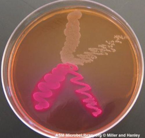
Media: the food we grow microbes on
To study or identify a microbe, we usually have to grow it in the lab on a culture medium — a nutrient broth or a firm gel. Solid media are set with agar, a jelly extracted from seaweed that microbes cannot digest and that stays firm at body temperature. Media come in different types for different jobs. A general-purpose (all-purpose) medium supports a broad range of organisms. An enriched medium adds extra nutrients — blood, serum — to grow fastidious pathogens. A selective medium contains ingredients that suppress unwanted organisms so only the target grows. A differential medium contains something — a dye, a sugar — that makes different organisms look different, so you can tell them apart at a glance. Many real plates, like MacConkey agar, are both selective and differential at once.
| Media type | What it does | Example |
|---|---|---|
| General-purpose | Grows a wide variety of microbes | Nutrient / tryptic soy agar |
| Enriched | Extra nutrients for fastidious organisms | Blood agar, chocolate agar |
| Selective | Inhibits some microbes so the target grows | MacConkey (Gram-negatives), MSA (staph) |
| Differential | Makes organisms look visibly different | Blood agar (hemolysis), MacConkey (lactose) |
| Transport | Keeps a specimen viable on the way to the lab | Anaerobic & swab transport systems |
Colonies and the pure culture
When a single bacterial cell is placed on solid agar and left to grow, it divides again and again in place until millions of its descendants pile up into a visible mound called a colony. Because every cell in that colony came from one original cell, a colony is essentially a clone — a pure culture of one organism, uncontaminated by others. Getting there is the whole point of the streak-plate technique: an inoculating loop drags the sample across the plate in a diminishing pattern so that, by the final streaks, individual cells are separated far enough apart to each grow into their own isolated colony. Pick one well-separated colony, and you have your pure culture to identify and test.
Counting what grew
Colonies are also how we count microbes. Because we assume each colony arose from one original cell (or one clump), lab counts are reported as colony-forming units (CFU) rather than exact cell numbers. This is more than bookkeeping: a urine culture growing a high CFU count of a single organism points to a true urinary tract infection, while a low count or a mix of many organisms often signals contamination from collection. Reading a culture report — the organism, the CFU count, and the sensitivities — is a daily bedside skill.
- Agar solidifies media; media may be general-purpose, enriched, selective, differential, or transport.
- Selective media suppress unwanted microbes; differential media make organisms look different.
- A colony is the visible clone of one original cell; an isolated colony gives a pure culture.
- Counts are reported as colony-forming units (CFU), and good specimen collection is essential to a meaningful result.
The chapter in one breath
- Microbes need carbon, an energy source, and nutrients; all human pathogens are chemoheterotrophs, and fastidious ones need enriched media.
- Oxygen is fuel for some and poison for others: obligate aerobes, obligate anaerobes, facultative anaerobes, microaerophiles, and aerotolerant anaerobes, with survival hinging on catalase and SOD.
- Anaerobic infections hide in low-oxygen tissue and need special specimen handling.
- Human pathogens are mesophiles (optimum ~37°C) and mostly neutrophiles; salt and sugar preserve food by osmosis, and Listeria still grows in the fridge.
- Bacteria double by binary fission, producing exponential growth through lag, log, stationary, and death phases.
- Antibiotics work best on actively dividing log-phase cells, and cultures should be drawn before antibiotics.
- We grow microbes on media (general, enriched, selective, differential, transport), isolate a colony for a pure culture, and report counts as CFU.
ReferenceWords worth knowing
A quick reference for the terms in this chapter — skim it now, or circle back whenever a word trips you up.
- Aerotolerant anaerobe
- A microbe that never uses oxygen but is not harmed by it, growing evenly in the presence or absence of air.
- Agar
- A gel from seaweed used to solidify culture media; microbes cannot digest it and it stays firm at body temperature.
- Autotroph
- An organism that builds its own organic molecules from inorganic carbon dioxide.
- Binary fission
- The asexual division of one bacterial cell into two identical daughter cells.
- Capnophile
- A microbe that grows best in an atmosphere enriched with carbon dioxide.
- Catalase
- An enzyme that detoxifies hydrogen peroxide by splitting it into water and oxygen.
- Chemoheterotroph
- An organism that obtains both carbon and energy from organic compounds; the category of all human pathogens.
- Colony-forming unit (CFU)
- A count of viable microbes based on the number of colonies grown, assuming each arose from one cell or clump.
- Differential medium
- A medium that makes different organisms look visibly different, often through color changes.
- Facultative anaerobe
- A microbe that uses oxygen when it is available but can grow by fermentation without it.
- Fastidious
- Describes a microbe with demanding nutritional needs that requires enriched media to grow.
- Generation time
- The time it takes one cell to divide into two; also called doubling time.
- Growth curve
- The plot of a bacterial population over time through lag, log, stationary, and death phases.
- Halophile
- A microbe that requires a high-salt environment to grow.
- Heterotroph
- An organism that must obtain carbon from organic compounds made by other organisms.
- Mesophile
- A microbe whose optimum growth temperature is moderate (~20–40°C); the group that includes human pathogens.
- Microaerophile
- A microbe that requires oxygen but only at levels lower than normal air.
- Obligate aerobe
- A microbe that requires oxygen to grow.
- Obligate anaerobe
- A microbe that is killed by oxygen and grows only in its absence.
- Pure culture
- A culture containing a single kind of microbe, typically obtained from one isolated colony.
- Selective medium
- A medium containing ingredients that inhibit unwanted organisms so the target can grow.
- Thermophile
- A microbe whose optimum growth temperature is high (~50–60°C), found in hot springs and compost.
Check yourself
Microbial Genetics & How Resistance Spreads
Every antibiotic-resistant infection you will ever meet at the bedside begins as a change in a microbe's DNA. A single altered gene, copied millions of times overnight, is the difference between a drug that cures and a drug that does nothing. This chapter opens up the genetics behind that story: how bacteria store and use their genetic code, how that code mutates, how bacteria pass useful genes sideways to one another — even across species — and how all of this adds up to the spread of antibiotic resistance through hospitals and communities. Understanding these mechanisms is what turns "finish your antibiotics" from a rule you recite into a rule you can explain.
By the end of this chapter you can
- Describe the structure of DNA and explain how a gene is expressed through the central dogma (replication, transcription, translation).
- Explain why several major antibiotic classes work by targeting replication, transcription, or translation.
- Define mutation and distinguish point mutations (silent, missense, nonsense) from frameshift mutations.
- Identify common mutagens and explain how spontaneous mutation seeds antibiotic resistance.
- Compare transformation, transduction, and conjugation as forms of horizontal gene transfer.
- Explain what plasmids are and how R plasmids carry and spread resistance genes.
- Describe how selective pressure and clinical practice drive the spread of resistance, and state the nurse's role in slowing it.
Section 1DNA, genes & the central dogma
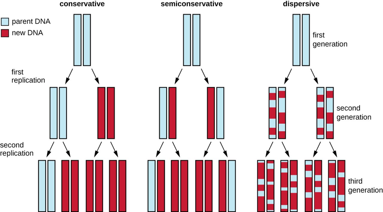
The molecule that runs the cell
Everything a microbe can do — build a cell wall, digest a sugar, resist a drug — is written in its DNA (deoxyribonucleic acid). DNA is a long double-stranded molecule shaped like a twisted ladder, the famous double helix. The rungs of the ladder are pairs of nucleotide bases, and they pair by a strict rule: adenine (A) with thymine (T), and guanine (G) with cytosine (C). Because the pairing is fixed, one strand is always a perfect template for rebuilding the other — a feature that matters enormously when the cell needs to copy itself.
A gene is simply a stretch of DNA that carries the instructions for one product, usually a protein. The complete set of an organism's genetic material is its genome. Most bacteria keep their genome as a single circular chromosome floating in the cytoplasm (they have no nucleus), often alongside small extra loops of DNA called plasmids — the little rings we will meet again in Section 4, because that is exactly where many resistance genes ride.
Copying the code: replication
Before a bacterium divides, it must make a complete second copy of its DNA so each daughter cell gets a full set of instructions. This is DNA replication. The double helix unzips into two single strands, and an enzyme called DNA polymerase reads each old strand and builds a new partner strand across from it, following the A–T, G–C rule. The result is two identical double helices where there was one. Because each new molecule keeps one old strand and one new strand, replication is called semiconservative. Bacteria replicate fast — some divide every 20 minutes — and that speed is the engine behind everything in this chapter: fast copying means fast spreading of any gene, including a resistance gene.
From gene to protein: transcription & translation
Having the instructions is not the same as carrying them out. To use a gene, the cell runs the central dogma: DNA → RNA → protein. First comes transcription, where an enzyme called RNA polymerase reads a gene and writes a working copy of it as a strand of messenger RNA (mRNA). Then comes translation, where a ribosome reads the mRNA three bases at a time — each three-base codon specifying one amino acid — and links amino acids into a protein. The finished protein folds up and goes to work as an enzyme, a structural part, or a pump. Change the DNA and you change the protein; change the protein and you change what the microbe can do.
| Step | Input → output | Key enzyme/machine | Antibiotic that blocks it |
|---|---|---|---|
| Replication | DNA → two identical DNAs | DNA polymerase, DNA gyrase | Fluoroquinolones (block gyrase) |
| Transcription | DNA → mRNA | RNA polymerase | Rifampin (blocks RNA polymerase) |
| Translation | mRNA → protein | Ribosome | Aminoglycosides, tetracyclines, macrolides |
- DNA stores instructions as base pairs (A–T, G–C); a gene codes for one product, and the whole set is the genome.
- Replication copies DNA so each daughter cell gets a full genome; bacteria do this fast.
- The central dogma is DNA → (transcription) → mRNA → (translation) → protein.
- Many antibiotics work by blocking replication, transcription, or translation in bacteria specifically.
Section 2Mutation
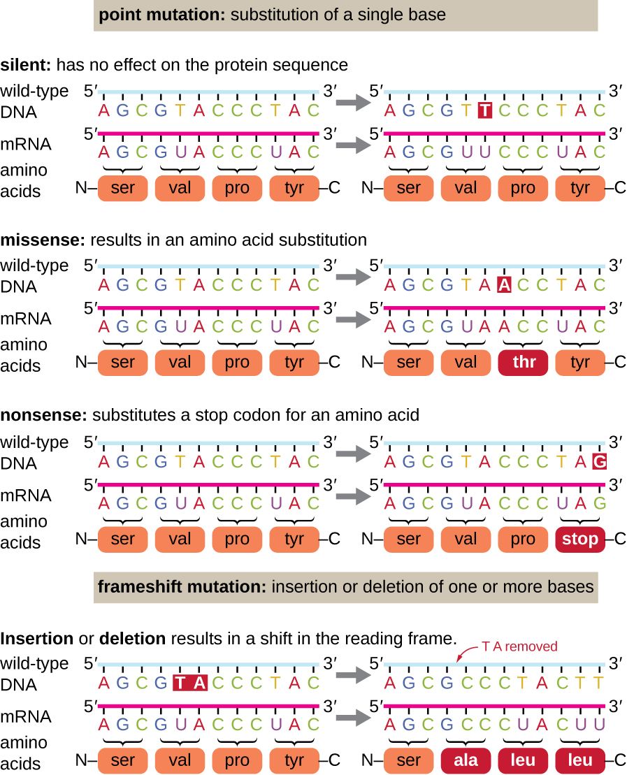
What a mutation is
A mutation is any change in the DNA sequence. Most mutations are copying errors: DNA polymerase is remarkably accurate, but it is not perfect, and when a bacterium divides billions of times, rare mistakes are guaranteed. A mutation may do nothing, may harm the microbe, or — occasionally — may hand it a brand-new ability. Whether a mutation matters depends on how it changes the protein a gene builds, which is why the type of mutation is worth knowing.
Types of mutation
The simplest change is a point mutation, where a single base is swapped for another. Its consequences vary. A silent mutation changes a codon but still codes for the same amino acid, so the protein is unchanged. A missense mutation changes one amino acid, which may subtly or seriously alter the protein — this is the classic route to a drug target that no longer binds its drug. A nonsense mutation turns an amino-acid codon into a "stop" signal, cutting the protein short and usually breaking it.
More disruptive is the frameshift mutation, where a base is inserted or deleted. Because the ribosome reads mRNA in fixed groups of three, adding or removing one base shifts the entire reading frame downstream, garbling every codon after the change. Frameshifts almost always destroy the protein completely.
Mutagens
Mutations that happen on their own during replication are spontaneous. Their rate rises sharply in the presence of a mutagen — an agent that damages DNA or causes copying errors. Chemical mutagens include certain reactive molecules and base analogs that slip into DNA in place of a normal base. Radiation is a powerful physical mutagen: ultraviolet (UV) light fuses adjacent thymine bases into thymine dimers that distort the helix, while X-rays and gamma rays break strands outright. The same DNA damage that causes mutations, at high enough doses, simply kills the cell — which is why radiation is used for microbial control. Be precise about which kind: ionizing radiation (gamma, electron beam) penetrates packaging and is a true sterilization method for heat-sensitive supplies such as sutures, gloves, and plastic syringes. UV light does not penetrate — it is blocked by glass, plastic, dust, organic soil, and any shadowed surface, and it does not reliably kill endospores — so UV is used for surface and air disinfection only, never sterilization.
| Mutation | What changes | Effect on protein |
|---|---|---|
| Silent (point) | One base; codon still means the same amino acid | None |
| Missense (point) | One base; codon now means a different amino acid | One amino acid changed — may alter function (e.g., a drug target) |
| Nonsense (point) | One base; codon becomes a "stop" | Protein cut short — usually nonfunctional |
| Frameshift | Base inserted or deleted | Reading frame shifts — protein usually destroyed |
- A mutation is any change in the DNA sequence; most arise as rare replication errors.
- Point mutations swap one base: silent (no change), missense (one amino acid changed), nonsense (premature stop).
- Frameshift mutations (insertion/deletion) shift the reading frame and usually wreck the protein.
- Mutagens — chemicals, UV, X-rays — raise the mutation rate; spontaneous mutation is the raw material of resistance.
Section 3Horizontal gene transfer
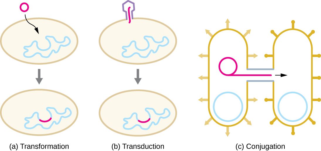
Sideways, not just downward
Mutation is slow and random. If it were the only way bacteria gained new genes, resistance would spread far more gradually than it does. But bacteria have a second, much faster trick: they trade genes sideways. Vertical gene transfer is the ordinary inheritance of genes from a parent cell to its daughters. Horizontal gene transfer (HGT) is the movement of genetic material between separate cells — even between different species — that are not parent and offspring. HGT is why a resistance gene that arose in one organism can turn up months later in an entirely unrelated one. There are three routes.
Transformation, transduction, conjugation
Transformation is the uptake of naked DNA from the environment. When a bacterium dies and breaks apart, it spills its DNA; a nearby "competent" cell can pull those fragments across its membrane and stitch them into its own genome. If the fragment carried a resistance gene, the recipient is now resistant — without ever meeting the original owner alive.
Transduction uses a bacterial virus (a bacteriophage) as an accidental delivery truck. When a phage infects a bacterium and assembles new virus particles, it sometimes packages a piece of the host's DNA by mistake. When that phage goes on to infect another bacterium, it injects the stray host gene along with itself, and the new host may keep it.
Conjugation is the most efficient of the three and the one that matters most for resistance. Two bacteria connect through a bridge called a sex pilus, and a copy of a plasmid — often an entire packet of resistance genes — is passed from donor to recipient. It is the closest thing bacteria have to direct sharing, and it can move genes between species that would otherwise never mix.
| Mechanism | How DNA moves | Vehicle | Clinical relevance |
|---|---|---|---|
| Transformation | Cell takes up free DNA from a dead microbe | Naked environmental DNA | Spreads genes released by lysed bacteria |
| Transduction | A virus carries bacterial DNA to a new cell | Bacteriophage | Moves genes, including some toxins & resistance |
| Conjugation | Direct cell-to-cell transfer through a pilus | Sex pilus + plasmid | Major driver of multidrug-resistance spread |
- Vertical transfer is parent-to-daughter inheritance; horizontal gene transfer (HGT) moves genes between separate cells, even across species.
- Transformation = uptake of free DNA; transduction = delivery by a bacteriophage; conjugation = direct transfer via a pilus.
- Conjugation is the biggest driver of multidrug-resistance spread because it moves whole plasmids of genes at once.
- HGT lets resistance jump between unrelated organisms far faster than mutation alone.
Section 4Plasmids & resistance genes
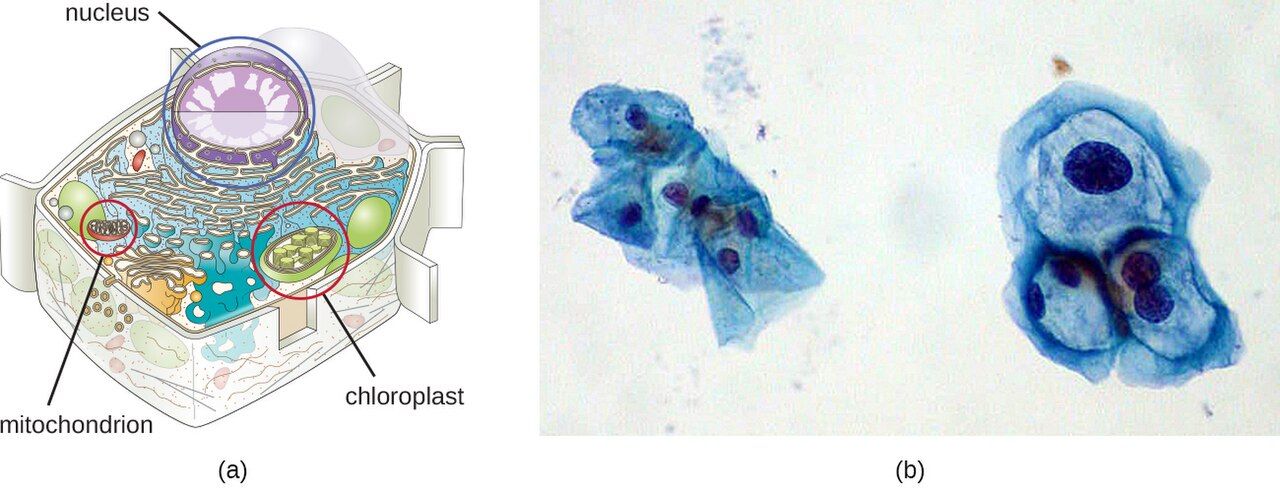
Little rings of optional DNA
Besides its main chromosome, a bacterium often carries one or more plasmids — small, circular loops of DNA that sit apart from the chromosome (they are extrachromosomal). Plasmids are copied by the cell and passed to daughter cells, but they are not strictly required for survival. Instead they carry "bonus" genes that come in handy under stress: genes for using an unusual nutrient, for producing a toxin, or — the ones that keep infection-control nurses up at night — genes for surviving antibiotics.
R plasmids: resistance to go
A plasmid that carries one or more antibiotic-resistance genes is called a resistance plasmid, or R plasmid. R plasmids are dangerous for two reasons. First, a single plasmid can carry resistance genes to several different antibiotics at once, so acquiring it in one step makes a bacterium multidrug-resistant (MDR). Second, R plasmids often carry their own conjugation machinery, so they are built to copy themselves into neighboring cells. Put those two facts together and you have a mobile, self-spreading package of multidrug resistance — a major reason resistant organisms like VRE and the multidrug-resistant gram-negatives are behind so many hospital outbreaks. (Not every resistant organism works this way: in MRSA, for instance, the key methicillin-resistance gene sits on the chromosome, not a plasmid.)
Jumping genes that build the package
How does a plasmid come to hold so many resistance genes? Often through transposons — nicknamed "jumping genes" — short DNA segments that can cut or copy themselves from one location and paste into another, including from a chromosome onto a plasmid. Related elements called integrons can capture resistance gene "cassettes" and line them up together. Over time these shuffling elements assemble multidrug-resistance plasmids gene by gene, ready to be shared by conjugation.
| Feature | Chromosome | Plasmid |
|---|---|---|
| Size | Large; the main genome | Small loop of extra DNA |
| Essential? | Yes — carries the genes for survival | No — carries optional/"situational" genes |
| Copied & inherited? | Yes, with each division | Yes, and can also be shared by conjugation |
| Typical cargo | Core metabolism, structure | Resistance, toxins, special nutrients |
- Plasmids are small, circular, extrachromosomal loops of DNA that carry optional but useful genes.
- R plasmids carry antibiotic-resistance genes, often to several drugs at once (multidrug resistance).
- R plasmids frequently carry conjugation machinery, so they copy themselves into neighboring cells.
- Transposons and integrons shuffle resistance genes onto plasmids, building the multidrug package.
Section 5How resistance spreads
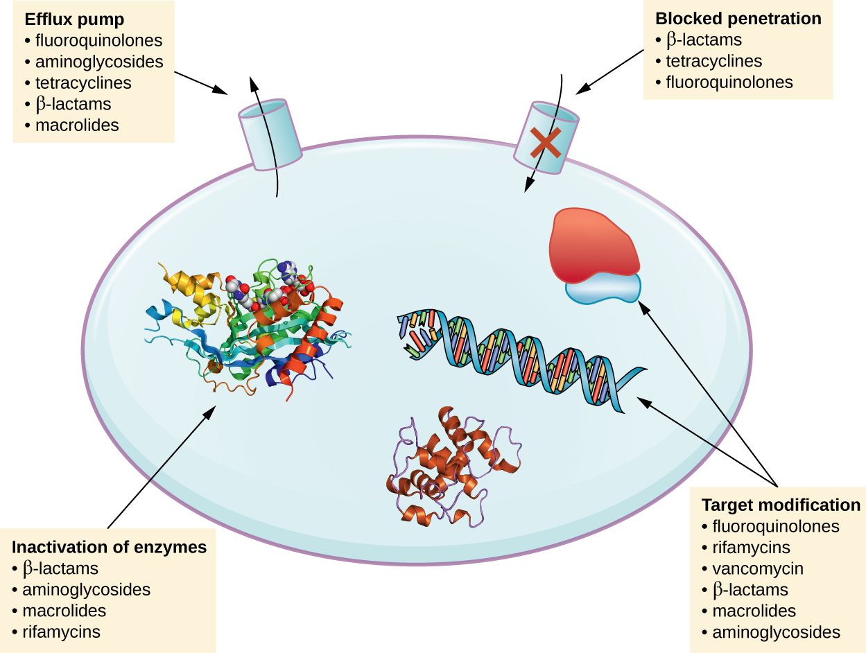
Selective pressure: the drug does the choosing
Antibiotics do not create resistance — they select for it. Picture a population of bacteria in which, by chance, a few cells already carry a resistance gene (from mutation or from a plasmid). Give an antibiotic and the susceptible cells die, but the resistant few survive and multiply, quickly repopulating the site with an all-resistant community. This is selective pressure: the drug clears the field for exactly the organisms it cannot kill. Every dose of antibiotic anywhere — appropriate or not — nudges the microbial world a little further toward resistance, which is why unnecessary antibiotic use is not a harmless "just in case."
Where the spreading happens
Resistant organisms spread the same way any microbe spreads — mostly by contact, and in healthcare that means hands, equipment, and surfaces moving flora from patient to patient. Several forces accelerate it: overprescribing and prescribing antibiotics for viral illnesses; patients stopping a course early, which kills the weak and leaves the tough survivors behind; heavy antibiotic use in agriculture, which seeds resistant organisms into the food supply and environment; and international travel, which carries resistant strains around the globe in days. The genetics from the earlier sections is what makes each of these routes so potent: once a resistance gene exists on a mobile plasmid, contact spread and conjugation do the rest.
The nurse at the center of it
Slowing resistance is not mainly a laboratory problem — it is a bedside one, and the nurse sits at the center. Hand hygiene and contact precautions stop resistant organisms from moving between patients. Antimicrobial stewardship — questioning whether an antibiotic is truly needed, pushing for cultures before starting drugs, narrowing broad-spectrum therapy once the organism is known, and stopping antibiotics that are no longer indicated — reduces the selective pressure in the first place. And patient teaching that explains why to finish the full course, why leftover antibiotics should never be shared, and why a cold will not respond to antibiotics turns the patient into a partner rather than a driver of the problem.
| Driver of spread | What happens | Nursing countermeasure |
|---|---|---|
| Contact transmission | Resistant flora ride hands, equipment, surfaces between patients | Hand hygiene; contact precautions; dedicated equipment |
| Overprescribing | Antibiotics given when not needed (e.g., for viruses) select for resistance | Stewardship; culture first; question the indication |
| Incomplete courses | Stopping early leaves the toughest survivors to repopulate | Teach adherence; explain the "why" |
| Broad-spectrum overuse | Wide kill flattens flora and selects widely for resistance | De-escalate to a narrow agent once the organism is known |
- Selective pressure: antibiotics do not create resistance, they select for the few cells that already have it, then those cells take over.
- Resistance spreads mainly by contact, amplified by overprescribing, incomplete courses, agricultural use, and travel.
- The genetics of Sections 1–4 (mutation, HGT, R plasmids) is what makes each route so fast.
- Hand hygiene, contact precautions, stewardship, and patient teaching are the nurse's levers against resistance.
The chapter in one breath
- DNA stores a microbe's instructions as base pairs; a gene is expressed through the central dogma — DNA replicated, transcribed to mRNA, translated to protein.
- Several antibiotic classes work by blocking replication, transcription, or translation in bacteria specifically.
- A mutation changes the DNA sequence; point mutations (silent, missense, nonsense) swap one base, frameshifts shift the reading frame, and mutagens raise the rate.
- Spontaneous mutation seeds resistance, which is why serious infections like TB are treated with multiple drugs at once.
- Horizontal gene transfer — transformation, transduction, conjugation — moves genes sideways between cells, even across species.
- R plasmids are mobile loops of DNA carrying resistance to several drugs at once, spread efficiently by conjugation.
- Selective pressure from antibiotic use plus contact spread drives resistance through healthcare and the community; hand hygiene, stewardship, and teaching are the nurse's countermeasures.
ReferenceWords worth knowing
A quick reference for the terms in this chapter — skim it now, or circle back whenever a word trips you up.
- Central dogma
- The flow of genetic information in a cell: DNA → RNA → protein (replication, transcription, translation).
- Codon
- A group of three mRNA bases that specifies one amino acid during translation.
- Conjugation
- Horizontal gene transfer by direct cell-to-cell contact through a sex pilus, often moving a plasmid.
- DNA (deoxyribonucleic acid)
- The double-stranded molecule that stores genetic instructions as paired bases (A–T, G–C).
- Frameshift mutation
- A mutation from inserting or deleting a base, which shifts the reading frame and usually destroys the protein.
- Gene
- A stretch of DNA that codes for one product, usually a protein.
- Genome
- The complete set of an organism's genetic material.
- Horizontal gene transfer (HGT)
- Movement of genetic material between separate cells (not parent to offspring), even across species.
- Missense mutation
- A point mutation that changes one amino acid in a protein; may alter its function.
- Mutagen
- A physical or chemical agent (e.g., UV light, X-rays, certain chemicals) that raises the mutation rate.
- Mutation
- Any change in the DNA sequence.
- Nonsense mutation
- A point mutation that turns an amino-acid codon into a stop codon, cutting the protein short.
- Plasmid
- A small, circular, extrachromosomal loop of DNA that carries optional genes and can be shared between cells.
- Point mutation
- A mutation that swaps a single base; may be silent, missense, or nonsense.
- R plasmid (resistance plasmid)
- A plasmid carrying one or more antibiotic-resistance genes, often to several drugs at once.
- Replication
- The copying of DNA before cell division, producing two identical double helices.
- Selective pressure
- The way antibiotic use favors the survival and multiplication of already-resistant cells.
- Transcription
- Copying a gene from DNA into messenger RNA by RNA polymerase.
- Transduction
- Horizontal gene transfer in which a bacteriophage carries bacterial DNA to a new cell.
- Transformation
- Horizontal gene transfer in which a cell takes up free DNA released by a dead microbe.
- Translation
- Building a protein from an mRNA sequence at the ribosome, one codon at a time.
- Transposon
- A "jumping gene" — a DNA segment that can move between locations, including onto plasmids.
Check yourself
Controlling Microbes — Sterilization, Disinfection & Asepsis
Every time you clean your hands, wipe down a bedrail, open a sterile dressing kit, or hang an IV, you are practicing microbial control — the deliberate work of killing microbes or holding their numbers down so they cannot harm your patient. The trouble is that the words for this work are used loosely in everyday speech and precisely in the clinical world, and the gap between "clean," "disinfected," and "sterile" is exactly where infections slip through. This chapter sorts out that vocabulary, walks through the physical and chemical tools we use to kill microbes, explains why killing follows a predictable curve rather than an on/off switch, and finishes at the bedside with the two kinds of asepsis every nurse practices by hand, many times a shift. Master this, and infection prevention stops being a list of rules and becomes something you understand.
By the end of this chapter you can
- Define and rank sterilization, disinfection, antisepsis, and sanitization, and use the -cidal vs -static suffixes correctly.
- Compare the major physical methods of control — moist and dry heat, the autoclave, radiation, and filtration — and match each to what it can and cannot sterilize.
- Explain how the autoclave achieves sterilization and how its success is verified.
- Describe the major families of chemical disinfectants and antiseptics and how they kill microbes.
- Interpret the microbial death curve, the meaning of D-value, and why sterility is defined statistically.
- List the factors that make microbial killing faster or slower, and rank microbes by their resistance to control.
- Distinguish medical (clean) from surgical (sterile) asepsis and apply the rules of a sterile field.
Section 1The vocabulary of control
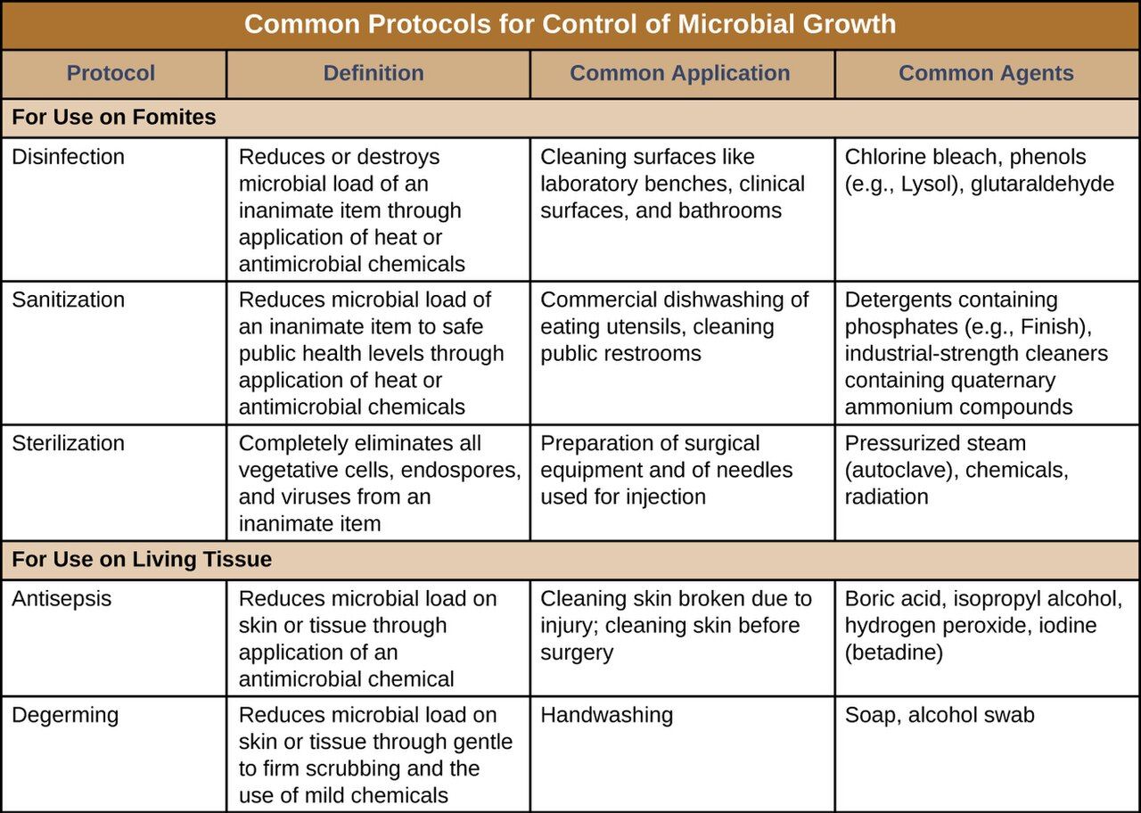
A ladder, not a light switch
The single most important idea in this chapter is that "getting rid of germs" is not one thing — it is a ladder of increasingly complete removal, and each rung has a precise name and a precise job. At the top sits sterilization: the complete destruction or removal of all microbial life, including the toughest of all targets, bacterial endospores. A sterile object has nothing living on it, full stop. One step down is disinfection, which destroys most pathogens on a non-living surface but does not reliably kill endospores. Because it is not absolute, disinfection comes in levels — high, intermediate, and low — depending on how much it kills. The distinction matters because much of what we do at the bedside is disinfection, not sterilization, and knowing the difference tells you which tasks are "good enough" and which demand the sterile item from central supply.
On skin versus on surfaces
The next two terms are constantly confused because they do similar work in different places. Antisepsis is the removal of microbes from living tissue — skin, a mucous membrane, a surgical site. The chemical used is an antiseptic, and because it goes on a living patient it must be gentle enough not to injure tissue. Disinfection, by contrast, happens on inanimate objects, and the chemical is a disinfectant, which can be far harsher because no living cells have to survive it. This is why you scrub a patient's skin with chlorhexidine or an alcohol swab (antiseptics) but wipe the counter with a bleach or quaternary-ammonium solution (disinfectants) — and why pouring a surface disinfectant into a wound is a genuine mistake, not a shortcut. A close cousin is degerming, the mechanical scrubbing away of microbes (the alcohol wipe before an injection does this), and sanitization, which simply lowers microbial counts to a "safe" public-health level rather than eliminating them — the standard for restaurant dishwashers and drinking glasses, not for anything that enters a patient.
Reading the suffixes
Microbiology labels its weapons with two suffixes you must be able to read on sight. An agent ending in -cidal (bactericidal, virucidal, fungicidal, sporicidal) kills the target outright. An agent ending in -static (bacteriostatic, fungistatic) only stops it from multiplying — the microbes are still alive, held in check, and will resume growing if the agent is removed or the patient's immune defenses fail. That difference is not academic: a bacteriostatic drug depends on a working immune system to finish the job, which is why the distinction resurfaces when we discuss antimicrobial drugs and immunocompromised patients.
| Term | What it does | Kills endospores? | Used on |
|---|---|---|---|
| Sterilization | Destroys or removes all microbial life | Yes | Instruments, IV fluids, implants |
| Disinfection | Destroys most pathogens (high/intermediate/low level) | Not reliably | Inanimate surfaces & equipment |
| Antisepsis | Reduces microbes on living tissue | No | Skin, mucous membranes, wounds |
| Degerming | Mechanically removes microbes | No | Skin (e.g., pre-injection swab) |
| Sanitization | Lowers counts to a "safe" public level | No | Dishes, food surfaces, floors |
- Sterilization alone removes all life including endospores; disinfection (surfaces) and antisepsis (living tissue) do not.
- Disinfectant = harsh chemical for objects; antiseptic = gentle chemical for skin and tissue.
- Sanitization only reduces counts to a safe level; degerming is mechanical removal.
- -cidal kills; -static merely halts multiplication and relies on host defenses.
Section 2Physical methods — heat, the autoclave, radiation & filtration
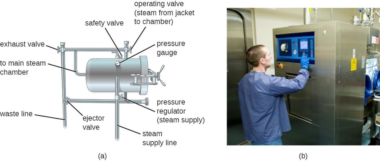
Heat: the oldest and most reliable tool
Heat kills by denaturing proteins and enzymes and by disrupting membranes, and it comes in two forms with very different power. Moist heat — steam and boiling water — is far more effective than dry heat because water conducts energy efficiently and drives protein coagulation quickly (think of how fast an egg white sets in boiling water versus a dry oven). Dry heat, delivered by a hot-air oven or an open flame, works too but needs much higher temperatures and longer times to accomplish the same kill. Two classic benchmarks describe heat's effect: the thermal death point (the lowest temperature that kills all microbes in a suspension in ten minutes) and the thermal death time (the time needed to kill them at a given temperature). Note one important limit: ordinary boiling at 100°C does not sterilize — it kills vegetative cells and most viruses but leaves hardy endospores alive, which is precisely the problem the autoclave was invented to solve. A gentler cousin, pasteurization, uses brief, mild heat not to sterilize but to lower the count of pathogens and spoilage organisms in milk and other liquids while preserving flavor.
The autoclave: steam under pressure
The autoclave is the single most important sterilizing device in health care, and it solves the boiling-water problem with a simple trick: raising the pressure raises the boiling point, so steam can be heated well past 100°C. A standard cycle holds saturated steam at 121°C under about 15 pounds per square inch (psi) of pressure for roughly 15–20 minutes — conditions that reliably destroy even endospores. The active ingredient is the moist heat of the steam, not the pressure itself; the pressure is only there to make the hotter steam possible. Because a load only counts as sterile if the steam actually reached every surface at temperature for long enough, autoclaving is always verified, never assumed. Chemical indicators — the striped autoclave tape and pouch dots that change color — confirm a load was exposed to heat, but the gold standard is a biological indicator: a sealed vial of Geobacillus stearothermophilus endospores run through the cycle and then incubated. If those famously heat-resistant spores are dead, everything else in the load is too.
Radiation and filtration: when heat would ruin the product
Plenty of medical items — plastic syringes, IV tubing, sutures, some fluids — would melt, warp, or degrade in an autoclave, so we sterilize them another way. Radiation splits into two types. Ionizing radiation (gamma rays, X-rays, electron beams) carries enough energy to smash DNA and generate destructive free radicals; it penetrates deeply, sterilizes in the cold, and is used industrially for prepackaged disposables and some foods. Nonionizing radiation, chiefly ultraviolet (UV) light, is weaker: it fuses adjacent thymine bases into dimers that block DNA replication, but it barely penetrates — it cannot pass through glass, plastic, or even a layer of dust — so its use is limited to disinfecting surfaces and air in biosafety cabinets and empty rooms (and it is a genuine hazard to the eyes and skin of anyone in the room). Finally, filtration physically removes microbes rather than killing them, by straining a fluid or gas through pores too small for cells to pass. A membrane filter with 0.2 µm pores sterilizes heat-sensitive liquids such as certain antibiotics, vitamins, and IV additives, and HEPA filters do the same for air, keeping operating rooms and protective-isolation rooms clean.
| Method | How it kills / removes | Sterilizes? | Typical use |
|---|---|---|---|
| Autoclave (moist heat) | Steam at 121°C, 15 psi denatures proteins | Yes (spores too) | Surgical instruments, glassware, media |
| Dry heat / flame | Oxidation & denaturation at high temp | Yes (long/hot) | Glassware, inoculating loops |
| Boiling (100°C) | Denatures proteins | No — spores survive | Home disinfection of vegetative cells |
| Ionizing radiation | Breaks DNA, forms free radicals | Yes | Disposable plastics, sutures, foods |
| UV (nonionizing) | Thymine dimers block replication | Surface only | Air & surfaces in cabinets/rooms |
| Membrane filtration | Physically strains out cells (0.2 µm) | Yes (fluids) | Heat-labile fluids; HEPA for air |
- Moist heat beats dry heat; ordinary boiling does not sterilize because endospores survive.
- The autoclave (121°C, ~15 psi, ~15 min) sterilizes with pressurized steam and is verified by a Geobacillus spore biological indicator.
- Ionizing radiation sterilizes heat-sensitive disposables; UV only disinfects surfaces and air and does not penetrate.
- Filtration (0.2 µm membranes, HEPA) removes microbes from heat-labile fluids and from air.
Section 3Chemical methods — disinfectants & antiseptics
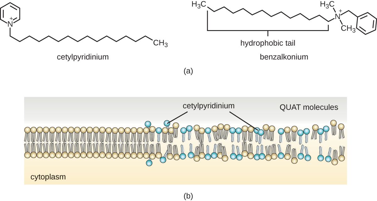
How chemicals kill
Chemical control agents work by a handful of mechanisms, and most fall into two big buckets: they denature proteins (unfolding the enzymes a microbe needs to live) or they disrupt the plasma membrane (dissolving its lipids so the cell leaks and dies). Some go further and damage nucleic acids or oxidize cell components. Whether a given chemical is called a disinfectant or an antiseptic depends on where it is used, not on the molecule itself — several agents, such as iodine and the quats, serve as both at different concentrations. Two practical properties decide whether a chemical is useful in the hospital: its use-dilution (the concentration at which it actually works, since more concentrated is not always better — 70% alcohol kills better than 100% because it needs water to denature protein) and its behavior in the presence of organic matter like blood and pus, which can neutralize many agents on contact.
The chemical families you will meet
You do not need to memorize every compound, but you should recognize the major families and where each shines. Alcohols (isopropyl and ethanol, best at 60–90%) denature proteins and dissolve membranes; they are fast, cheap skin antiseptics and surface wipes, but they evaporate quickly and do not kill spores. Chlorhexidine is a gentle, long-lasting skin antiseptic that binds to the skin and keeps working for hours — the reason it is used for surgical scrubs and central-line site care. Iodophors (iodine complexed with a carrier, e.g., povidone-iodine/Betadine) are broad-spectrum skin preps for surgery and injections. Halogens also include chlorine (household bleach, sodium hypochlorite), a powerful, cheap disinfectant that is one of the few agents that reliably kills Clostridioides difficile spores on surfaces. Hydrogen peroxide and other peroxygens oxidize cell components and are useful for surfaces and some equipment. Quaternary ammonium compounds (quats) are low-toxicity detergent-like disinfectants for everyday surface cleaning (though weak against some resistant organisms). Finally, glutaraldehyde (with a long contact time) and the gas ethylene oxide can achieve sterilization of delicate, heat-sensitive equipment — but they are toxic and demand strict handling. Ortho-phthalaldehyde (OPA) belongs beside them chemically but not in capability: it is a fast-acting high-level disinfectant with poor sporicidal activity, cleared for high-level disinfection of endoscopes, not as a sterilant.
Matching the agent to the risk
Because no single chemical does everything, hospitals classify items by how much microbial killing they need, a scheme known as the Spaulding classification. Critical items enter sterile tissue or the bloodstream (surgical instruments, catheters, implants) and must be sterilized. Semicritical items touch mucous membranes but not sterile tissue (endoscopes, respiratory equipment) and require at least high-level disinfection. Noncritical items touch only intact skin (blood-pressure cuffs, bedrails, stethoscopes) and need only low- or intermediate-level disinfection. This one framework quietly governs a huge amount of bedside and central-supply practice: it is the reason a scope gets a different reprocessing protocol than a bedrail.
| Agent family | Mechanism | Kills spores? | Typical use |
|---|---|---|---|
| Alcohols (60–90%) | Denature protein, dissolve membrane | No | Skin antiseptic, surface wipes, hand rub |
| Chlorhexidine | Disrupts membrane; persists on skin | No | Surgical scrub, line-site antisepsis |
| Iodophors (povidone-iodine) | Oxidize / halogenate cell components | No (routine use) | Pre-op skin prep, injection sites |
| Chlorine (bleach) | Strong oxidizer | Yes (surfaces) | Surface disinfection, C. diff rooms |
| Hydrogen peroxide | Oxidizes cell components | Yes (high conc.) | Surface disinfection, equipment, vaporized room decontamination |
| Quats | Detergent-like membrane disruption | No | Routine surface cleaning |
| Glutaraldehyde / ethylene oxide | Alkylate proteins & DNA | Yes (long contact) | Sterilizing heat-sensitive equipment |
| Ortho-phthalaldehyde (OPA) | Alkylates proteins | No — poor sporicide | High-level disinfection of endoscopes (not sterilization) |
- Chemical agents mostly denature proteins or disrupt membranes; the same molecule can be a disinfectant on surfaces and an antiseptic on skin.
- Alcohols and chlorhexidine are workhorse skin antiseptics; bleach handles spores on surfaces; glutaraldehyde and ethylene oxide sterilize heat-sensitive equipment, while OPA is a high-level disinfectant only.
- Use-dilution matters — 70% alcohol out-kills 100% because it needs water.
- The Spaulding classification (critical / semicritical / noncritical) matches the level of killing to how the item is used.
Section 4The microbial death curve & what changes the odds
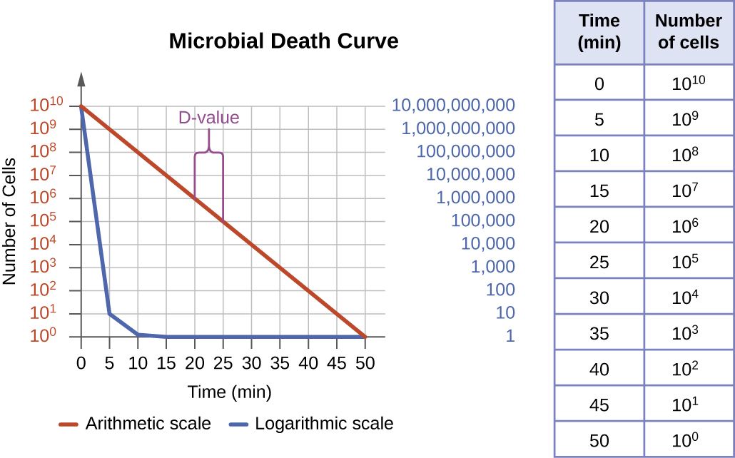
Death is logarithmic, not all-at-once
It is tempting to imagine that a disinfectant kills every microbe the instant it is applied, but that is not how killing works. Under a lethal agent, a microbial population dies logarithmically — a constant percentage dies in each equal unit of time, not a constant number. If a chemical kills 90% of the cells in the first minute, it kills 90% of the survivors in the next minute, and 90% of those in the third, and so on. Plotted on a log scale, this makes a straight downward line: the microbial death curve. The practical consequence is profound: because you are always killing a fraction and never the last stubborn cell instantly, you can drive the number vanishingly low but you can never mathematically reach absolute zero in finite time. That is why sterility is defined statistically, as a probability — a sterility assurance level of one survivor in a million (10−6) — rather than as a literal guarantee of nothing alive.
The D-value
The steepness of that line is captured by a single number, the decimal reduction time or D-value: the time required, at a given temperature or agent concentration, to kill 90% of the population — that is, to drop it by one power of ten (one "log"). A microbe or a process with a short D-value is killed quickly; a long D-value means a tough target or a weak method. The D-value is the bridge between the death curve and real protocols: if you know a starting count and the D-value, you can calculate how long a treatment must run to reach your target survival level, which is exactly how sterilization cycles and canning times are set.
What speeds up or slows down the kill
The same agent can kill in seconds or fail entirely depending on conditions, and a nurse who knows the variables can spot when a control measure is being undermined. Killing goes faster with higher temperature, higher concentration or intensity, and longer contact time — the last of which is why every disinfectant label lists a wet contact time that the surface must stay visibly wet to work. Killing goes slower, or fails, when the microbial load is enormous (more cells take longer to clear), when the microbes are inherently resistant, when a protective biofilm shields them, and — crucially at the bedside — when organic matter such as blood, pus, feces, or biofilm is present, because it physically blocks the agent and chemically inactivates many of them. Microbes themselves also fall in a predictable resistance ranking, from the nearly indestructible prions and bacterial endospores down through mycobacteria (their waxy wall makes tuberculosis hard to kill) and non-enveloped viruses, to the easiest targets of all: ordinary vegetative bacteria and fragile enveloped viruses like influenza and HIV.
| Factor | Effect on killing | Why |
|---|---|---|
| Time (contact time) | More time → more kill | Each interval removes another fraction (log) |
| Temperature / concentration | Higher → faster kill | Speeds denaturation and reaction rate |
| Number of microbes | More cells → slower to clear | Same % killed each interval — larger start, longer to finish |
| Microbe type | Spores & mycobacteria resist | Endospore coats and waxy walls block agents |
| Organic matter | Present → killing fails | Blood/pus shield and inactivate the agent |
| Biofilm | Present → strong resistance | Matrix blocks penetration; cells shielded |
- Microbes die logarithmically — a constant fraction per unit time — so absolute zero is never guaranteed and sterility is a probability (a 10−6 assurance level).
- The D-value is the time to kill 90% (one log) of a population and sets how long a process must run.
- Killing speeds up with more time, heat, and concentration; it slows with a big microbial load, resistant organisms, biofilm, and organic matter.
- Resistance ranks prions & endospores (toughest) → mycobacteria → non-enveloped viruses → vegetative bacteria & enveloped viruses (easiest).
Section 5Asepsis at the bedside — medical vs surgical
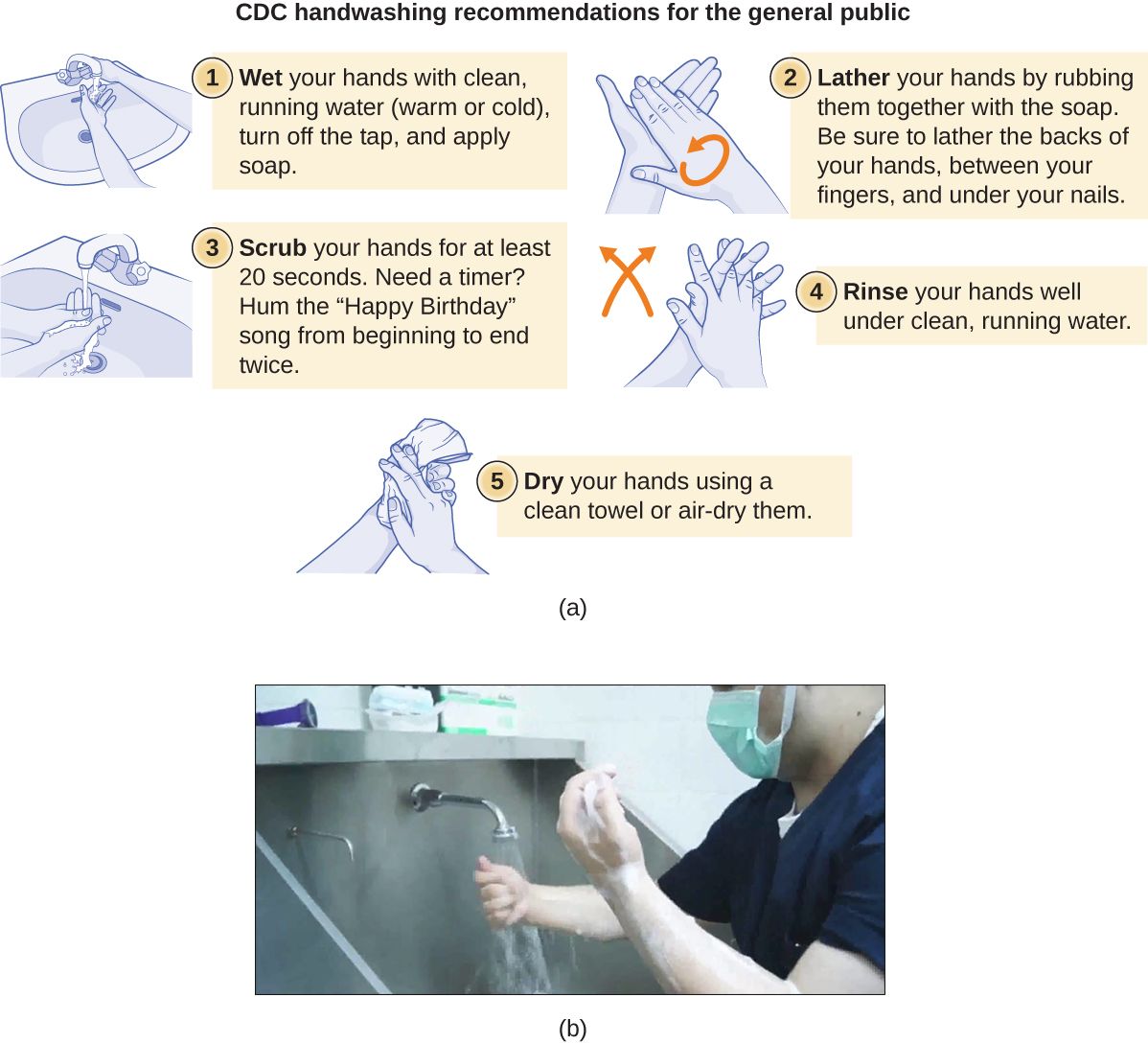
Two kinds of asepsis
Asepsis simply means the absence of contamination by pathogens, and at the bedside it is practiced at two different levels. Medical asepsis — also called clean technique — aims to reduce the number and spread of microbes and to break the chain of transmission. It is the everyday level: hand hygiene, gloves, gowns, cleaning shared equipment, and the standard precautions you use with every patient regardless of diagnosis. It does not require sterility; it requires cleanliness and consistency. Surgical asepsis — sterile technique — aims for the total absence of microbes and is used whenever you breach the body's natural barriers: inserting a urinary or central catheter, starting a sterile dressing change on a deep wound, or assisting in the operating room. The mental shift from "clean" to "sterile" is exactly the ladder from Section 1, now applied with gloved hands.
Clean technique: the everyday defense
Medical asepsis rests on a small number of habits that, done reliably, prevent an enormous share of infections. Hand hygiene is first among them — performed before and after every patient contact, before a clean task, after body-fluid exposure, and after touching the patient's surroundings (the familiar moments of hand hygiene). Alcohol-based hand rub is the default because it is fast and effective; soap and water are reserved for visibly soiled hands and for spore-formers like C. diff. Around this core sit gloves and gowns to protect against splashes, safe handling of linens and waste, disinfection of shared devices between patients, and the transmission-based precautions layered on top of standard precautions when a patient carries a specific organism. None of this is glamorous, and all of it works.
Sterile technique: the rules of the field
Surgical asepsis is governed by a set of blunt, non-negotiable rules that exist because a sterile field is only sterile until proven otherwise. Once a sterile package is opened, a field is established, and from that moment: only sterile items may touch sterile items; a 1-inch border around the drape is considered contaminated; anything held below waist level or out of your line of sight is considered contaminated, because you cannot vouch for it; you never reach across or turn your back on the field; and any moisture that wicks through the drape (strike-through) contaminates it, because microbes travel through the wet path. Sterile gloves and gowns extend the field to your hands and front. The underlying philosophy is honest humility: when in doubt about whether something is still sterile, you treat it as contaminated and start over — a re-drape is cheap, a bloodstream infection is not.
| Feature | Medical asepsis (clean) | Surgical asepsis (sterile) |
|---|---|---|
| Goal | Reduce number & spread of microbes | Eliminate all microbes |
| Standard | Clean | Sterile |
| Typical tasks | Hand hygiene, gloves/gowns, cleaning | Catheter insertion, sterile dressings, OR |
| Hand practice | Handwash or alcohol rub | Surgical hand scrub, sterile gloves |
| If contaminated | Re-clean and continue | Discard field and restart |
- Medical asepsis (clean technique) reduces and contains microbes — hand hygiene, gloves, gowns, cleaning — and is used with every patient.
- Surgical asepsis (sterile technique) eliminates all microbes and is used whenever a natural barrier is breached.
- A sterile field is contaminated by anything nonsterile, a 1-inch border, items below the waist or out of sight, and moisture strike-through.
- The governing rule is "when in doubt, it's contaminated" — discard and restart rather than gamble.
The chapter in one breath
- Microbial control is a ladder: sterilization (all life, including spores) → disinfection (surfaces) → antisepsis (living tissue) → sanitization (safe reduction).
- -cidal agents kill; -static agents only stop multiplication and rely on the host's immune system.
- Physical methods: moist heat beats dry heat; the autoclave (121°C, 15 psi) sterilizes and is verified by a Geobacillus spore indicator; radiation and 0.2 µm filtration handle heat-sensitive items.
- Chemical methods denature proteins or disrupt membranes; alcohols and chlorhexidine prep skin, bleach kills surface spores, and the Spaulding classification matches killing level to how an item is used.
- Microbes die logarithmically; the D-value is the time to kill 90%, and sterility is a statistical assurance level, never an absolute.
- Killing is slowed by big loads, resistant organisms, biofilm, and especially organic matter — so always clean before you disinfect.
- At the bedside, medical asepsis (clean) is the everyday default and surgical asepsis (sterile) is used whenever a barrier is breached — and when in doubt, it's contaminated.
ReferenceWords worth knowing
A quick reference for the terms in this chapter — skim it now, or circle back whenever a word trips you up.
- Antisepsis
- The reduction or removal of microbes from living tissue, using an antiseptic.
- Antiseptic
- A chemical gentle enough to apply to living skin or tissue, e.g., alcohol or chlorhexidine.
- Asepsis
- The absence of contamination by pathogens; practiced as medical (clean) or surgical (sterile) technique.
- Autoclave
- A device that sterilizes with pressurized saturated steam, typically 121°C at ~15 psi for ~15 minutes.
- Bactericidal / bacteriostatic
- Agents that respectively kill bacteria (-cidal) or merely stop them multiplying (-static).
- Biological indicator
- A vial of heat-resistant spores run through a cycle to confirm true sterilization.
- D-value (decimal reduction time)
- The time needed to kill 90% (one log) of a microbial population under set conditions.
- Degerming
- The mechanical removal of microbes from skin, e.g., scrubbing before an injection.
- Disinfection
- Destruction of most pathogens on an inanimate surface; does not reliably kill endospores.
- Endospore
- A dormant, extremely resistant bacterial structure that is the toughest target of microbial control.
- Filtration
- Physical removal of microbes by straining fluid or air through pores too small for cells (e.g., 0.2 µm, HEPA).
- Medical asepsis
- "Clean technique" that reduces the number and spread of microbes; the everyday level of care.
- Microbial death curve
- The logarithmic (straight-line, on a log scale) decline of a population under a lethal agent.
- Pasteurization
- Brief mild heating that lowers pathogen and spoilage counts in liquids without sterilizing.
- Sanitization
- Reduction of microbial counts to a level judged safe for public health, e.g., dishes and food surfaces.
- Spaulding classification
- A scheme rating items critical, semicritical, or noncritical to set the required level of processing.
- Sterility assurance level (SAL)
- The statistical probability of a survivor after sterilization, conventionally one in a million (10−6).
- Sterilization
- The complete destruction or removal of all microbial life, including endospores.
- Surgical asepsis
- "Sterile technique" aiming for the total absence of microbes; used when barriers are breached.
- Use-dilution
- The working concentration at which a chemical agent is effective (more is not always better).
Check yourself
Antimicrobial Drugs
Few discoveries have saved more lives than the antibiotic. In the span of a single lifetime, infections that once emptied hospital wards — pneumonia, sepsis, a wound gone bad — became, for the most part, curable. That victory rests on one elegant idea: a bacterium is built differently from you, and a well-chosen drug can exploit that difference to kill the microbe while leaving your patient unharmed. This chapter unpacks how antimicrobials work, how we pick the right one, and why bacteria are fighting back. Along the way you will see that the nurse is not a bystander to antibiotic therapy but one of its most important stewards — deciding when cultures are drawn, whether a dose lands on time, and whether the whole enterprise keeps working for the next patient.
By the end of this chapter you can
- Explain selective toxicity and why it makes antimicrobial therapy possible.
- Define the therapeutic index and connect a narrow index to the need for drug-level monitoring.
- Name the five bacterial targets antibacterial drugs attack and give a drug class for each.
- Distinguish bactericidal from bacteriostatic and narrow- from broad-spectrum drugs.
- Describe how susceptibility testing (MIC, Kirby–Bauer, Etest) guides drug choice.
- Summarize the four main resistance mechanisms and how resistance genes spread.
- Explain what MRSA is and why it matters at the bedside.
- State the goals of antibiotic stewardship and the nurse's concrete role in it.
Section 1What makes a good antimicrobial
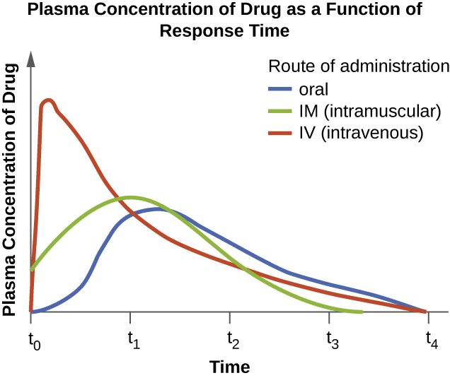
Selective toxicity: hurt the microbe, spare the patient
The reason we can pour a poison into a sick person and call it medicine is a principle named selective toxicity: a good antimicrobial harms the microbe far more than it harms the host. This is possible because a bacterium is a prokaryote — it is built from parts your own cells simply do not have. It wraps itself in a rigid cell wall made of peptidoglycan; your cells have no wall. Its ribosomes (the machines that build proteins) are a different size and shape from yours. It makes its own folic acid from scratch, while you get folate from your diet. Each of those differences is a handle a drug can grab. Aim at a structure the microbe has and the patient lacks, and you get a cure without collateral damage.
Selective toxicity is a matter of degree, not perfection. The drugs with the widest safety margin tend to be those that attack the cell wall — the penicillins and cephalosporins — precisely because human cells have no equivalent target to injure. Drugs that hit something bacteria and humans share, even a little, are more likely to cause the toxicities nurses watch for: an aminoglycoside can damage the kidney and the inner ear, and an antifungal that targets the fungal cell membrane can also disturb the patient's own membranes because both are built from sterols. The closer a microbe is to us biologically — a fungus, a protozoan, a human cell hijacked by a virus — the harder selective toxicity becomes, which is exactly why antibacterials are gentler than most antifungals and antivirals.
The therapeutic index: how much room for error
Every drug has a dose that helps and a higher dose that harms. The therapeutic index puts a number on the gap between them — roughly, the toxic dose divided by the effective dose. A large index means a comfortable margin: you would have to overshoot badly before the patient got hurt, which is why penicillin can be given in generous doses with little worry. A small or narrow therapeutic index means the helpful dose and the harmful dose sit uncomfortably close together, so the drug must be dosed carefully and, often, measured directly in the blood.
This is where the graph above becomes a bedside tool. The goal is to keep the drug's concentration inside the therapeutic window — high enough to kill bacteria, low enough to spare the patient. For narrow-index antimicrobials such as vancomycin and the aminoglycosides (gentamicin, tobramycin), we do not guess. We draw blood levels: a trough just before the next dose to confirm the drug has not built up to toxic levels, and sometimes a peak to confirm it climbed high enough to work.
| Drug | Therapeutic index | What that means for nursing |
|---|---|---|
| Penicillins | Wide | Large safety margin; routine levels not needed |
| Cephalosporins | Wide | Well tolerated; watch for allergy, not toxicity |
| Vancomycin | Narrow | Monitor trough levels; watch kidney function |
| Aminoglycosides | Narrow | Monitor peak & trough; risk of kidney & ear toxicity |
- Selective toxicity — hitting a target the microbe has and the host lacks — is what makes antimicrobial therapy possible.
- The beta-lactams (penicillins, cephalosporins) are the safest drugs because human cells have no wall to injure — but vancomycin also targets the wall and is still nephrotoxic and narrow-index, so target alone does not guarantee safety.
- The therapeutic index is the gap between the helpful dose and the harmful one.
- Narrow-index drugs (vancomycin, aminoglycosides) need blood-level monitoring; timing of the draw is everything.
Section 2How the drugs work
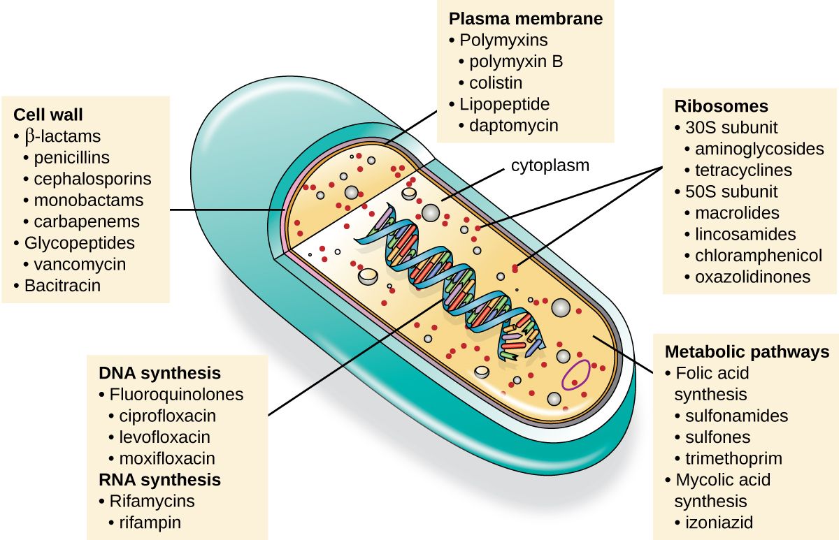
Five ways to kill a bacterium
For all their variety, antibacterial drugs attack a bacterium in only a handful of places. Learn these five targets and you can slot almost any antibiotic you meet into a mental map — and, more usefully, predict its selective toxicity and its side effects.
1. Cell wall synthesis. The peptidoglycan wall is the bacterium's pressure vessel; without it the cell bursts. Penicillins, cephalosporins, and vancomycin block wall construction, so growing bacteria lyse. Because human cells have no wall, this is the most selectively toxic target of all. Drugs in the penicillin and cephalosporin families share a chemical ring called a beta-lactam, which is why they are grouped as beta-lactam antibiotics.
2. Protein synthesis. Bacterial ribosomes (built from 30S and 50S subunits) differ from the human 40S/60S version, so a drug can jam the bacterial machine while sparing yours. Aminoglycosides and tetracyclines hit the 30S subunit; macrolides (azithromycin, erythromycin) and clindamycin hit the 50S. The match is imperfect, however — your mitochondria carry bacteria-like ribosomes, which helps explain some of these drugs' toxicities.
3. Nucleic acid synthesis. Some drugs block the enzymes bacteria use to copy and unwind DNA. The fluoroquinolones (ciprofloxacin, levofloxacin) inhibit bacterial DNA gyrase and topoisomerase, enzymes shaped differently from the human versions.
4. Metabolic (folate) pathways. Bacteria must synthesize folic acid themselves; you eat yours. Sulfonamides and trimethoprim starve the bacterium of folate by blocking two steps of that pathway — a target you simply do not possess.
5. Cell membrane. A few drugs punch holes in the bacterial membrane, causing the cell's contents to leak out. These tend to be more toxic, because your cells have membranes too; they are reserved for stubborn infections.
Kill it or just stop it: -cidal vs -static
Drugs also differ in how thoroughly they act. A bactericidal drug kills the bacterium outright (penicillins, aminoglycosides, fluoroquinolones). A bacteriostatic drug merely halts its growth (tetracyclines, macrolides, sulfonamides), then leaves the finishing blow to the patient's own immune system. In a healthy person, static is often enough. In a patient whose immune defenses are down — a neutropenic chemotherapy patient, someone with endocarditis or meningitis — the team usually reaches for a bactericidal drug, because there is no reliable immune backup to mop up survivors.
| Target | Example drug classes | Why it is selective |
|---|---|---|
| Cell wall synthesis | Penicillins, cephalosporins, vancomycin | Human cells have no cell wall |
| Protein synthesis (ribosome) | Aminoglycosides, tetracyclines, macrolides | Bacterial ribosome (70S) differs from human (80S) |
| Nucleic acid synthesis | Fluoroquinolones | Bacterial DNA enzymes differ from human ones |
| Folate metabolism | Sulfonamides, trimethoprim | Humans get folate from diet; bacteria make it |
| Cell membrane | Polymyxins, daptomycin | Least selective — reserved for resistant cases |
- Antibacterials hit five targets: cell wall, ribosome, nucleic acids, folate metabolism, and cell membrane.
- Beta-lactams (penicillins, cephalosporins) attack the cell wall — the safest, most selective target.
- Bactericidal drugs kill; bacteriostatic drugs stall growth and rely on the immune system to finish.
- Immunocompromised patients usually need a bactericidal drug.
Section 3Spectrum and choosing a drug
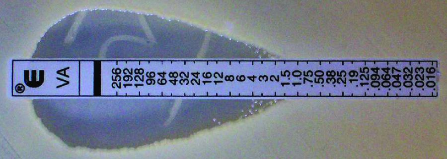
Narrow versus broad spectrum
A drug's spectrum of activity is simply the range of bacteria it works against. A narrow-spectrum drug hits only a few kinds of bacteria; a broad-spectrum drug hits many. Broad sounds better, and in a crisis it can be lifesaving — but breadth has a cost. A broad-spectrum antibiotic clear-cuts the patient's normal flora along with the pathogen, opening the door to a superinfection such as Clostridioides difficile colitis or a yeast overgrowth, and every unnecessary exposure nudges the whole bacterial population toward resistance. The goal, whenever the culprit is known, is the narrowest drug that will do the job.
Empiric first, then definitive
Cultures take time, and a septic patient cannot wait a day or two for results. So therapy usually happens in two stages. Empiric therapy is the educated first guess: based on the likely source, the local resistance patterns, and how sick the patient is, the team starts a broad-spectrum drug immediately — but only after cultures are drawn. Once the lab reports what grew and what it is susceptible to, the team switches to definitive therapy, ideally narrowing (or de-escalating) to a focused drug. This is why the order in which things happen at the bedside matters so much: cultures first, then antibiotics.
Susceptibility testing: proving the drug will work
How does the lab know which drug to recommend? By testing the patient's own isolate against a panel of antibiotics. The key number is the minimum inhibitory concentration (MIC) — the lowest drug concentration that stops the bacterium from growing. Several methods get at it. In the classic Kirby–Bauer (disk-diffusion) test, paper disks soaked in different antibiotics sit on a lawn of bacteria; the larger the clear zone of inhibition around a disk, the more susceptible the organism — though disk diffusion sorts the isolate into susceptible, intermediate, or resistant rather than reading out an exact MIC. The Etest strip in the figure refines this into an actual MIC value, and automated broth microdilution systems do the same in tiny wells. The result comes back to the chart as S (susceptible), I (intermediate), or R (resistant) — a report the nurse reads alongside the team when a patient is not improving on the current drug.
| Narrow-spectrum | Broad-spectrum | |
|---|---|---|
| Bacteria covered | Few, targeted | Many |
| Effect on normal flora | Spares most of it | Wipes out much of it |
| Superinfection risk | Lower | Higher (C. diff, yeast) |
| Resistance pressure | Lower | Higher |
| Best used when | The pathogen is known | Empirically, while awaiting cultures |
- Spectrum is the range of bacteria a drug covers; narrow is preferred once the pathogen is known.
- Broad-spectrum drugs invite superinfection and resistance — use them empirically, then narrow.
- Empiric therapy is the first guess; definitive therapy follows the culture and de-escalates.
- The MIC is the lowest concentration that inhibits growth; results are reported as S / I / R.
Section 4How bacteria fight back
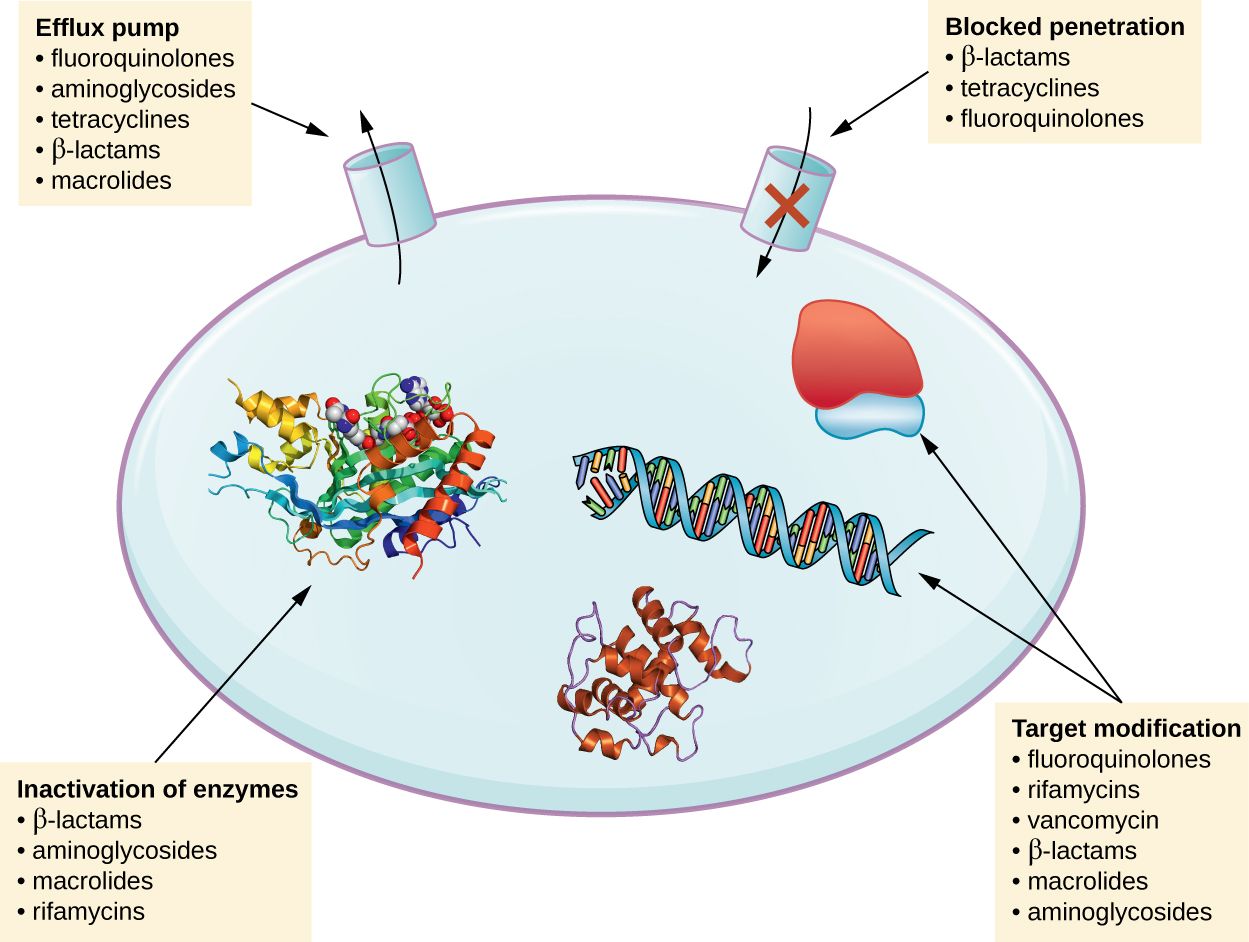
Four ways to survive the drug
Resistance is not the drug “wearing off.” It is evolution in fast-forward: a drug kills the susceptible bacteria and leaves the rare survivor that happened to carry a defense, and that survivor's descendants inherit the trick. There are four main defenses, and the figure lays them out.
Destroy the drug. The most famous example is beta-lactamase, an enzyme that snips open the beta-lactam ring of penicillins before it can work. This is why some penicillins are combined with a beta-lactamase inhibitor (as in amoxicillin–clavulanate) to protect the drug.
Change the target. If the drug can no longer recognize its binding site, it cannot act. This is the trick behind MRSA: the bacterium acquires an altered penicillin-binding protein that beta-lactams cannot grip.
Pump the drug out. Efflux pumps in the membrane actively bail the antibiotic back out before it reaches a lethal concentration inside the cell.
Keep the drug out. By altering the porins or the permeability of its outer membrane, a bacterium can simply reduce how much drug gets in at all.
How resistance spreads — and why it spreads so fast
Resistance can arise by spontaneous mutation, but its terrifying speed comes from horizontal gene transfer: bacteria pass resistance genes to one another, even across species, on mobile loops of DNA called plasmids. A single plasmid can carry resistance to several drug classes at once, so one exposure can select for a strain resistant to antibiotics it never even met. Every unnecessary antibiotic anywhere — in a clinic, a farm, a fish tank — adds to the selective pressure that favors these survivors.
The named threats: MRSA and friends
MRSA — methicillin-resistant Staphylococcus aureus — is resistant to essentially all beta-lactam antibiotics, which knocks out the safest, most familiar drugs and forces the team toward vancomycin and its relatives. It spreads readily in hospitals and communities, so patients known to carry it are often placed on contact precautions. It is not alone: VRE (vancomycin-resistant enterococci), ESBL-producing and carbapenem-resistant gram-negatives, and multidrug-resistant tuberculosis all crowd the same worrying list. The common thread is that our shelf of reliable drugs is shrinking faster than we are refilling it.
| Mechanism | What the bacterium does | Example |
|---|---|---|
| Drug inactivation | Makes an enzyme that destroys the drug | Beta-lactamase vs penicillins |
| Target modification | Alters the drug's binding site | MRSA's altered binding protein |
| Efflux pump | Pumps the drug back out | Tetracycline resistance |
| Reduced uptake | Keeps the drug from entering | Altered porins in gram-negatives |
- Bacteria resist drugs four ways: destroy the drug, change the target, pump it out, or keep it out.
- Resistance genes spread fast by horizontal gene transfer on plasmids, driven by selective pressure.
- MRSA resists all beta-lactams by altering the drug's binding site, forcing a switch to vancomycin.
- Contact precautions and hand hygiene contain resistant organisms on the unit.
Section 5Stewardship and the nurse
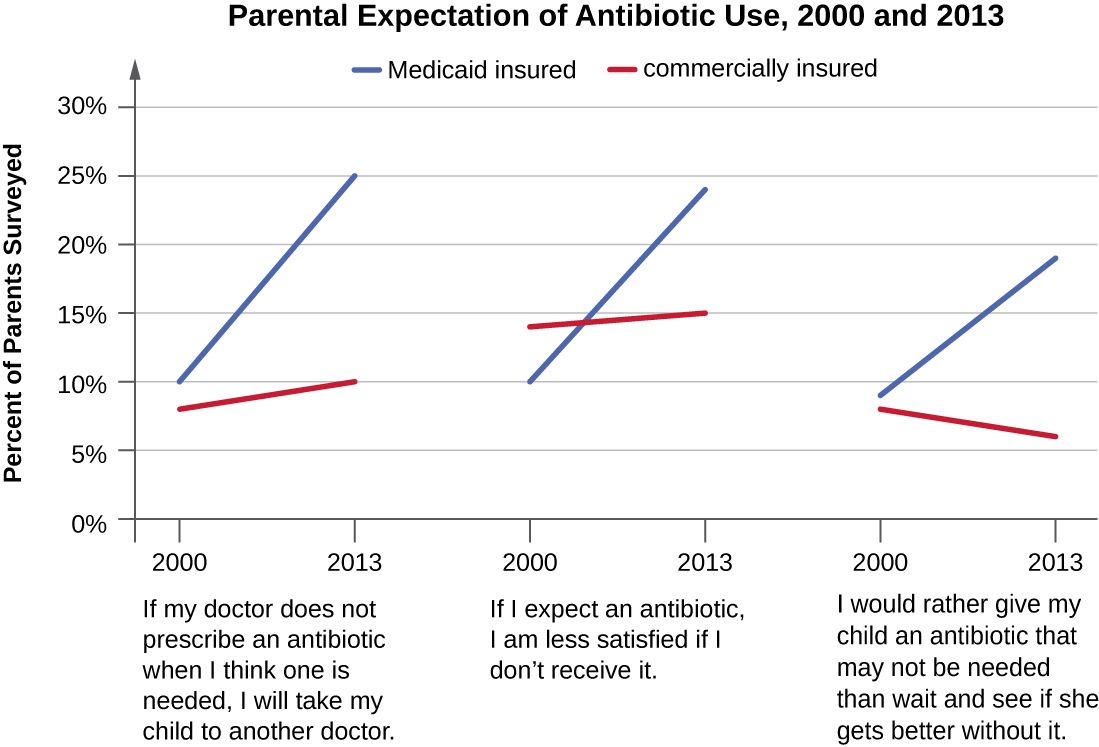
What stewardship means
Antibiotic stewardship is the coordinated effort to use these drugs only when they help, and to use them well. It is often summarized as the five rights of antimicrobials: the right drug, at the right dose, by the right route, for the right duration, in the right patient. The purpose is twofold — better outcomes for the patient in front of you (fewer side effects, less C. diff, shorter stays) and protection of the drugs themselves for the patients who come after. Because resistance is a shared resource being spent, stewardship is genuinely a public-health duty, not just a cost-control exercise.
The nurse is a steward — here is how
Stewardship is often described as a physician-and-pharmacist activity, but the nurse touches nearly every point of it. You are the one who draws cultures before the first dose, who gives antibiotics on time so blood levels stay effective, and who monitors the patient for both improvement and toxicity. You are positioned to ask the questions that trim unnecessary drug use: Can this IV antibiotic switch to an oral one now that the patient is eating and improving? Is this still needed on day 5, or is it just still ordered? Are two drugs covering the same bug? And you are the last check on the allergy history — clarifying a vague “penicillin allergy” so the patient is not pushed onto a broader, more toxic alternative for no reason.
Adherence and what you teach the patient
Stewardship reaches beyond the hospital, and here patient teaching is the tool. Explain that antibiotics do nothing for colds, flu, and most sore throats — and that demanding them “just in case” carries real risk with no benefit. Teach patients to take the drug exactly as prescribed, to never save or share leftover antibiotics, and to never use someone else's. The old blanket rule to “always finish every pill” is being refined — some infections do better with shorter courses — so the honest, durable message is simpler and stronger: take it the way it was prescribed, and call before stopping early or doubling up.
| Stewardship goal | What the nurse does |
|---|---|
| Right diagnosis | Collect cultures before the first antibiotic dose |
| Right timing | Give doses on schedule to keep levels effective |
| Right duration | Prompt the team to review or de-escalate ongoing orders |
| Right route | Flag when a stable, eating patient could switch IV → oral |
| Right patient | Verify and clarify the allergy history |
| Prevention | Hand hygiene, timely device removal, patient teaching |
- Stewardship = right drug, dose, route, duration, and patient — for the patient now and for future patients.
- The nurse steward draws cultures first, gives doses on time, and watches for toxicity.
- Advocate to de-escalate, switch IV to oral, and clarify allergies.
- Teach adherence: take as prescribed, never share or save antibiotics, and don't expect them for viral illness.
The chapter in one breath
- Selective toxicity — attacking a microbe-only target — is why antimicrobials cure without poisoning the patient.
- The therapeutic index is the safety margin; narrow-index drugs like vancomycin and aminoglycosides need blood-level monitoring.
- Antibacterials hit five targets: cell wall, ribosome, nucleic acids, folate metabolism, and cell membrane.
- Drugs are bactericidal or bacteriostatic, and narrow- or broad-spectrum; narrow and cidal have their place, and so does empiric breadth.
- Susceptibility testing (MIC via Kirby–Bauer, Etest, or microdilution) reports S / I / R to guide definitive therapy — after cultures are drawn.
- Bacteria resist by destroying, dodging, ejecting, or excluding the drug, and spread resistance fast via plasmids; MRSA is the headline example.
- Stewardship protects both the patient and the drugs, and the nurse is a frontline steward — cultures first, doses on time, de-escalation advocated, adherence taught.
ReferenceWords worth knowing
A quick reference for the terms in this chapter — skim it now, or circle back whenever a word trips you up.
- Adherence
- Taking a medication exactly as prescribed — correct dose, timing, and duration.
- Antibiotic stewardship
- The coordinated effort to use antimicrobials only when needed and to use them well, protecting both the patient and future effectiveness.
- Bactericidal
- Describes a drug that kills bacteria outright.
- Bacteriostatic
- Describes a drug that halts bacterial growth, leaving the immune system to finish.
- Beta-lactam
- The chemical ring shared by penicillins and cephalosporins; the part a beta-lactamase attacks.
- Beta-lactamase
- A bacterial enzyme that destroys beta-lactam antibiotics, a common resistance mechanism.
- Broad-spectrum
- Active against many kinds of bacteria; convenient empirically but harder on normal flora.
- De-escalation
- Narrowing therapy from a broad drug to a focused one once cultures identify the pathogen.
- Efflux pump
- A membrane transporter that pumps an antibiotic back out of the cell before it can act.
- Empiric therapy
- Best-guess antibiotic treatment started before culture results are available.
- Horizontal gene transfer
- The passing of genes (including resistance genes) between bacteria, often on plasmids.
- Kirby–Bauer test
- Disk-diffusion susceptibility test; zone-of-inhibition size reflects susceptibility.
- Minimum inhibitory concentration (MIC)
- The lowest drug concentration that prevents visible bacterial growth.
- MRSA
- Methicillin-resistant Staphylococcus aureus; resistant to all beta-lactams via an altered binding protein.
- Narrow-spectrum
- Active against only a few kinds of bacteria; preferred once the pathogen is known.
- Selective toxicity
- Harming the microbe far more than the host by targeting a structure the microbe has and the host lacks.
- Spectrum of activity
- The range of bacterial types a given antimicrobial can affect.
- Superinfection
- A new infection (e.g., C. diff or yeast) that arises after a drug disrupts the normal flora.
- Susceptibility testing
- Laboratory testing of a patient's isolate against antibiotics, reported as S, I, or R.
- Therapeutic index
- The safety margin between a drug's effective dose and its toxic dose.
- Trough level
- The drug concentration measured just before the next dose, used to avoid toxic accumulation.
Check yourself
How Microbes Cause Disease
A pathogen does not make your patient sick by magic. It follows a script — get in, hold on, multiply, do damage, and dodge the immune system long enough to spread to the next host. Every step of that script is powered by a specific molecular tool, and every tool is a target that nursing care, drugs, and vaccines are built to block. This chapter takes apart the machinery of disease: what makes one microbe a killer and another a harmless bystander, how germs breach the body's borders and anchor themselves, the toxins and enzymes they use to wreck tissue, the disguises they wear to stay hidden, and the predictable stages an infection passes through from the moment of exposure to recovery. Understand the script, and you understand why we do almost everything we do at the bedside.
By the end of this chapter you can
- Distinguish pathogenicity from virulence, and explain what virulence factors are.
- Interpret ID₅₀ and LD₅₀ as measures of how infectious or deadly a microbe is.
- Name the major portals of entry and explain why the same microbe can be harmless at one portal and lethal at another.
- Describe how adhesins let pathogens attach, and why attachment is a drug and vaccine target.
- Compare exotoxins and endotoxin in source, chemistry, potency, and clinical effect.
- Explain how capsules, antigenic variation, and intracellular hiding let microbes evade host defenses.
- List the five stages of infection and identify when a patient is most contagious.
- Connect each step of the disease process to a specific nursing or public-health intervention.
Section 1Pathogenicity and virulence
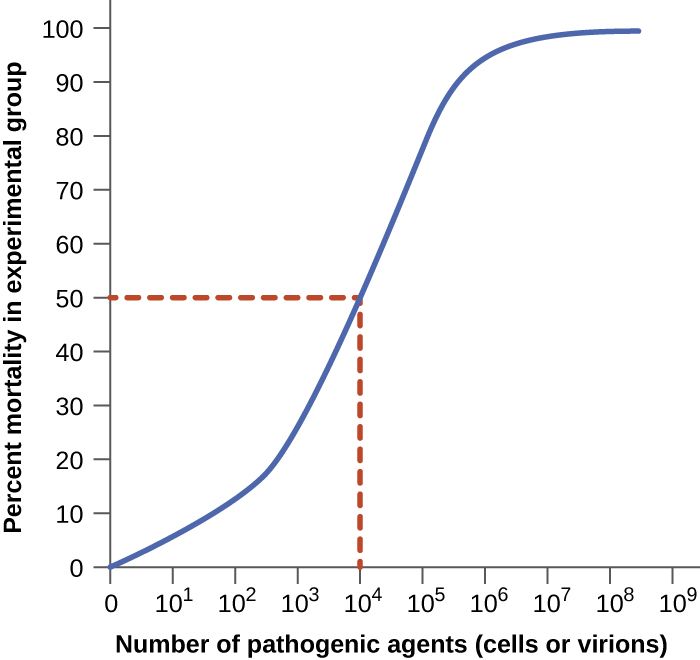
Two words that are not the same
In everyday speech "pathogenic" and "virulent" get used interchangeably, but microbiology draws a sharp line between them, and the distinction is worth keeping straight. Pathogenicity is the ability of a microbe to cause disease at all — a yes-or-no property. Virulence is the degree of that ability — how much harm, how fast, at how small a dose. Two organisms can both be pathogens while sitting at opposite ends of the virulence scale: Clostridium botulinum is staggeringly virulent (on the order of a nanogram of its toxin per kilogram of body weight can kill), while a common cold rhinovirus is a genuine pathogen that rarely does more than make you miserable for a week.
It also matters whose pathogen we mean. A primary pathogen can cause disease in an otherwise healthy person with intact defenses — influenza, Salmonella, Mycobacterium tuberculosis. An opportunistic pathogen only causes disease when it gets an opening: a breached barrier, wiped-out normal flora, or a weakened host. Much of what a nurse guards against — catheter infections, ventilator pneumonia, Candida overgrowth — is opportunistic disease caused by organisms that would be harmless in a healthy person.
Virulence factors: the toolkit of harm
What makes one strain more virulent than another comes down to virulence factors — the specific genes and molecules that let a microbe establish itself and cause damage. These are not vague qualities; they are concrete structures and secreted products, and the whole rest of this chapter is essentially a tour of them. They fall into a few functional buckets: tools for attaching (adhesins), tools for spreading through tissue (invasins and enzymes), tools for damaging the host (toxins), and tools for evading defenses (capsules, antigenic variation). Because each factor is a distinct molecule, each one is a potential target — an antibody a vaccine can raise, an enzyme a drug can inhibit, a toxin an antitoxin can neutralize.
Measuring virulence: ID₅₀ and LD₅₀
Virulence can be quantified, which is how we compare organisms objectively. The ID₅₀ (median infectious dose) is the number of microbes needed to establish infection in half of an exposed population; the LD₅₀ (median lethal dose) is the amount of a microbe or toxin needed to kill half of them. A lower number means a more virulent agent, because it takes fewer organisms to do the damage. Shigella has an ID₅₀ of only 10 to 100 cells — which is exactly why it spreads so easily person-to-person and why hand hygiene is decisive — whereas Vibrio cholerae needs roughly a hundred million organisms, so it usually takes contaminated water to deliver a big enough dose.
| Term | What it measures | Low number means |
|---|---|---|
| Pathogenicity | Whether a microbe can cause disease (yes/no) | — |
| Virulence | How severe the disease it causes | — |
| ID₅₀ | Dose that infects 50% of a population | Very infectious (spreads at low dose) |
| LD₅₀ | Dose that kills 50% of a population | Very deadly (lethal at low dose) |
- Pathogenicity is the ability to cause disease; virulence is how much harm it does.
- Primary pathogens sicken healthy hosts; opportunistic pathogens need a breach, wiped-out flora, or a weakened host.
- Virulence factors are the specific molecules for attaching, spreading, damaging, and evading — each one a drug or vaccine target.
- ID₅₀ and LD₅₀ quantify virulence; a lower value means a more dangerous organism.
Section 2Getting in and attaching
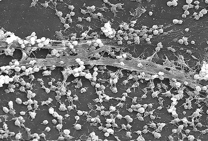
Portals of entry
A microbe cannot cause infection until it reaches tissue it can colonize, and it gets there through a portal of entry — a specific route into the body. The great majority of pathogens enter through the mucous membranes of the respiratory tract (by far the most common — influenza, tuberculosis, the common cold), the gastrointestinal tract (Salmonella, hepatitis A, norovirus), the genitourinary tract (gonorrhea, chlamydia), or the conjunctiva of the eye. Others enter through the skin, which is normally an excellent barrier but is breachable through cuts, bites, burns, and surgical wounds. A special case is the parenteral route, where an organism is deposited directly into deeper tissue or the bloodstream — a needlestick, an IV line, a puncture wound — bypassing the surface defenses entirely.
The clinical punchline is that the same organism can be harmless at one portal and dangerous at another. Escherichia coli is a peaceful resident of the colon, but delivered to the bladder it causes a UTI, and reaching the bloodstream it can cause fatal sepsis. Every invasive device a nurse manages — a urinary catheter, a central line, an endotracheal tube — is essentially an artificial portal of entry that hands a microbe a shortcut past the body's borders. That is why device insertion is done sterile and why prompt removal is one of the most reliable infection-prevention moves there is.
| Portal of entry | Route | Example diseases |
|---|---|---|
| Respiratory tract | Inhaled droplets/aerosols | Influenza, TB, COVID-19, colds |
| GI tract | Ingested (fecal–oral, food, water) | Salmonellosis, hepatitis A, norovirus |
| Genitourinary tract | Sexual contact, ascending | Gonorrhea, chlamydia, UTIs |
| Skin / mucous breaks | Cuts, bites, burns, wounds | Cellulitis, tetanus, rabies |
| Parenteral | Directly into tissue/blood | Line infections, hepatitis B, HIV |
Adhesion: sticking around
Getting in is not enough — a microbe that cannot anchor itself is swept away by mucus, urine flow, peristalsis, or the tide of tears and saliva. So the next step is attachment, accomplished by adhesins: surface molecules (often on fimbriae, pili, or in the capsule and glycocalyx) that bind like a key to specific receptor molecules on host cells. This binding is precise and often explains a pathogen's tropism — why a given microbe infects one tissue and not another. When many organisms adhere and then encase themselves in a slimy matrix, they form a biofilm, a community that is dramatically harder for antibiotics and immune cells to penetrate. Biofilms on catheters, prosthetic joints, and heart valves are a major reason device-associated infections are so stubborn.
Because attachment is a mandatory first move, it is a favorite target for prevention. Some vaccines work by raising antibodies against a pathogen's adhesins so the microbe can never gain a foothold, and part of what hand hygiene, catheter care, and oral care in ventilated patients accomplish is physically removing organisms before they establish a biofilm.
Infectious dose
How many organisms does it actually take? That is the infectious dose, captured numerically by the ID₅₀ from Section 1. It depends on both the microbe's virulence factors and the host's defenses, which is why an exposure that does nothing to a healthy adult can seed a serious infection in an immunocompromised or very young patient. A pathogen armed with strong adhesins and a good evasion toolkit needs far fewer organisms to succeed, which loops back to why low-dose organisms demand such strict precautions.
- Pathogens enter through portals of entry: respiratory, GI, GU, conjunctiva, skin breaks, and the parenteral route.
- The same microbe can be harmless at one portal and lethal at another (E. coli: colon vs. bladder vs. blood).
- Adhesins anchor microbes to specific host receptors; clumped, matrix-encased communities become drug-resistant biofilms.
- Invasive devices are artificial portals of entry — sterile insertion and early removal are core nursing defenses.
Section 3Causing damage: exotoxins and endotoxin
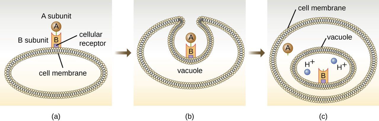
Exotoxins: precision weapons
Much of the harm a pathogen does comes from toxins, and the more dramatic of the two families is the exotoxin. Exotoxins are proteins that bacteria manufacture and secrete, produced by both gram-positive and gram-negative organisms. Being proteins, they are exquisitely specific and often astonishingly potent — the toxins of tetanus, botulism, and diphtheria are among the most lethal substances known. They come in three broad styles. A-B toxins have two parts: a B (binding) subunit that latches onto a target cell and an A (active) subunit that enters and sabotages a specific internal process — diphtheria toxin halts protein synthesis, cholera toxin drives the massive fluid loss of rice-water diarrhea, and botulinum and tetanus toxins block neurotransmitter release to paralyze muscle. Membrane-disrupting toxins punch holes in host-cell membranes; these include the hemolysins that lyse red cells and the leukocidins that kill white cells. Superantigens short-circuit the immune system, triggering a huge, indiscriminate release of cytokines that produces the shock of toxic shock syndrome.
Two clinically useful facts follow from exotoxins being proteins. First, they are usually heat-labile — destroyed by cooking — which is why proper food handling prevents many toxin-mediated illnesses. Second, a protein toxin can be chemically inactivated into a toxoid that no longer harms but still provokes protective antibodies; the tetanus and diphtheria components of routine vaccines are toxoids, and pre-formed antitoxin antibodies can be given to neutralize circulating toxin in an acute poisoning.
Endotoxin: the gram-negative alarm
The second toxin is not secreted and not a protein. Endotoxin is the lipid A portion of lipopolysaccharide (LPS), a fixed component of the outer membrane of gram-negative bacteria. It is only released when the bacterial cell is damaged or dies — including when antibiotics lyse large numbers of organisms. Any single molecule of endotoxin is far weaker than a potent exotoxin, but the body's reaction to it is powerful and generic: fever, vasodilation with falling blood pressure, and widespread activation of clotting. When a bloodstream infection floods the body with endotoxin, that reaction becomes septic shock — dangerously low blood pressure and the microclotting of disseminated intravascular coagulation (DIC). Unlike a protein exotoxin, endotoxin is heat-stable and cannot be made into a toxoid, so there is no vaccine against it.
| Feature | Exotoxin | Endotoxin |
|---|---|---|
| Chemistry | Protein, secreted | Lipid A of LPS (outer membrane) |
| Source | Gram-positive & gram-negative | Gram-negative only |
| Released | Actively secreted by living cell | Only when the cell is damaged/lysed |
| Potency | Very high; specific effect | Lower per molecule; general effect |
| Heat stability | Usually heat-labile | Heat-stable |
| Toxoid / vaccine? | Yes (tetanus, diphtheria) | No |
| Typical effect | Specific (paralysis, diarrhea, etc.) | Fever, hypotension, shock, DIC |
- Exotoxins are secreted proteins — potent, specific, often heat-labile, and convertible to protective toxoids.
- Exotoxin styles: A-B toxins, membrane-disrupting hemolysins/leukocidins, and superantigens.
- Endotoxin is lipid A of LPS from gram-negative cells, released on lysis; it is heat-stable and has no toxoid.
- A flood of endotoxin drives fever, hypotension, septic shock, and DIC.
Section 4Evading the body's defenses
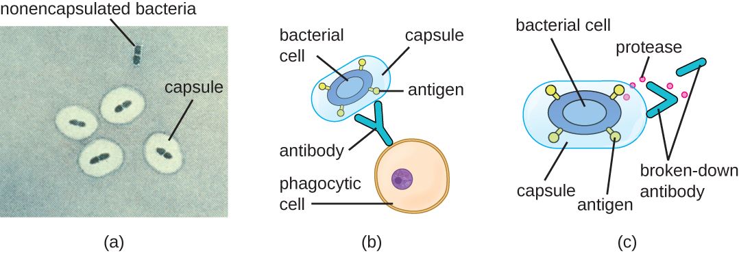
Capsules: the invisibility cloak
Getting in, holding on, and doing damage still isn't enough if the immune system mops the microbe up. So successful pathogens carry tools for immune evasion, and the classic one is the capsule — a thick, slippery polysaccharide layer around the cell. A capsule works two ways: it is physically hard for a phagocyte to grip and engulf, and it hides the surface molecules that antibodies and complement would otherwise latch onto. The capsule is the master virulence factor of several of the most feared pathogens — Streptococcus pneumoniae, Neisseria meningitidis, Haemophilus influenzae type b, and Klebsiella pneumoniae. It is no accident that our capsular vaccines — against S. pneumoniae, N. meningitidis, and H. influenzae type b — are aimed squarely at the capsule: teach the immune system to recognize that coat and the invisibility cloak stops working. (Klebsiella pneumoniae depends just as heavily on its capsule, but no vaccine against it is licensed; candidates remain investigational.)
Antigenic variation: the costume change
Another strategy is to keep changing the disguise. In antigenic variation, a microbe alters the surface molecules the immune system has learned to recognize, so that yesterday's antibodies no longer fit. Influenza does this every year through small mutations (antigenic drift) and occasionally through big reassortments (antigenic shift) — which is why the vaccine is reformulated annually and why a truly novel shift can cause a pandemic. Neisseria gonorrhoeae constantly reshuffles the proteins on its pili, one reason gonorrhea produces no lasting immunity and can reinfect the same person repeatedly. The trypanosomes of African sleeping sickness and the Borrelia of relapsing fever run the same trick, producing waves of illness as each new variant outruns the antibody response.
Hiding and sabotage
Pathogens have still more moves. Some become intracellular, living inside host cells where circulating antibodies cannot reach them — Mycobacterium tuberculosis famously survives inside the very macrophages sent to destroy it, and Listeria and Salmonella do the same. Others secrete IgA proteases that chop up the antibodies guarding mucous membranes, or coagulase that walls the organism off inside a clot of the host's own fibrin (a Staphylococcus aureus specialty). Some coat themselves in host proteins to look like "self" — that is antigenic masking, as when S. aureus protein A grabs the tail of IgG. Distinguish it from molecular mimicry, in which the microbe's own antigens happen to resemble host tissue, so the antibodies made against the microbe cross-react with the patient — streptococcal M protein resembling cardiac myosin in rheumatic fever is the classic example. Each of these explains a clinical reality: TB requires months of therapy because the bug is hidden inside cells; staph abscesses often need drainage because antibiotics cannot penetrate the walled-off pocket.
| Evasion strategy | How it works | Example |
|---|---|---|
| Capsule | Blocks phagocytosis; hides surface antigens | S. pneumoniae, N. meningitidis |
| Antigenic variation | Changes surface molecules to dodge antibodies | Influenza, N. gonorrhoeae |
| Intracellular hiding | Lives inside host cells, out of antibody reach | M. tuberculosis, Listeria |
| IgA protease | Destroys mucosal antibodies | N. gonorrhoeae, H. influenzae |
| Coagulase | Walls the microbe inside a fibrin clot | Staphylococcus aureus |
- Capsules block phagocytosis and hide antigens; capsular vaccines target exactly this.
- Antigenic variation changes surface molecules so antibodies no longer fit (flu, gonorrhea).
- Intracellular hiding keeps microbes out of antibody reach and forces long therapy (TB).
- Coagulase and abscess formation are why "source control" — drainage or device removal — is often essential.
Section 5The stages of an infection
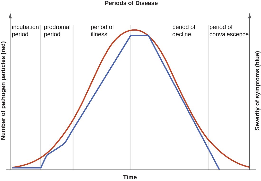
Five predictable stages
Most acute infections unfold along the same timeline, and knowing the stages tells you what to expect and when a patient is most dangerous to others. The incubation period runs from the moment of exposure to the first symptoms; the microbe is multiplying but the patient feels fine, and this interval can be hours (food poisoning) or years (HIV, TB). Next comes the prodromal period, a short phase of vague, non-specific complaints — mild fever, fatigue, general achiness — as the microbial load and the immune response both build. The period of illness is the peak: the microbe is most numerous, the signs and symptoms specific to the disease are at their worst, and this is when the patient is sickest and, untreated, most at risk. Then the period of decline arrives as the immune system (helped by treatment) gains the upper hand and symptoms recede, though the patient is weakened and vulnerable to secondary infections. Finally the convalescence period is recovery, when the body repairs itself and strength returns.
Why the stages matter at the bedside
The practical payoff is understanding communicability — when a patient can spread the infection — because it does not line up neatly with how sick they look. For many illnesses, shedding begins in the prodromal stage or even late incubation, meaning a person is contagious before they feel truly ill and before anyone suspects infection. Measles is the clean example: it is most contagious through the 3–4 day catarrhal prodrome, which is why a patient can seed an outbreak in a waiting room while feeling only "a little off." Influenza shows a milder version — shedding starts about a day before symptoms but peaks in the first one to two days of illness itself. Just as important, some patients keep shedding organisms into the decline and convalescence stages, and a few become long-term carriers who feel completely well yet remain a source — the reason isolation precautions are timed to the organism's shedding pattern, not merely to how bad the patient's symptoms are right now.
| Stage | What is happening | Symptoms |
|---|---|---|
| Incubation | Microbe multiplying after exposure | None yet |
| Prodromal | Load and immune response building | Mild, vague (fatigue, low fever) |
| Illness | Microbe at peak; disease fully expressed | Most severe, disease-specific |
| Decline | Immune system gaining control | Subsiding; patient weakened |
| Convalescence | Recovery and tissue repair | Resolving; strength returns |
- Acute infections move through incubation → prodromal → illness → decline → convalescence.
- Symptoms peak during the period of illness, when the microbe is most numerous.
- Communicability often begins in the prodromal stage — patients spread infection before they seem sick.
- Some patients shed during decline/convalescence or become long-term carriers, so isolation is timed to shedding, not just symptoms.
The chapter in one breath
- Pathogenicity is whether a microbe causes disease; virulence is how much harm it does, measured by ID₅₀ and LD₅₀ (lower = more dangerous).
- Virulence factors — molecules for attaching, spreading, damaging, and evading — are the concrete tools of disease and the targets of drugs and vaccines.
- Microbes enter through portals of entry (respiratory, GI, GU, skin, parenteral) and anchor with adhesins; invasive devices are artificial portals nurses control.
- Exotoxins are potent secreted proteins (A-B toxins, hemolysins, superantigens) that can be turned into protective toxoids.
- Endotoxin is lipid A of LPS from gram-negative cells, released on lysis, driving fever, hypotension, septic shock, and DIC.
- Pathogens evade defenses with capsules, antigenic variation, and intracellular hiding — explaining vaccines, annual flu shots, and the need for drainage or long therapy.
- Infections pass through five stages, and patients are often most contagious in the prodromal phase, before they look sick.
ReferenceWords worth knowing
A quick reference for the terms in this chapter — skim it now, or circle back whenever a word trips you up.
- Adhesin
- A surface molecule (often on fimbriae or pili) that binds a pathogen to specific receptors on host cells so it is not washed away.
- Antigenic variation
- A pathogen's ability to change its surface molecules so previously formed antibodies no longer recognize it.
- Biofilm
- A matrix-encased community of adherent microbes that resists antibiotics and immune cells; common on catheters and implants.
- Capsule
- A thick polysaccharide layer around a cell that blocks phagocytosis and hides surface antigens.
- Carrier
- A person who harbors and can transmit a pathogen while showing no symptoms.
- Convalescence period
- The final stage of infection, during which the body recovers and repairs.
- Endotoxin
- The lipid A portion of LPS in the gram-negative outer membrane; released on cell lysis, causing fever, hypotension, and shock.
- Exotoxin
- A secreted protein toxin, often potent and specific; producible by gram-positive and gram-negative bacteria.
- ID₅₀ (median infectious dose)
- The number of microbes needed to infect 50% of an exposed population; a lower value means a more infectious organism.
- Incubation period
- The interval from exposure to the first appearance of symptoms.
- LD₅₀ (median lethal dose)
- The dose of microbe or toxin that kills 50% of a population; a lower value means a deadlier agent.
- Lipopolysaccharide (LPS)
- A molecule of the gram-negative outer membrane whose lipid A component is endotoxin.
- Opportunistic pathogen
- A microbe that causes disease only when defenses are breached, normal flora are lost, or the host is weakened.
- Pathogenicity
- The ability of a microbe to cause disease.
- Portal of entry
- The route by which a pathogen enters the body (respiratory, GI, GU, skin, or parenteral).
- Primary pathogen
- A microbe that can cause disease in a healthy host with intact defenses.
- Prodromal period
- An early stage of mild, non-specific symptoms as microbial load and immune response build.
- Superantigen
- An exotoxin that triggers a massive, non-specific cytokine release, producing shock.
- Toxoid
- A chemically inactivated exotoxin that no longer harms but still induces protective antibodies; used in vaccines.
- Virulence
- The degree of harm a pathogen is capable of causing.
- Virulence factor
- A specific molecule or structure that helps a microbe attach, spread, damage tissue, or evade defenses.
Check yourself
Infection & Epidemiology
A single sick patient is a story about one person. A ward full of them is a story about a system — how a microbe found a home, slipped out of one body, traveled, and settled into the next. This chapter zooms out from the individual infection to the pattern: the six-link chain of infection that every outbreak follows, the reservoirs microbes hide in, the routes they travel, and the language epidemiologists use to count and track disease across a population. Then it zooms back in to the bedside, where the most dangerous infections a patient can catch are often the ones we hand them — and where breaking a single link in that chain is the most powerful thing a nurse does all shift.
By the end of this chapter you can
- Name the six links in the chain of infection and give a nursing action that breaks each one.
- Distinguish the major types of reservoir — human, animal, and environmental — and explain what a carrier is.
- Compare the five modes of transmission: contact, droplet, airborne, vehicle, and vector.
- Define incidence and prevalence and explain why they answer different questions.
- Distinguish endemic, epidemic (outbreak), and pandemic disease.
- Identify the four major healthcare-associated infections (CAUTI, CLABSI, SSI, VAP) and the bundle that prevents each.
- Explain standard precautions and the three categories of transmission-based precautions, and choose the right one for a given organism.
Section 1The chain of infection
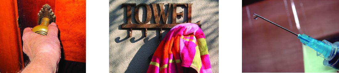
Why think in a chain
Infection is never a single event. For a microbe to make a new person sick, a whole sequence of things has to line up: the microbe must exist, it must be living somewhere, it must get out of that somewhere, travel by some route, get into a new person, and find that person vulnerable enough to take hold. Epidemiologists picture this sequence as the chain of infection — six links that must all connect for transmission to succeed. The reason this model matters so much to nurses is its most hopeful feature: break any one link and the whole chain fails. You do not have to sterilize the world; you just have to interrupt the microbe at the weakest, most reachable point.
The six links
Read the chain in order and you can see where a nurse stands in it. The infectious agent is the microbe itself. Its reservoir is the place it normally lives and multiplies — a person, an animal, the soil, a sink drain. The portal of exit is how it leaves the reservoir (respiratory droplets, stool, blood, wound drainage). The mode of transmission is the route it travels (touch, droplets, air, a shared object, an insect). The portal of entry is how it gets into the next person — often the very same routes running in reverse, plus the artificial doorways we create with catheters, IV lines, and surgical incisions. Finally, the susceptible host is a person whose defenses are low enough to be colonized and infected. Notice how many links live on the nurse's side of the bed.
| Link | What it is | Nursing action that breaks it |
|---|---|---|
| Infectious agent | The pathogen (bacterium, virus, fungus, parasite) | Appropriate antimicrobials; treat & identify the organism |
| Reservoir | Where the microbe lives & multiplies | Clean the environment; treat carriers; remove standing water & soiled linen |
| Portal of exit | How it leaves the reservoir | Cover coughs; contain drainage & body fluids; dressings over wounds |
| Mode of transmission | The route to a new host | Hand hygiene; PPE; isolation; disinfect shared equipment |
| Portal of entry | How it enters the next person | Aseptic technique; catheter & line care; keep skin intact; remove devices early |
| Susceptible host | A person with lowered defenses | Vaccinate; nutrition; glycemic control; treat underlying illness |
- Transmission requires six links to connect: agent → reservoir → portal of exit → mode of transmission → portal of entry → susceptible host.
- Breaking any single link stops the infection — you don't have to break them all.
- The mode of transmission link is the one nurses control most directly, and hand hygiene is its master key.
- Devices (catheters, lines, tubes) create new portals of entry that don't exist in nature.
Section 2Reservoirs & transmission
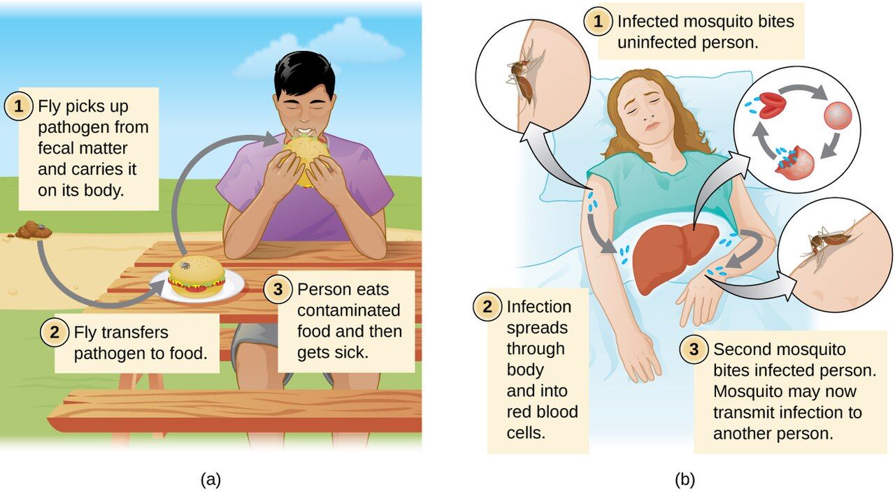
Where microbes hide: reservoirs
A reservoir is the habitat where a pathogen normally lives, grows, and multiplies, waiting for a chance to spread. Reservoirs come in three kinds. Human reservoirs include people who are visibly sick, but also — and more dangerously — carriers, people who harbor and shed a microbe without symptoms. "Typhoid Mary" (Mary Mallon) infected roughly 50 people — three of whom died — across the households and institutions she cooked for, all while feeling perfectly well; today, an asymptomatic staff member colonized with MRSA can do something similar. Animal reservoirs give us zoonoses — diseases that jump from animals to humans, such as rabies, Lyme disease, and salmonellosis. Environmental reservoirs are non-living: soil harbors Clostridium tetani and fungal spores; water harbors Legionella, which famously breeds in cooling towers and warm hospital plumbing.
How microbes travel: the five modes
Once a microbe exits its reservoir it needs a route to the next host, and there are five to know. Contact is the most common: direct contact means skin-to-skin or body-fluid transfer (a handshake, a kiss, sex), while indirect contact runs through a contaminated object called a fomite — a bed rail, a stethoscope, a shared glucometer. Droplet transmission carries large respiratory droplets a short distance (classically within about 1–2 meters, roughly 3–6 feet) when someone coughs, sneezes, or talks; the droplets are too heavy to float and fall quickly. Airborne transmission is the dangerous cousin: tiny droplet nuclei stay suspended in the air for long periods and travel on air currents, so tuberculosis, measles, and varicella can infect someone who was never near the patient. Vehicle transmission uses a shared medium — food, water, blood, or IV fluids — to reach many people at once (a single contaminated batch causes an outbreak). Vector transmission uses a living carrier, usually an arthropod: mechanically (a fly tracking microbes on its feet) or biologically (a mosquito in which the pathogen actually multiplies, as with malaria).
| Mode | How it spreads | Classic example | Precaution |
|---|---|---|---|
| Contact (direct) | Skin-to-skin or fluid-to-fluid | MRSA, scabies, herpes | Gloves & gown; hand hygiene |
| Contact (indirect) | Via a contaminated fomite | C. difficile, norovirus, RSV | Disinfect shared equipment |
| Droplet | Large droplets, short range (<2 m) | Influenza, pertussis, meningococcus | Surgical mask; ~2 m spacing |
| Airborne | Tiny nuclei suspended & drifting | Tuberculosis, measles, varicella | N95; negative-pressure room (add gown & gloves for varicella) |
| Vehicle | Shared food, water, blood, fluids | Salmonella, hepatitis A, HIV | Food/water safety; blood screening |
| Vector | Living carrier (usually an arthropod) | Malaria, Lyme, West Nile | Repellent; nets; vector control |
- A reservoir is where a microbe lives; reservoirs are human (including symptom-free carriers), animal (zoonoses), or environmental (soil, water).
- The five modes are contact (direct/indirect via a fomite), droplet, airborne, vehicle, and vector.
- Droplets fall within ~2 m (mask); airborne nuclei drift (N95 + negative-pressure room).
- Vehicle transmission can infect many people from one contaminated source — the signature of a point-source outbreak.
Section 3Epidemiology basics
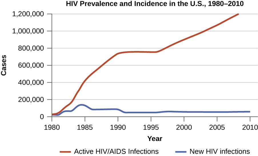
Counting disease: incidence and prevalence
Epidemiology is the study of how disease is distributed in populations and what drives that distribution — the science behind every outbreak investigation and every public-health decision. Its two most fundamental numbers are easy to mix up. Incidence is the number of new cases that appear in a population during a set period of time; it measures how fast a disease is arriving. Prevalence is the total number of cases — new and old — present at a given moment; it measures how much disease is out there right now. A brief, intense illness like influenza has high incidence during flu season but low prevalence at any snapshot, because people recover quickly. A chronic condition like HIV has low incidence but high and climbing prevalence, because cases accumulate and persist. Two more terms round out the vocabulary: morbidity is the amount of illness in a population, and mortality is the amount of death.
Patterns over place and time: endemic, epidemic, pandemic
Epidemiologists also describe the pattern a disease shows. A disease is endemic when it is constantly present in a region at a fairly steady, expected level — the common cold is endemic everywhere; malaria is endemic in parts of the tropics. An epidemic (often called an outbreak when it is smaller or more local) is a rise in cases above the expected baseline for that place and time. When an epidemic crosses countries and continents to affect a large part of the world, it is a pandemic — COVID-19 being the defining example of this generation. A disease that appears only occasionally and irregularly, with no steady pattern, is called sporadic. These are not just labels: the moment case counts climb above the endemic baseline, public-health systems switch from routine surveillance into outbreak response.
| Term | Meaning | Example |
|---|---|---|
| Incidence | New cases in a period | 1,200 new flu cases this week |
| Prevalence | All current cases at a moment | 38 million people living with HIV |
| Sporadic | Occasional, irregular cases | Tetanus in a vaccinated country |
| Endemic | Steady, expected background level | Malaria in tropical regions |
| Epidemic / outbreak | Cases above the expected baseline | A measles outbreak in a school |
| Pandemic | Epidemic spanning countries/continents | COVID-19, 1918 influenza |
- Incidence counts new cases over time; prevalence counts all existing cases at a moment.
- Short, curable illnesses have high incidence but low prevalence; chronic ones have low incidence but high prevalence.
- Endemic = steady baseline; epidemic/outbreak = above baseline; pandemic = epidemic across many countries.
- Morbidity is illness; mortality is death — both are core public-health measures.
Section 4Healthcare-associated infections
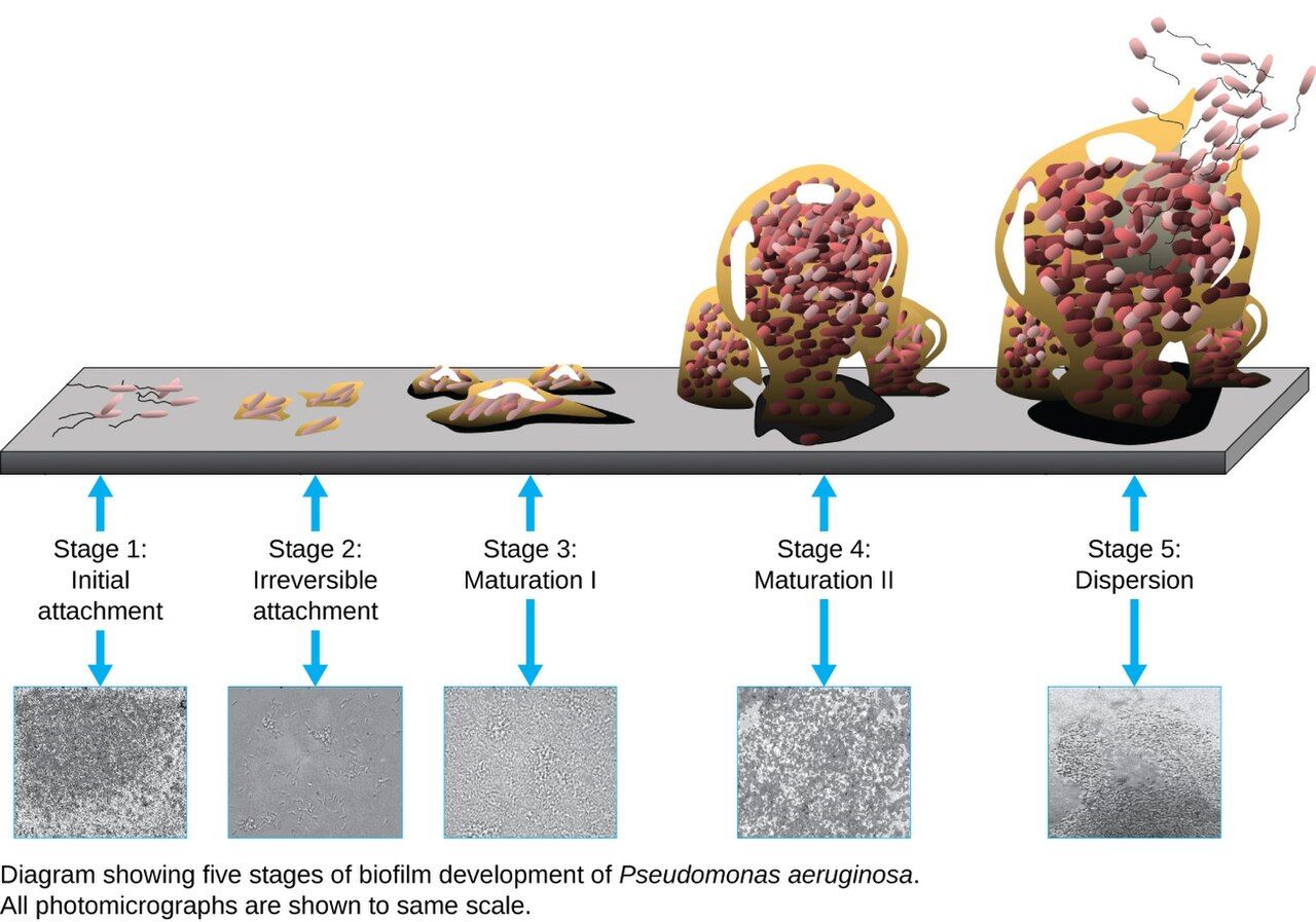
The infections we hand to patients
A healthcare-associated infection (HAI) is an infection a patient acquires while receiving care — one they did not have, and were not incubating, on admission. HAIs are common: on any given day, roughly 1 in 31 hospitalized patients in the United States has at least one. They are also, for the most part, preventable, which is exactly why they are tracked, reported, and taken so seriously. Most of the big HAIs share a villain: the biofilm. When we thread a catheter into a bladder or a line into a vein, microbes colonize the plastic and build a slimy, drug-resistant biofilm along its surface — a fortress antibiotics and immune cells struggle to breach. Every indwelling device is both a portal of entry and a biofilm scaffold, which is why the guiding rule is blunt: if the device isn't needed, it comes out.
The big four — and the bundles that stop them
Four HAIs dominate. CAUTI (catheter-associated urinary tract infection) follows the indwelling urinary catheter — the single most-used, and most over-used, device in the hospital. CLABSI (central-line–associated bloodstream infection) is less common but far deadlier, because it delivers microbes straight into the bloodstream. SSI (surgical-site infection) develops in a wound after an operation. VAP (ventilator-associated pneumonia) strikes patients whose breathing tube bypasses the natural filters of the upper airway. For each, prevention isn't one trick but a bundle — a small set of evidence-based steps done together, every time, with nothing skipped. Bundles work because HAIs are chains too, and a bundle breaks several links at once.
| HAI | Device / cause | Prevention bundle (nursing core) |
|---|---|---|
| CAUTI | Indwelling urinary catheter | Insert only if truly needed; sterile insertion; keep bag below bladder; remove ASAP |
| CLABSI | Central venous line | Full-barrier insertion; chlorhexidine skin prep; scrub the hub; daily line-necessity review |
| SSI | Surgical incision | Timely pre-op antibiotics; clippers (not razors); glucose & normothermia; sterile dressing care |
| VAP | Endotracheal tube / ventilator | Head of bed 30–45°; minimize sedation with a daily sedation interruption & readiness-to-extubate assessment; regular oral care with toothbrushing (routine chlorhexidine is no longer recommended); early enteral nutrition; change the circuit only when visibly soiled |
- An HAI is acquired during care; about 1 in 31 inpatients has one on any given day, and most are preventable.
- The big four are CAUTI, CLABSI, SSI, and VAP — three of the four are device-associated.
- Biofilms on indwelling devices shelter microbes from drugs and immune defenses.
- Bundles — and early device removal — are the proven way to drive HAI rates down.
Section 5Breaking the chain
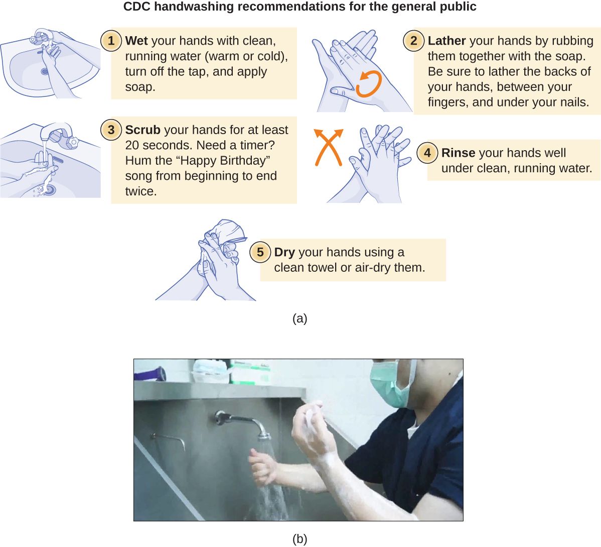
Standard precautions: the floor for everyone
Infection control rests on a two-tier system. The base tier is standard precautions, and its founding assumption is deliberately paranoid: treat the blood and body fluids of every patient as potentially infectious, regardless of diagnosis, because you cannot tell by looking who is carrying what. Standard precautions apply to all patients, all the time, and include hand hygiene before and after every contact, PPE (gloves, gown, mask, or eye protection) whenever you might contact body fluids, respiratory hygiene / cough etiquette, safe injection practices (one needle, one syringe, one time), and safe handling of soiled equipment and linens. This tier alone, done faithfully, breaks the transmission link for the majority of hospital microbes.
Transmission-based precautions: adding a tier for known threats
When a patient is known or suspected to carry a highly transmissible organism, you add a second tier layered on top of standard precautions. There are three categories, matched to the three high-risk modes of transmission. Contact precautions (gown and gloves; dedicated equipment) counter organisms spread by touch and fomites — MRSA, VRE, C. difficile, RSV. Droplet precautions (surgical mask within ~2 m) counter influenza, pertussis, and meningococcal disease. Airborne precautions (an N95 respirator and a negative-pressure room with the door closed) counter the drifting-nuclei diseases — tuberculosis, measles, varicella. A useful memory hook for the airborne set is "My Chicken Hez TB": Measles, Chickenpox (varicella), Herpes zoster (disseminated), TB. Note one wrinkle in that list: varicella and disseminated zoster require Airborne plus Contact precautions, because the vesicle fluid is infectious by direct touch and on linens and dressings — so you add gown and gloves. TB and measles are airborne only. Staff without evidence of immunity to measles or varicella should not enter those rooms at all. Choosing the wrong tier — or none — leaves the chain intact.
| Precaution | PPE / room | Example organisms |
|---|---|---|
| Standard (all patients) | Hand hygiene; PPE as risk dictates | Assume all blood/body fluids infectious |
| Contact | Gown + gloves; dedicated equipment | MRSA, VRE, C. difficile, RSV, scabies |
| Droplet | Surgical mask within ~2 m; private room | Influenza, pertussis, N. meningitidis |
| Airborne | N95 + negative-pressure room, door closed — add gown and gloves for varicella and disseminated zoster | Tuberculosis, measles (airborne only); varicella and disseminated zoster (airborne plus contact) |
- Standard precautions apply to every patient and treat all body fluids as infectious — hand hygiene is their core.
- Transmission-based precautions add a tier: Contact (gown/gloves), Droplet (mask), Airborne (N95 + negative-pressure room; add gown and gloves for varicella or disseminated zoster).
- Match the precaution to the mode: airborne threats need an N95, not a surgical mask.
- Spore formers like C. difficile require soap-and-water handwashing and bleach cleaning — alcohol gel won't do.
The chapter in one breath
- The chain of infection has six links — agent, reservoir, portal of exit, transmission, portal of entry, susceptible host — and breaking any one stops transmission.
- Reservoirs are human (including symptom-free carriers), animal (zoonoses), or environmental (soil, water).
- The five modes of transmission are contact (direct/indirect via a fomite), droplet, airborne, vehicle, and vector; droplet and airborne need different masks.
- Incidence counts new cases; prevalence counts all current cases — they answer different questions.
- Disease patterns run endemic (steady) → epidemic/outbreak (above baseline) → pandemic (across continents).
- HAIs — CAUTI, CLABSI, SSI, VAP — affect ~1 in 31 inpatients, ride on biofilm-coated devices, and fall with bundles and early device removal.
- Standard precautions protect against every patient; transmission-based precautions (Contact, Droplet, Airborne) add protection against known threats.
ReferenceWords worth knowing
A quick reference for the terms in this chapter — skim it now, or circle back whenever a word trips you up.
- Airborne precautions
- Isolation for pathogens spread by droplet nuclei; requires an N95 respirator and a negative-pressure room (TB, measles, varicella) — varicella and disseminated zoster also need contact precautions.
- Carrier
- A person who harbors and can transmit a pathogen without showing symptoms.
- CAUTI
- Catheter-associated urinary tract infection; the most common device-associated HAI.
- Chain of infection
- The six-link sequence — agent, reservoir, portal of exit, transmission, portal of entry, susceptible host — required for an infection to spread.
- CLABSI
- Central-line–associated bloodstream infection; less common but highly lethal.
- Contact precautions
- Gown, gloves, and dedicated equipment for organisms spread by touch or fomites (MRSA, VRE, C. difficile).
- Droplet precautions
- A surgical mask within close range for pathogens spread by large respiratory droplets (influenza, pertussis).
- Endemic
- Constantly present in a population at a steady, expected level.
- Epidemic (outbreak)
- A rise in cases above the expected baseline for a place and time.
- Epidemiology
- The study of how disease is distributed in populations and what determines that distribution.
- Fomite
- An inanimate object that carries a pathogen from one host to another (bed rail, stethoscope, glucometer).
- Healthcare-associated infection (HAI)
- An infection acquired while receiving care, not present or incubating on admission.
- Incidence
- The number of new cases arising in a population over a defined period.
- Pandemic
- An epidemic that spreads across countries and continents to affect much of the world.
- Portal of entry
- The route by which a pathogen enters a new host (respiratory tract, broken skin, a catheter).
- Portal of exit
- The route by which a pathogen leaves its reservoir (coughing, stool, blood, wound drainage).
- Prevalence
- The total number of existing cases in a population at a given moment.
- Reservoir
- The habitat where a pathogen normally lives and multiplies: human, animal, or environmental.
- Standard precautions
- Baseline infection control applied to all patients, treating all body fluids as potentially infectious.
- Susceptible host
- A person whose defenses are low enough to allow a pathogen to take hold.
- Vector
- A living carrier — usually an arthropod — that transmits a pathogen mechanically or biologically.
- Zoonosis
- A disease transmitted from animals to humans (rabies, Lyme disease, salmonellosis).
Check yourself
Innate Immunity
Long before your body knows the name of an invader, it is already fighting back. Innate immunity is the defense you were born with — a set of barriers, cells, and chemical alarms that respond within seconds to minutes, the same way every time, to almost anything that doesn't belong. It doesn't need to have met the microbe before, and it doesn't remember it afterward. From the skin that keeps microbes out, to the neutrophils that swarm a wound, to the fever that makes your patient miserable at 3 a.m., innate immunity is the fast, blunt, always-on first responder of the immune system. This chapter walks through its layers in the order the body meets an invader — and shows you why so much of nursing is really the art of protecting, and reading, these defenses.
By the end of this chapter you can
- Contrast innate and adaptive immunity by speed, specificity, and memory.
- Describe the first-line defenses — skin, mucous membranes, and normal flora — and how each keeps microbes out.
- Identify the major phagocytic cells and walk through the steps of phagocytosis.
- Explain the inflammatory response and relate the four cardinal signs to what is happening in the tissue.
- Describe how pyrogens reset the hypothalamic thermostat to produce a fever, and weigh its benefits and dangers.
- Summarize the outcomes of the complement cascade — opsonization, inflammation, and cytolysis.
- Explain how interferons protect cells from viral infection.
- Connect each of these defenses to concrete nursing assessments at the bedside.
Section 1First line: barriers
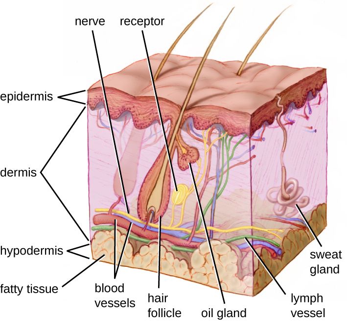
Two immune systems, one goal
Your body defends itself with two overlapping systems. Innate immunity is present from birth, responds within seconds to minutes, and treats all invaders with the same non-specific weapons — it has no memory, so the tenth exposure gets the same response as the first. Adaptive immunity (the subject of the next chapter) is slower to start, exquisitely specific to one particular microbe, and it remembers, which is why a second exposure is met faster and harder. Innate immunity buys the days that adaptive immunity needs to gear up. In practice they work as one team, but learning them separately makes each far easier to understand.
The immune system is often taught as three lines of defense, and innate immunity supplies the first two. The first line is the set of physical, chemical, and biological barriers that stop microbes from ever entering — the focus of this section. The second line is the internal response that kicks in once something breaches a barrier: phagocytes, inflammation, fever, and antimicrobial proteins (Sections 2–5). Adaptive immunity is the third line.
Skin and mucous membranes
The skin is the single largest organ of the body and the most important physical barrier there is. Its outer layer, the epidermis, is built of tightly packed cells stuffed with keratin and stacked into dead, shingled sheets that are constantly shed — taking any attached microbes with them. Most bacteria simply cannot penetrate intact skin. This is exactly why every break in the skin matters clinically: an IV site, a surgical incision, a pressure injury, or a simple scratch is a door in the wall, and the nurse's job of keeping that door clean and dressed is frontline infection control.
Where the body opens to the outside — the respiratory, digestive, urinary, and reproductive tracts — the barrier is not dry skin but a mucous membrane. These surfaces are kept moist and coated with mucus, a sticky gel that traps microbes before they can reach the cells beneath. In the airway, cells carrying tiny hair-like cilia beat in waves to sweep the trapped debris up and out of the lungs — the so-called mucociliary escalator. Tears, saliva, and the flushing action of urine physically wash microbes away. Understanding this tells you why an intubated patient, whose tube bypasses the mucociliary escalator, is at such high risk for pneumonia, and why we encourage coughing, deep breathing, and adequate hydration.
Chemical and microbial reinforcements
Barriers are not just walls; they are hostile chemical environments. The skin secretes sebum (oily, acidic) and sweat, keeping the surface at a low, unfriendly pH. Lysozyme, an enzyme found in tears, saliva, and mucus, chews apart bacterial cell walls. Stomach acid is brutally low in pH, and the vagina is kept acidic by resident lactobacilli. On top of all this sits the normal flora — the harmless microbes that already occupy the skin and mucosal surfaces. By taking up space and consuming nutrients, they crowd out would-be pathogens, a protection called competitive exclusion. Wipe out that flora — with broad-spectrum antibiotics, for instance — and opportunists like Candida or Clostridioides difficile move into the vacancy.
| First-line defense | Type | How it stops microbes |
|---|---|---|
| Skin (epidermis) | Physical | Keratinized, dry, shedding cell layers block entry |
| Mucous membranes | Physical | Trap microbes in mucus; cilia and flushing remove them |
| Sebum & sweat | Chemical | Low pH and fatty acids inhibit growth |
| Lysozyme | Chemical | Enzyme that digests bacterial cell walls |
| Stomach acid / vaginal acidity | Chemical | Low pH kills or inhibits many microbes |
| Normal flora | Biological | Competitive exclusion — crowd out pathogens |
- Innate immunity is fast, non-specific, and has no memory; adaptive is slow, specific, and remembers.
- The first line is physical, chemical, and microbial barriers that keep invaders out.
- Skin and mucous membranes are the physical walls; lysozyme, acid, and sebum are the chemical defenses.
- Normal flora protect by competitive exclusion; any break in the skin is a breach that nursing care must cover.
Section 2Second line: cells
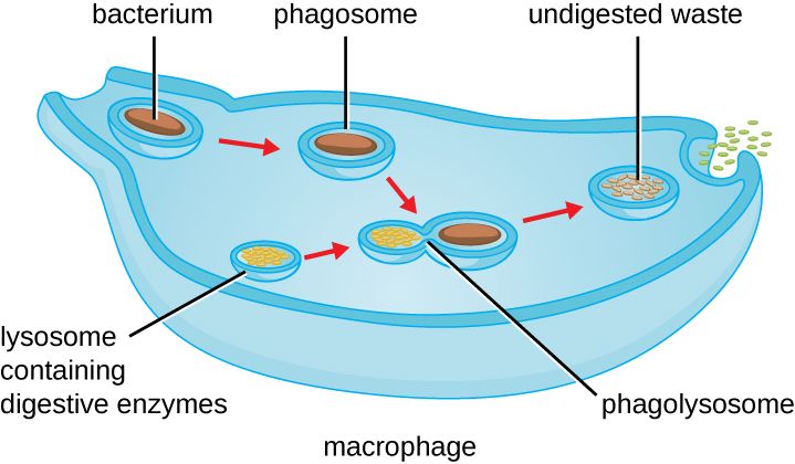
The white blood cell responders
Once a microbe slips past the barriers, it meets the cellular second line — the white blood cells, or leukocytes. These are made in the bone marrow and patrol the blood and tissues. Two of them do most of the eating, and they are the cells you must know. Neutrophils are the most numerous white cell and the first to arrive at an infection — fast, aggressive, short-lived front-line soldiers. Macrophages (from circulating monocytes that mature after leaving the blood) are the larger, longer-lived cleanup crew; they patrol the tissues, engulf microbes and debris, and help kick off other immune responses. A microbe-eating cell of either kind is called a phagocyte, and the act of eating is phagocytosis (Greek for "cell eating").
These cells don't reach a wound by accident. Chemical signals released at the site of infection — from damaged cells, from the microbes themselves, and from complement (Section 5) — draw phagocytes toward the trouble, a directed migration called chemotaxis. To get out of the bloodstream, circulating white cells first stick to the vessel wall (margination) and then squeeze between the cells of the vessel lining into the tissue, a maneuver called diapedesis (or extravasation).
How a phagocyte recognizes an enemy
How does a phagocyte tell "self" from "invader"? It carries pattern-recognition receptors (PRRs) — sensors that latch onto molecular signatures shared by whole classes of microbes but absent from human cells, called pathogen-associated molecular patterns (PAMPs). A classic PAMP is the lipopolysaccharide (LPS) in the outer membrane of Gram-negative bacteria. Because these patterns are broad, one receptor recognizes many microbes — the hallmark of innate, non-specific immunity. Recognition is made even easier when a microbe is coated with tags called opsonins (antibodies or the complement protein C3b); this coating, opsonization, is like painting a target "eat me" and dramatically speeds engulfment.
Phagocytosis, step by step
The killing sequence is worth memorizing because it recurs throughout immunology. (1) Chemotaxis — the phagocyte migrates toward the microbe. (2) Adherence — it binds the microbe, helped by opsonins. (3) Ingestion — the cell membrane wraps around the microbe and pinches off an internal bubble called a phagosome. (4) Fusion — the phagosome merges with a lysosome (full of digestive enzymes) to form a phagolysosome. (5) Killing and digestion — enzymes, reactive oxygen (the "respiratory burst"), and low pH destroy the microbe. (6) Exocytosis — the indigestible remains are dumped back out. The spent neutrophils, dead microbes, and tissue fluid left behind are what we recognize as pus.
| Cell | Comes from | Role |
|---|---|---|
| Neutrophil | Bone marrow (granulocyte) | First responder; most abundant WBC; fast, short-lived phagocyte |
| Monocyte → macrophage | Bone marrow; matures in tissue | Large, long-lived phagocyte; cleanup and immune signaling |
| Dendritic cell | Bone marrow | Phagocyte that alerts adaptive immunity |
| Natural killer (NK) cell | Lymphoid line | Kills virus-infected and tumor cells (not classic phagocytosis) |
| Eosinophil | Bone marrow (granulocyte) | Attacks parasites; role in allergy |
- Neutrophils are the first, most numerous phagocytes; macrophages (from monocytes) are the long-lived cleanup phagocytes.
- Phagocytes reach infection by chemotaxis, margination, and diapedesis.
- They recognize microbes by PRRs binding PAMPs, and engulf opsonized microbes fastest.
- Phagocytosis: chemotaxis → adherence → ingestion (phagosome) → fusion (phagolysosome) → digestion → exocytosis; leftover debris is pus.
Section 3Inflammation
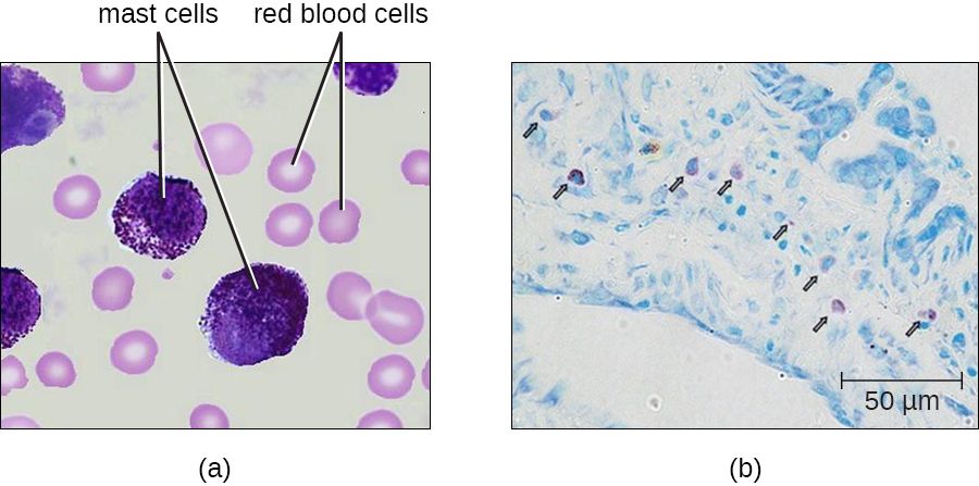
The four cardinal signs
Inflammation is the tissue's coordinated reaction to injury or infection, and it produces four classic signs that have been recognized since antiquity: redness, heat, swelling, and pain (in Latin, rubor, calor, tumor, dolor). A fifth is often added: loss of function (functio laesa). Far from being the enemy, these signs are the visible proof that the second line has arrived and is working. When you assess a wound, an IV site, or a joint, you are reading the four cardinal signs directly — they are the everyday language of infection assessment.
What is actually happening in the tissue
Each sign has a mechanism. When cells are injured or microbes are detected, resident mast cells and damaged tissue release chemical mediators — above all histamine, along with prostaglandins, bradykinin, and cytokines. These do two things. First, they cause vasodilation, widening the blood vessels so more blood floods the area — that extra warm blood is the redness and the heat. Second, they increase vascular permeability, making the vessel walls leaky so fluid, proteins, and cells pour out into the tissue — that accumulation of fluid (edema) is the swelling. The pressure of that swelling on nerve endings, along with mediators like prostaglandins and bradykinin that sensitize those nerves, produces the pain. The whole point is to deliver phagocytes and clotting proteins to the site and to wall the infection off with a mesh of fibrin so it cannot spread.
| Cardinal sign | Latin | What causes it |
|---|---|---|
| Redness | rubor | Vasodilation — more blood flowing in |
| Heat | calor | Warmth of that increased blood flow |
| Swelling | tumor | Fluid leaking from leaky vessels (edema) |
| Pain | dolor | Pressure on nerves plus pain-sensitizing mediators |
| Loss of function | functio laesa | Swelling and pain limit use of the part |
Acute, chronic, and when inflammation turns dangerous
Acute inflammation is short-lived and resolves as the threat is cleared. When a threat can't be cleared, inflammation becomes chronic: macrophages persist and may wall off the invader in a ball of immune cells called a granuloma (classically in tuberculosis). Localized inflammation is protective — but when the same mediators spill into the bloodstream system-wide, the result can be catastrophic. In sepsis, a body-wide inflammatory response to infection causes widespread vasodilation and leaky vessels, dropping the blood pressure and starving organs of oxygen. The very tools that heal a local wound, unleashed everywhere at once, can kill.
- The four cardinal signs are redness, heat, swelling, and pain (plus loss of function).
- Mast cells and injured tissue release histamine and other mediators that cause vasodilation and increased vascular permeability.
- Inflammation delivers phagocytes and walls off infection with fibrin.
- Localized inflammation heals; systemic inflammation (sepsis) is life-threatening.
Section 4Fever
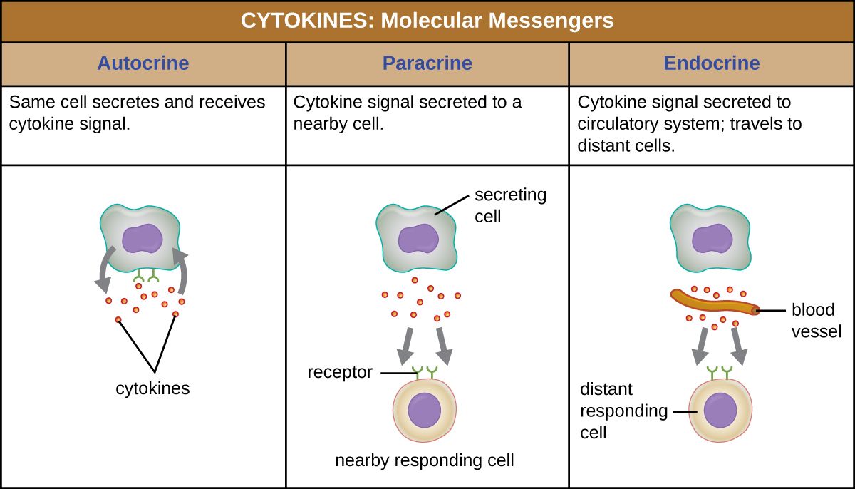
Resetting the thermostat
Fever (pyrexia) is an abnormally high core body temperature, and it is not the infection getting the upper hand — it is a deliberate, regulated defense. Your temperature is controlled by a "thermostat" in the hypothalamus, which normally holds the body near 37°C (98.6°F). Fever happens when substances called pyrogens raise the hypothalamic set-point. There are two kinds. Exogenous pyrogens come from outside the body — most famously the LPS of Gram-negative bacteria (also called endotoxin). Endogenous pyrogens are made by the patient's own immune cells: the cytokines interleukin-1 (IL-1), IL-6, and tumor necrosis factor (TNF), released by macrophages when they meet a microbe. These pyrogens drive the production of prostaglandin E₂ in the hypothalamus, which is what actually pushes the set-point up.
Here is the key idea: once the set-point is raised, the body isn't "overheating" — it now believes 39°C is the correct temperature and works to reach it. That is why the onset of a fever brings shivering and a feeling of cold: muscles contract to generate heat and blood vessels constrict to conserve it, all to climb toward the new, higher target.
The three phases of a fever
A fever runs a predictable course, and recognizing the phase changes what you do. In the onset (chill phase), the set-point jumps; the patient feels cold, shivers, and has cool, pale skin as the body drives its temperature up. In the plateau (stadium), the body has reached the new set-point — the patient feels warm, flushed, and dry, and the temperature holds steady at its elevated level. In defervescence (the flush or crisis), pyrogens fall and the set-point drops back to normal; now the body is "too hot," so it sweats and flushes to shed the excess heat. Reading these phases is practical. A shivering patient is climbing toward a set-point the hypothalamus has already chosen, and external warmth will not push the temperature past it — so cover the patient, add blankets, and cut the drafts to stop the shivering, because shivering thermogenesis sharply raises oxygen consumption, metabolic rate, and cardiac workload in exactly the cardiac, respiratory, and septic patients who can least afford it. What you must not do in the chill phase is apply cooling: tepid sponging or a cooling blanket while the patient is still shivering fights the set-point and makes the shivering worse. Cooling measures belong to the plateau and defervescence phases; antipyretics work differently, by lowering the set-point itself.
| Phase | Set-point | What you see | Nursing note |
|---|---|---|---|
| Onset (chill) | Rising | Shivering, cold, pale, "goosebumps" | Temp is climbing; warm the patient and add blankets to stop the shivering — do not cool now |
| Plateau (stadium) | Reached (high) | Warm, flushed, dry skin | Watch hydration; temp is holding high |
| Defervescence (flush) | Falling | Sweating, flushed, feels hot | Replace fluids lost in sweat |
Friend or foe?
A moderate fever helps: higher temperatures slow the growth of many pathogens, speed up the enzymes of the immune response, and enhance the activity of phagocytes and other defenders. That is why routinely erasing every low fever with antipyretics is not always wise — the fever is doing work. But fever also has costs and limits. It raises metabolic demand, the heart rate, and fluid loss; in young children a rapid rise can trigger a febrile seizure; and a very high temperature — roughly above 40–41°C (104–106°F) — begins to denature the body's own proteins and becomes a medical emergency. Antipyretics such as acetaminophen and NSAIDs work by blocking prostaglandin production, lowering the set-point back down.
- Fever is a regulated rise in the hypothalamic set-point, not loss of control.
- Exogenous pyrogens (e.g., LPS) and endogenous pyrogens (cytokines IL-1, IL-6, TNF) act via prostaglandin E₂.
- Phases: onset (chills, climbing) → plateau (steady, high) → defervescence (sweating, falling).
- Moderate fever aids defense; very high fever (>40°C) is dangerous, and some patients can't mount one at all.
Section 5Complement & interferons
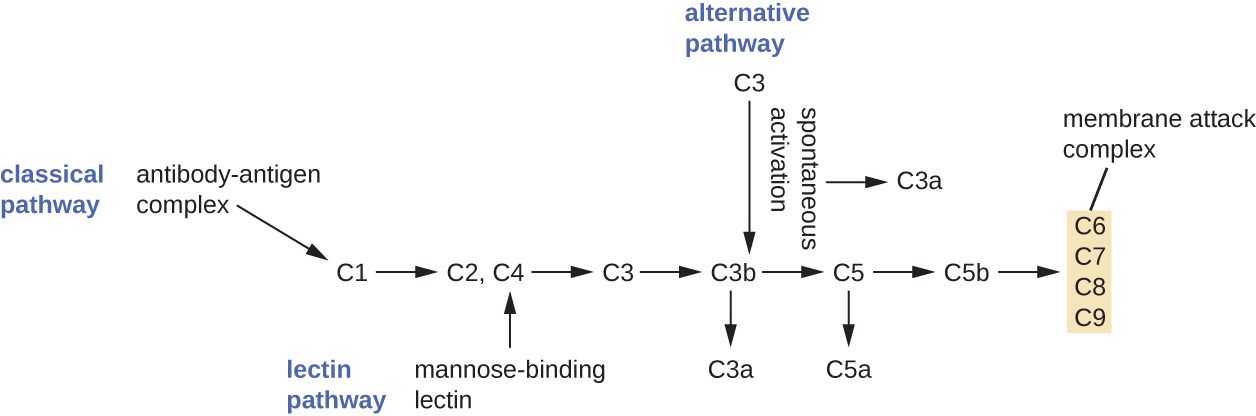
Complement: a cascade of ready-made proteins
Complement is a set of about 30 blood proteins, made mostly by the liver, that circulate in an inactive form and wait. When triggered, they activate one another in sequence — a cascade, where each activated protein cuts and activates the next, amplifying the signal at every step. Three pathways can set it off: the classical pathway (triggered by antibodies bound to a microbe), the alternative pathway (triggered directly by microbial surfaces), and the lectin pathway (triggered by carbohydrates on microbes). All three converge on the central protein, C3, which is split into C3a and C3b — the hinge of the whole system.
From there, complement produces three life-saving outcomes. Opsonization: C3b coats the microbe as an opsonin, tagging it so phagocytes grab it far more easily (linking this section straight back to phagocytosis). Inflammation: the fragments C3a and C5a are anaphylatoxins and chemotactic factors — they trigger mast cells to release histamine and they call phagocytes to the scene. Cytolysis: the terminal proteins assemble into a ring called the membrane attack complex (MAC), which punches a hole in the microbe's membrane so its contents leak out and it bursts. Together, these are sometimes remembered as the three jobs of complement: tag, recruit, and destroy.
| Complement outcome | Key players | What it does |
|---|---|---|
| Opsonization | C3b | Coats the microbe so phagocytes engulf it faster |
| Inflammation | C3a, C5a | Anaphylatoxins — trigger histamine release and attract phagocytes |
| Cytolysis | MAC (C5b–C9) | Forms pores that lyse the microbe |
Interferons: the antiviral alarm
Barriers, phagocytes, and complement are formidable against bacteria — but a virus hides inside a host cell, where those defenses can't reach. For viruses, innate immunity has a different tool: interferons (IFNs), a family of cytokines named because they "interfere" with viral replication. When a cell is infected by a virus, it releases type I interferons (IFN-α and IFN-β). These don't save the dying cell that made them; instead they act as a warning to neighboring cells. A neighbor that receives the interferon signal switches on an antiviral state — it produces enzymes that degrade viral genetic material and shut down protein synthesis if a virus enters — so the virus finds no factory to hijack. A separate interferon, IFN-γ, mainly activates macrophages and bridges to adaptive immunity.
This matters clinically in two directions. The malaise, muscle aches, and fever of a viral illness like influenza are largely the effects of your own interferons working, not the virus directly. And because interferons are natural antivirals and immune modulators, purified versions are used as medications — for certain viral hepatitis infections, some cancers, and multiple sclerosis.
- Complement is a cascade of ~30 proteins; three pathways (classical, alternative, lectin) converge on C3.
- Its three outcomes are opsonization (C3b), inflammation (C3a/C5a), and cytolysis (the MAC).
- Interferons are antiviral cytokines: a virus-infected cell releases type I IFN to put neighboring cells into an antiviral state.
- Interferons cause much of the "sick" feeling of viral illness and are used as drugs.
The chapter in one breath
- Innate immunity is the fast, non-specific, memory-free defense you are born with; it buys time for adaptive immunity.
- The first line keeps microbes out: skin and mucous membranes, chemical defenses (lysozyme, acid, sebum), and normal flora.
- When barriers fail, phagocytes — neutrophils first, then macrophages — find microbes by chemotaxis and destroy them by phagocytosis.
- Inflammation (redness, heat, swelling, pain) comes from histamine-driven vasodilation and leaky vessels; systemic, it becomes sepsis.
- Fever is a raised hypothalamic set-point driven by pyrogens (LPS; cytokines IL-1/IL-6/TNF) via prostaglandin E₂ — helpful in moderation, dangerous when very high.
- Complement tags (opsonization), recruits (inflammation), and destroys (MAC); interferons warn neighboring cells to resist viruses.
- Nearly every innate defense maps onto a bedside assessment — skin integrity, the WBC differential, wound signs, temperature phase, and sepsis recognition.
ReferenceWords worth knowing
A quick reference for the terms in this chapter — skim it now, or circle back whenever a word trips you up.
- Antipyretic
- A drug (e.g., acetaminophen, an NSAID) that lowers fever by blocking prostaglandin production and resetting the hypothalamic set-point down.
- Chemotaxis
- The directed movement of a cell, such as a phagocyte, toward a chemical signal released at a site of infection.
- Complement
- A cascade of about 30 blood proteins that, when activated, cause opsonization, inflammation, and cell lysis.
- Cytokine
- A signaling protein released by cells to communicate; includes interleukins, interferons, and TNF.
- Diapedesis
- The squeezing of white blood cells between the cells of a vessel wall to enter infected tissue; also called extravasation.
- Fever (pyrexia)
- A regulated rise in core body temperature caused by pyrogens raising the hypothalamic set-point.
- Inflammation
- The tissue response to injury or infection, marked by redness, heat, swelling, and pain.
- Innate immunity
- The non-specific defenses present from birth that respond rapidly and without memory.
- Interferon (IFN)
- An antiviral cytokine; a virus-infected cell releases type I IFN to put neighboring cells into an antiviral state.
- Lysozyme
- An enzyme in tears, saliva, and mucus that digests bacterial cell walls.
- Macrophage
- A large, long-lived phagocyte derived from a monocyte; cleans up microbes and debris and signals other immune cells.
- Membrane attack complex (MAC)
- The ring of terminal complement proteins (C5b–C9) that forms pores and lyses a target cell.
- Neutrophil
- The most abundant white blood cell and the first phagocyte to arrive at an infection.
- Opsonization
- The coating of a microbe with tags (antibody or C3b) that make phagocytosis faster and easier.
- PAMP
- Pathogen-associated molecular pattern — a molecular signature (e.g., LPS) shared by many microbes and recognized by innate receptors.
- Pattern-recognition receptor (PRR)
- A receptor on innate immune cells that binds PAMPs to detect microbes.
- Phagocytosis
- The engulfment and destruction of a microbe by a phagocyte inside a phagolysosome.
- Pyrogen
- A fever-inducing substance; exogenous (e.g., LPS) or endogenous (cytokines such as IL-1, IL-6, TNF).
- Sepsis
- A dysregulated, body-wide inflammatory response to infection that can cause dangerously low blood pressure and organ failure.
- Vasodilation
- Widening of blood vessels; in inflammation it increases blood flow, causing redness and heat.
Check yourself
Adaptive Immunity & Immunization
Your innate defenses are fast, but they are blunt — they treat every microbe the same and they never learn. Adaptive immunity is the opposite: slow to start, exquisitely precise, and blessed with memory. It is the arm of the immune system that recognizes one specific microbe out of millions, mounts a tailored attack, and then remembers that microbe for years or a lifetime. This chapter follows the two great cell lineages that make it work — T cells and B cells — explains how antibodies are built and what they do, and shows why a second exposure is so much faster than the first. That last fact is the whole reason vaccines work, so we finish where nursing practice lives: the immunizations you will draw up, document, and defend to families.
By the end of this chapter you can
- Contrast innate and adaptive immunity, and name the three hallmarks of the adaptive response: specificity, memory, and self-tolerance.
- Define antigen and epitope, and explain what it means to distinguish self from non-self.
- Distinguish helper (CD4) from cytotoxic (CD8) T cells and describe the role of MHC molecules in showing antigen to them.
- Describe how a B cell becomes a plasma cell, and diagram the basic antibody structure.
- List the five antibody classes and the main jobs antibodies do — neutralization, opsonization, agglutination, and complement activation.
- Compare the primary and secondary antibody responses and explain immunologic memory.
- Classify immunity as active or passive and natural or artificial, and match each to a clinical example.
- Describe the major vaccine types and the nursing implications of live versus non-live vaccines.
Section 1The adaptive response
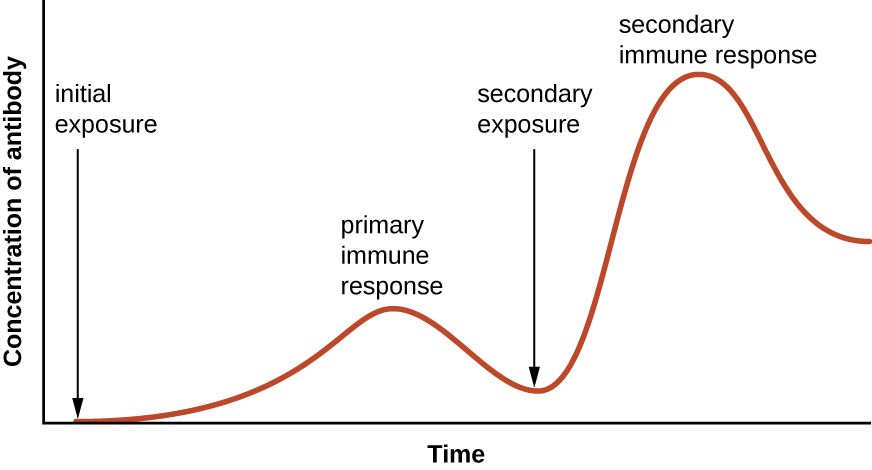
Two systems, one defense
Your immune system runs on two teams that work together. Innate immunity — skin, mucus, stomach acid, fever, phagocytes, and inflammation — is present from birth, responds within minutes to hours, and reacts the same way to almost any invader. It buys time. Adaptive immunity is the specialist team called in behind it. It takes days to gear up the first time, but it recognizes one particular microbe with extraordinary precision, and once the fight is over it keeps a record so that the next encounter is faster and stronger. Nearly everything protective about a vaccine, a childhood illness you never catch twice, and a positive antibody titer comes from this second system.
Adaptive immunity itself splits into two branches. The humoral (antibody-mediated) response is run by B lymphocytes, which secrete antibodies into blood and body fluids to attack microbes outside of cells — bacteria in the bloodstream, toxins, viruses in transit. The cell-mediated response is run by T lymphocytes, which handle threats inside cells, especially virus-infected cells and cancer cells. Both branches begin when the body detects something foreign.
Antigens, epitopes, and the meaning of “foreign”
An antigen is any molecule the immune system can recognize and respond to — usually a protein or polysaccharide on the surface of a microbe. Adaptive cells do not read the whole antigen at once; each one recognizes a small, specific patch called an epitope. A single bacterium carries many different epitopes, which is why one infection can trigger many different lymphocytes at the same time. The precision here is staggering: your body pre-builds millions of different lymphocytes, each tuned to a different epitope, before it has ever met the microbe. When a matching antigen shows up, the rare lymphocyte that fits it is selected and told to multiply — a process called clonal selection.
Specificity, memory, and tolerance
Three properties define adaptive immunity. Specificity means each response is aimed at one antigen and not its neighbors — measles immunity does nothing for mumps. Memory means the system remembers a past antigen and reacts faster and harder on re-exposure; this is the property vaccines exploit. Self-tolerance means the system learns, during development, to ignore the body's own molecules (self) while attacking foreign ones (non-self). Lymphocytes that would attack self are normally deleted or silenced early on. When that quality-control step fails, the immune system turns on the body — the basis of autoimmune disease such as type 1 diabetes, rheumatoid arthritis, and lupus.
| Feature | Innate immunity | Adaptive immunity |
|---|---|---|
| Present when? | From birth, always ready | Develops after exposure to an antigen |
| Speed | Minutes to hours | Days on first exposure |
| Specificity | Broad — same response to many microbes | Exquisite — one antigen at a time |
| Memory? | No — reacts the same every time | Yes — faster & stronger on re-exposure |
| Key cells | Phagocytes, NK cells, mast cells | T lymphocytes and B lymphocytes |
- Innate immunity is fast and non-specific; adaptive immunity is slower, highly specific, and remembers.
- Adaptive immunity has two arms: humoral (B cells, antibodies, extracellular threats) and cell-mediated (T cells, intracellular threats).
- An antigen is recognized at a small patch called an epitope; clonal selection multiplies the lymphocyte that fits.
- The three hallmarks are specificity, memory, and self-tolerance; loss of tolerance causes autoimmunity.
Section 2T cells
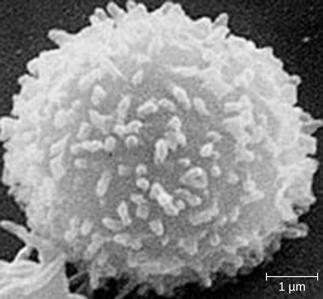
Where T cells come from and how they “see”
T lymphocytes are born in the bone marrow but finish their training in the thymus (the T is for thymus), where the dangerous ones — those that would attack self — are weeded out. A mature T cell cannot recognize a free-floating antigen the way an antibody can. It only reacts to antigen that has been chopped up and displayed on a special surface molecule called MHC (major histocompatibility complex). Think of MHC as a serving tray: a cell holds up a fragment of protein on its MHC, and a passing T cell inspects it. This is called antigen presentation, and it is the reason T cells can tell an infected cell from a healthy one.
There are two classes of tray. MHC class I is found on almost every nucleated cell in the body and displays fragments of whatever is being made inside — so a virus-infected cell involuntarily advertises the virus. MHC class II is found only on professional antigen-presenting cells (dendritic cells, macrophages, and B cells), which deliberately swallow microbes and display their pieces to recruit help.
Helper T cells — the conductors (CD4)
Helper T cells carry a surface marker called CD4 and read antigen presented on MHC class II. They do not kill anything directly; they orchestrate. When a helper T cell recognizes its antigen, it releases chemical messengers (cytokines) that switch on the rest of the response: they push B cells to make antibody, amplify cytotoxic T cells, and rev up macrophages. Because they sit at the center of the network, losing them is catastrophic — which is exactly what makes HIV so devastating.
Cytotoxic T cells — the assassins (CD8)
Cytotoxic T cells carry CD8 and read antigen on MHC class I. When one recognizes a cell displaying foreign (viral or tumor) protein on its class I tray, it delivers the kill: it punches holes in the target and triggers it to self-destruct (apoptosis), taking the virus factory down with it. This is the front line against viral infections and abnormal cells. A handy memory aid is the “rule of eight”: CD4 × MHC II = 8, and CD8 × MHC I = 8 — each T-cell type pairs with the MHC class whose number multiplies to eight.
| Feature | Helper T cell | Cytotoxic T cell |
|---|---|---|
| Surface marker | CD4 | CD8 |
| Reads antigen on | MHC class II | MHC class I |
| Main job | Coordinate & amplify the response via cytokines | Directly kill infected or abnormal cells |
| Best against | Directing all responses (works with B cells too) | Virus-infected cells, tumor cells |
| Clinical note | Target of HIV; count tracks immune status | Key defense in viral illness |
- T cells mature in the thymus and recognize antigen only when it is presented on MHC.
- Helper T cells (CD4) read MHC II and coordinate the response with cytokines; they do not kill directly.
- Cytotoxic T cells (CD8) read MHC I and destroy infected or abnormal cells.
- MHC I is on nearly all cells; MHC II is only on antigen-presenting cells. HIV depletes CD4 cells, crippling adaptive immunity.
Section 3B cells & antibodies
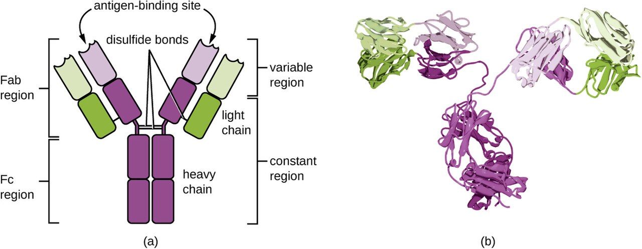
From B cell to antibody factory
B lymphocytes mature in the bone marrow (a helpful coincidence: B for bone marrow). Each B cell carries a unique receptor on its surface, and when that receptor binds its matching antigen — usually with a push from a helper T cell — the B cell activates and divides. Most of its offspring become plasma cells, short-lived factories that pump out thousands of identical antibodies per second. A smaller share become memory B cells that linger for years, ready for a repeat encounter (the subject of the next section). Antibodies are also called immunoglobulins (Ig), and they are the working product of the humoral response.
Antibody structure: the Y
Every antibody has the same basic shape: a Y built from four protein chains — two identical long heavy chains and two identical short light chains, held together by disulfide bonds. The two tips of the Y are the variable regions: their amino-acid sequence differs from antibody to antibody, giving each its unique antigen-binding site. The stem of the Y is the constant region, which is the same within a class and acts as a handle that other immune components — phagocytes and complement proteins — can grab. So one end of the molecule does the recognizing and the other end does the recruiting.
What antibodies actually do
Antibodies rarely destroy a microbe by themselves. Instead they tag and disable it in four main ways. In neutralization, antibodies coat a virus or toxin so it can no longer attach to host cells — this is how antitoxins and many vaccine antibodies protect. In opsonization, antibodies coat a microbe so phagocytes grab it more easily (the constant stem is the “handle”). In agglutination, antibodies cross-link many microbes into clumps that are easier to clear and can no longer spread. And in complement activation, clustered antibodies trigger the complement cascade, which can punch holes in bacterial membranes. The five classes below differ in their constant stems, and therefore in where they go and what they do.
| Class | Where / role | Clinical pearl |
|---|---|---|
| IgG | Most abundant in blood; long-term protection; crosses the placenta | Signals a past or resolved infection / established immunity; protects the newborn |
| IgM | First antibody made in a new infection; large, stays in blood | A positive IgM suggests a current or recent infection |
| IgA | Guards mucous membranes & secretions; found in breast milk | Passes protection to the breastfeeding infant's gut |
| IgE | Binds mast cells; drives allergy & parasite defense | The antibody behind hay fever and anaphylaxis |
| IgD | On the surface of naive B cells | Helps activate the B cell; least understood clinically |
- B cells mature in bone marrow; activated B cells become antibody-secreting plasma cells and long-lived memory B cells.
- An antibody is a Y: variable tips recognize antigen; the constant stem recruits phagocytes and complement.
- Antibodies work by neutralization, opsonization, agglutination, and complement activation.
- The five classes are IgG, IgM, IgA, IgE, IgD; remember IgM = new infection, IgG = past/lasting, IgA = mucosa/milk, IgE = allergy.
Section 4Primary vs secondary response
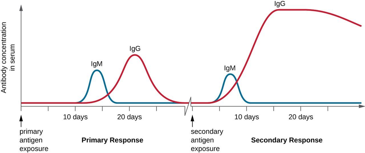
The primary response: slow and costly
The first time the body meets a new antigen, it must find the one rare lymphocyte that fits, activate it, and let it multiply before any meaningful defense appears. This primary response has a lag of roughly one to two weeks, produces a modest amount of antibody, and is led by IgM, the first-responder class, before IgG builds up. During that lag the patient is often sick — the immune system is fighting from behind. This delay is exactly why a brand-new infection can be dangerous and why an unvaccinated person has no head start.
Memory cells: the immune system's notebook
As the primary response resolves, it does not simply pack up. It leaves behind memory B cells and memory T cells — long-lived cells, sometimes surviving for decades, that carry the receptor for that one antigen. They circulate quietly, primed. Memory is the physical basis of immunity: it is what “having had” a disease or a vaccine actually means at the cellular level.
The secondary response: fast and fierce
When the same antigen returns, memory cells are already waiting, so the body skips most of the search-and-activate delay. The secondary response begins in hours to a few days, produces far more antibody, and is dominated by high-affinity IgG. Often the microbe is cleared before symptoms even appear — which is why you may never know you were re-exposed. Every booster shot deliberately triggers a secondary response to push antibody levels back up and refresh the memory pool.
| Feature | Primary response | Secondary response |
|---|---|---|
| Trigger | First exposure to the antigen | Re-exposure to the same antigen |
| Lag before antibody | ~1–2 weeks | Hours to a few days |
| Amount of antibody | Low | Much higher |
| Dominant class | IgM first, then IgG | Mostly high-affinity IgG |
| Driven by | Naive lymphocytes | Memory B and T cells |
- The primary response is slow (1–2 week lag), small, and IgM-first.
- Memory B and T cells persist for years and are the physical basis of lasting immunity.
- The secondary response is faster, larger, and IgG-dominant — often clearing the microbe before symptoms appear.
- Boosters and titers both work through this memory: one refreshes it, the other measures it.
Section 5Immunization & vaccines
Four kinds of immunity
Clinical immunity is sorted along two axes. The first is active versus passive: in active immunity the person's own immune system makes the antibody and builds memory (long-lasting); in passive immunity ready-made antibody is given to the person, protecting them immediately but only until that borrowed antibody breaks down (no memory). The second axis is natural versus artificial: natural means it happened through ordinary life, artificial means it was delivered deliberately by a clinician. Crossing the two axes gives four categories, and every immunization you handle fits one of them.
| Type | How it happens | Example | Lasting? |
|---|---|---|---|
| Natural active | You catch the illness and recover | Immunity after chickenpox | Yes — memory forms |
| Artificial active | A vaccine teaches your immune system | MMR, hepatitis B, tetanus vaccine | Yes — memory forms |
| Natural passive | Antibody given by nature | Mother's IgG across placenta; IgA in breast milk | No — wears off in months |
| Artificial passive | Pre-made antibody injected | Antivenom, rabies/tetanus immune globulin, RhoGAM | No — temporary |
How vaccines are built
All vaccines do the same job — show the immune system an antigen so it builds memory — but they differ in how they present it, and that difference drives the nursing precautions. Live attenuated vaccines (MMR, varicella, oral polio, intranasal flu) use a weakened but living microbe; they give strong, long-lasting immunity but are generally contraindicated in pregnancy and in immunocompromised patients because even a weakened live microbe can be risky. Inactivated (killed) vaccines (hepatitis A, injectable polio, most flu shots) use a dead microbe — safer, but often needing boosters. Subunit, conjugate, and toxoid vaccines (hepatitis B, Hib, pertussis, tetanus and diphtheria toxoids) use only a purified piece or an inactivated toxin, so they cannot cause the disease at all. mRNA and viral-vector vaccines deliver the genetic instructions for a single antigen so the body makes it briefly and learns to recognize it. None of the non-live types can give the patient the actual infection.
Herd immunity and the nurse's role
When a high enough share of a community is immune, a microbe can no longer find enough susceptible hosts to spread — this is herd immunity, and it shelters the few who cannot be vaccinated (newborns, the immunocompromised, those with true contraindications). Nurses are central to keeping that number high: you administer vaccines by the correct route, observe for the rare immediate reaction, document the lot number and site, and — often most important — you answer families' questions with accurate, calm information. Vaccine hesitancy is met at the bedside far more effectively than in a headline.
- Active immunity = the body makes its own antibody & memory (lasting); passive = borrowed antibody, immediate but temporary.
- Natural vs artificial is simply whether it happened through life or was delivered by a clinician — giving four categories.
- Vaccine types: live attenuated, inactivated, subunit/conjugate/toxoid, and mRNA/vector; only live types carry an infection risk in vulnerable hosts.
- Live vaccines are generally contraindicated in pregnancy and immunocompromise; herd immunity protects those who cannot be vaccinated.
The chapter in one breath
- Adaptive immunity is slow but specific and remembers; its arms are humoral (B cells/antibodies) and cell-mediated (T cells).
- Its hallmarks are specificity, memory, and self-tolerance; lost tolerance causes autoimmunity.
- T cells read antigen on MHC: helper (CD4) cells coordinate via cytokines; cytotoxic (CD8) cells kill infected cells. HIV destroys CD4 cells.
- B cells become plasma cells that secrete antibodies — Y-shaped, with variable tips that recognize and a constant stem that recruits.
- Antibodies work by neutralization, opsonization, agglutination, and complement; classes are IgG, IgM, IgA, IgE, IgD.
- The secondary response is faster and stronger than the primary thanks to memory cells — the basis of boosters and titers.
- Immunity is active/passive and natural/artificial; vaccines create artificial active immunity, and live vaccines are avoided in pregnancy and immunocompromise.
ReferenceWords worth knowing
A quick reference for the terms in this chapter — skim it now, or circle back whenever a word trips you up.
- Active immunity
- Protection produced by the person's own immune system making antibody and memory; long-lasting.
- Adaptive immunity
- The specific, memory-forming arm of the immune system, run by T and B lymphocytes.
- Antibody (immunoglobulin)
- A Y-shaped protein made by plasma cells that binds a specific antigen to tag or neutralize it.
- Antigen
- Any molecule the immune system can recognize and respond to; recognized at a small patch called an epitope.
- Antigen-presenting cell (APC)
- A cell (dendritic cell, macrophage, or B cell) that displays antigen on MHC class II to activate helper T cells.
- Attenuated vaccine
- A vaccine using a living but weakened microbe; strong immunity but avoided in pregnancy and immunocompromise.
- CD4 (helper) T cell
- A T cell that reads antigen on MHC II and coordinates the immune response with cytokines; target of HIV.
- CD8 (cytotoxic) T cell
- A T cell that reads antigen on MHC I and kills infected or abnormal cells.
- Clonal selection
- The process by which the one lymphocyte matching an antigen is chosen and multiplied.
- Epitope
- The small, specific portion of an antigen that a lymphocyte or antibody actually binds.
- Herd immunity
- Protection of a whole community, including the non-immune, when enough members are immune to stop spread.
- Humoral immunity
- The antibody-mediated branch of adaptive immunity, driven by B cells; fights extracellular threats.
- Immunologic memory
- The persistence of memory cells that make a repeat response faster and stronger.
- Memory cell
- A long-lived B or T lymphocyte that persists after infection and enables the secondary response.
- MHC (major histocompatibility complex)
- Surface molecules that display antigen fragments to T cells; class I on most cells, class II on APCs.
- Opsonization
- Coating a microbe with antibody so phagocytes engulf it more readily.
- Passive immunity
- Protection from ready-made antibody given to the person; immediate but temporary, with no memory.
- Plasma cell
- An activated B cell that secretes large amounts of antibody.
- Primary response
- The slow, modest, IgM-first reaction to a first exposure to an antigen.
- Secondary response
- The fast, large, IgG-dominant reaction to re-exposure, driven by memory cells.
- Self-tolerance
- The immune system's learned ability to ignore the body's own molecules; its failure causes autoimmunity.
- Toxoid vaccine
- A vaccine made from an inactivated bacterial toxin, e.g., tetanus and diphtheria toxoids.
Check yourself
Immune Disorders
The immune system is your patient's greatest protector — and, when it misfires, one of their most dangerous threats. Sometimes it overreacts to something harmless, like pollen or a dose of penicillin, and the response itself does the damage. Sometimes it turns against the body's own tissues. Sometimes it is too weak to defend at all. And sometimes, when we deliberately introduce blood or an organ from another person, it attacks the very help we are trying to give. This chapter walks through these problems — hypersensitivity, allergy and anaphylaxis, autoimmunity, immunodeficiency, and transfusion and transplant reactions — and shows you what each looks like at the bedside and what nursing actions keep patients safe.
By the end of this chapter you can
- Describe what hypersensitivity is and distinguish the four types (I–IV) by mediator, timing, and mechanism.
- Explain type I allergy from sensitization through IgE, mast-cell degranulation, and anaphylaxis.
- State why epinephrine is the first-line treatment for anaphylaxis and recognize the emergency.
- Explain how self-tolerance breaks down and name common autoimmune diseases and their targets.
- Distinguish primary (congenital) from secondary (acquired) immunodeficiency.
- Summarize the course of HIV/AIDS in terms of CD4 counts and opportunistic infections.
- Explain ABO and Rh blood typing and the cause of a hemolytic transfusion reaction.
- Distinguish the major kinds of graft and describe rejection and graft-versus-host disease.
Section 1Hypersensitivity: when defense becomes damage
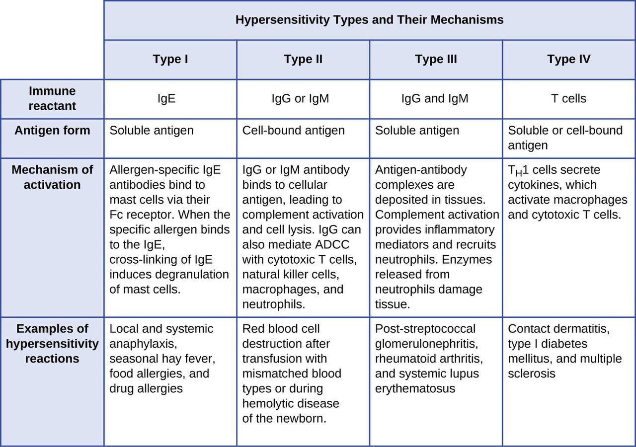
An immune response that harms the host
A healthy immune response is protective — it clears an invader and then quiets down. A hypersensitivity reaction is an immune response that is exaggerated, misdirected, or inappropriate, so that the response itself injures the body's own tissues. The trigger may be genuinely dangerous (a microbe), harmless (pollen, a food, a drug), or even part of the patient (their own cells). What all hypersensitivities share is that the damage comes from the immune system's activity, not directly from the trigger.
Almost every hypersensitivity requires a first, silent exposure called sensitization. On that first encounter the immune system does not cause symptoms; it simply learns the antigen and builds a stockpile of antibodies or primed T cells against it. The dramatic reaction happens on a later exposure, when that pre-built machinery fires. This is why a patient can take a drug uneventfully once and react violently the second time — and why "no reaction last time" never guarantees safety.
The four types (Gell and Coombs)
In the 1960s Gell and Coombs sorted hypersensitivities into four types by the immune mechanism behind them. The first three are driven by antibodies; the fourth is driven by T cells. A classic memory aid is ACID: type I Anaphylactic/allergic, type II Cytotoxic, type III Immune-complex, type IV Delayed. Types I–III happen within minutes to hours because antibodies are already present; type IV takes a day or more because T cells must be recruited to the site.
| Type | Also called | Mediator | Timing | Mechanism & example |
|---|---|---|---|---|
| I | Immediate / allergic / anaphylactic | IgE on mast cells | Seconds to minutes | Allergen cross-links IgE → mast-cell degranulation. Hay fever, food allergy, anaphylaxis |
| II | Cytotoxic | IgG / IgM vs. cell-surface antigen | Minutes to hours | Antibody binds a cell, marking it for destruction (complement, phagocytes). Hemolytic transfusion reaction, HDN, autoimmune hemolytic anemia |
| III | Immune-complex | IgG in antigen–antibody complexes | Hours | Soluble complexes deposit in vessels, joints, kidneys and inflame them. Serum sickness, SLE, some glomerulonephritis |
| IV | Delayed-type / cell-mediated | T cells (no antibody) | 1–3 days | Sensitized T cells release cytokines, recruit macrophages. Contact dermatitis (poison ivy, nickel), TB skin test |
Keep the antibody-versus-T-cell divide front of mind, because it explains the tempo you see clinically. If a patient breaks out in hives within minutes of a bee sting, you are watching type I. If a rash appears two days after a new topical cream, that slow onset points to type IV. Timing is a genuine diagnostic clue, not a trivia detail.
Why nurses care about all four
Each type maps to real bedside events. Type I is the drug allergy and the anaphylaxis you must be ready to treat in seconds. Type II is the mismatched blood transfusion you exist to prevent. Type III underlies flares of lupus and the joint and kidney damage of immune-complex disease. Type IV is the positive tuberculin skin test you read at 48–72 hours and the contact dermatitis a patient blames on their gloves or tape.
- Hypersensitivity is an immune response that damages the host's own tissues.
- Most reactions need a silent sensitization exposure first; the reaction fires on a later exposure.
- Types I–III are antibody-mediated (fast); type IV is T-cell-mediated (delayed 1–3 days).
- Remember ACID: Anaphylactic, Cytotoxic, Immune-complex, Delayed.
Section 2Allergy and anaphylaxis
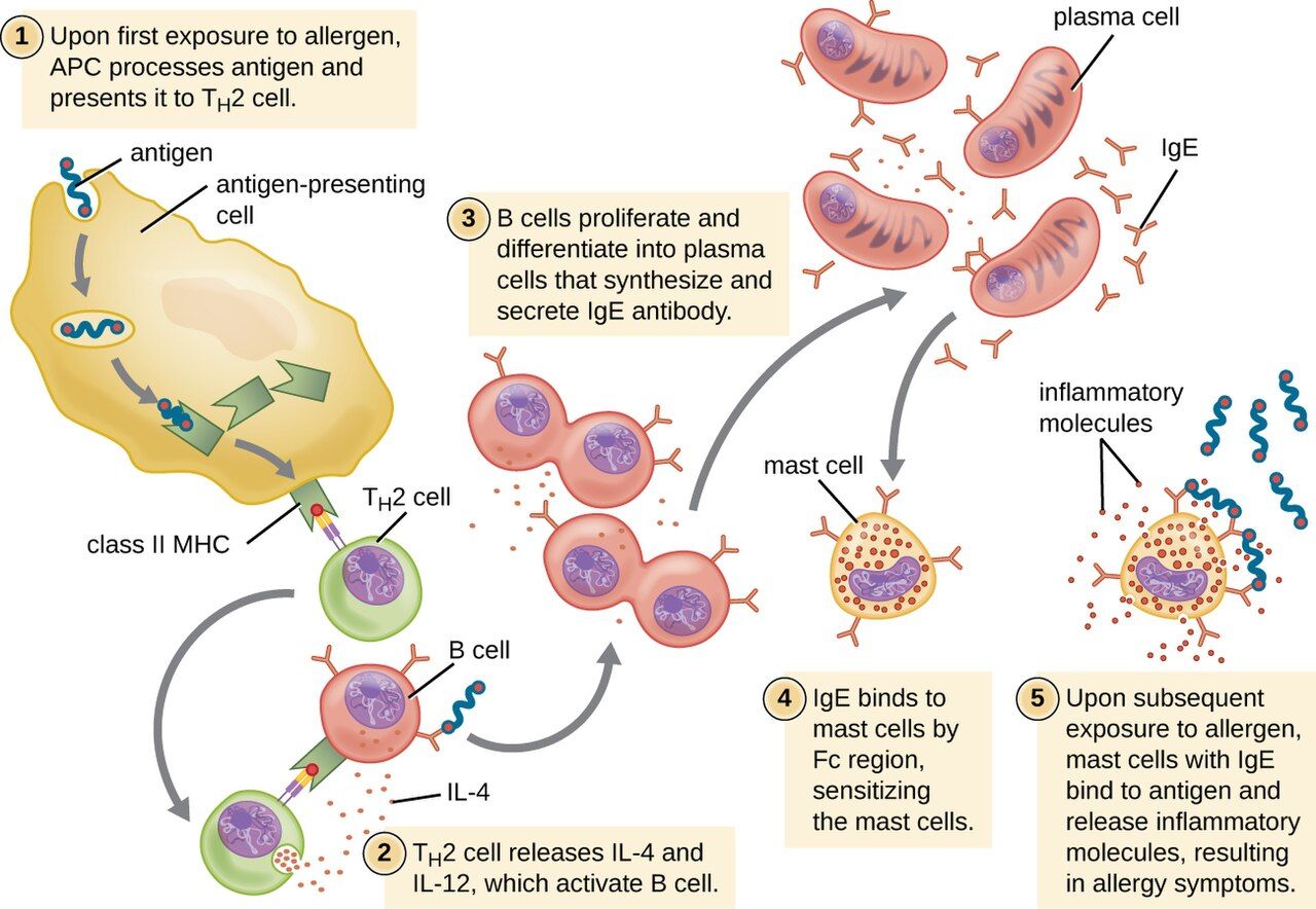
Sensitization first, reaction later
A type I reaction — what most people mean by "an allergy" — unfolds in two acts. During sensitization, a first exposure to an otherwise harmless allergen (pollen, peanut, penicillin, bee venom) is taken up and presented, and the immune system responds by making IgE antibody specific to it. That IgE then coats the surface of mast cells in the tissues and basophils in the blood, arming them. Nothing dramatic happens yet; the patient feels fine.
On the next exposure, the allergen bridges two adjacent IgE molecules on a mast cell — cross-linking them — and that is the trigger. Within seconds the mast cell undergoes degranulation, dumping preformed histamine and other mediators, and begins synthesizing leukotrienes and prostaglandins. Those mediators cause the familiar package: vasodilation (redness, warmth, dropping blood pressure), increased vascular permeability (swelling, hives), smooth-muscle contraction (bronchospasm, cramping), and increased secretions (runny nose, watery eyes).
From a local nuisance to a systemic emergency
Where the reaction happens decides how serious it is. A localized reaction stays near the point of contact: allergic rhinitis (hay fever), hives at a skin site, or asthma in the airways. A systemic reaction — anaphylaxis — occurs when mediators are released body-wide, usually because the allergen entered the bloodstream (an injected drug, an IV contrast agent, a sting, an ingested food). Anaphylaxis is a life-threatening emergency: widespread vasodilation causes blood pressure to crash (distributive shock), and airway swelling plus bronchospasm can choke off breathing within minutes.
| Feature | Localized allergy | Anaphylaxis (systemic) |
|---|---|---|
| Where mediators act | Near contact site | Whole body |
| Typical signs | Hives, itching, sneezing, wheeze | Hives plus hypotension, throat/airway swelling, stridor, wheeze, GI cramps, sense of doom |
| Main danger | Discomfort | Airway closure and shock — can kill in minutes |
| First-line treatment | Antihistamine; avoid trigger | Epinephrine, IM, immediately |
Recognizing and treating the emergency
The drug that reverses anaphylaxis is epinephrine, given intramuscularly into the outer thigh, and it must be given at the first sign — not after antihistamines, not after a wait-and-see. Epinephrine constricts blood vessels to restore blood pressure, relaxes airway smooth muscle to open breathing, and stabilizes mast cells. Antihistamines and corticosteroids are helpful adjuncts but are far too slow and too weak to be relied on alone. Alongside the drug, protect the airway, give oxygen, lay the patient flat with legs raised (unless breathing is easier sitting up), run IV fluids for the shock, and monitor closely — a biphasic reaction can return hours later even after the patient looks recovered.
Prevention is nursing work too. A documented allergy must be verified, banded, and honored on every medication pass; the single most common cause of a preventable drug reaction is a missed or ignored allergy history. For patients with severe allergies, teaching correct use of an epinephrine auto-injector — and to call emergency services and go to the hospital even after using it — is part of discharge.
- Type I allergy = sensitization (make IgE, arm mast cells) then a later reaction when allergen cross-links IgE.
- Mast-cell degranulation releases histamine, causing vasodilation, swelling, bronchospasm, and secretions.
- Anaphylaxis is systemic type I: airway swelling and distributive shock, potentially fatal in minutes.
- Treat anaphylaxis with IM epinephrine immediately; watch for a biphasic return.
Section 3Autoimmunity: the body against itself
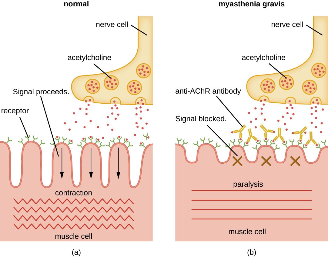
How self-tolerance breaks down
The immune system normally learns, early in life, to ignore the body's own molecules — a state called self-tolerance. Developing lymphocytes that react too strongly against self are deleted or disabled before they mature. Autoimmunity is the failure of that tolerance: self-reactive T cells or antibodies (autoantibodies) escape control and attack the body's own tissues, producing an autoimmune disease. The attack uses the same weapons as any hypersensitivity — often type II (autoantibodies against cell-surface targets), type III (immune complexes), or type IV (self-reactive T cells).
Why tolerance fails is usually a mix of factors. A genetic predisposition (many autoimmune diseases run in families and link to particular HLA/MHC types) sets the stage, and something in the environment — often an infection — pulls the trigger. One well-studied mechanism is molecular mimicry: a microbial antigen resembles a human protein closely enough that antibodies raised against the microbe also attack the body. The classic example is rheumatic fever, where antibodies against Streptococcus pyogenes cross-react with heart tissue. Autoimmune diseases also fall more often on women, and many wax and wane in flares and remissions.
Organ-specific versus systemic disease
It helps to sort autoimmune diseases by how much of the body they target. Organ-specific diseases aim autoantibodies or T cells at a single tissue — the thyroid, the pancreatic islets, the neuromuscular junction — so the damage and the symptoms are focused. Systemic diseases, like systemic lupus erythematosus, generate autoantibodies against widespread components (such as the cell nucleus), so they injure many organs at once: skin, joints, kidneys, blood, and heart.
| Disease | Target | Effect | Scope |
|---|---|---|---|
| Graves’ disease | TSH receptor (stimulating antibody) | Overactive thyroid (hyperthyroidism) | Organ-specific |
| Hashimoto thyroiditis | Thyroid tissue | Underactive thyroid (hypothyroidism) | Organ-specific |
| Type 1 diabetes mellitus | Pancreatic islet β-cells | No insulin production | Organ-specific |
| Myasthenia gravis | Acetylcholine receptor | Muscle weakness, ptosis | Organ-specific |
| Multiple sclerosis | CNS myelin sheath | Nerve conduction failure | Organ-specific (CNS) |
| Rheumatoid arthritis | Joint synovium | Symmetric joint inflammation, deformity | Systemic |
| Systemic lupus erythematosus | Nuclear antigens (anti-nuclear antibodies) | Skin, joints, kidney, blood, heart | Systemic |
Living with an autoimmune disease
There is no cure for most autoimmune diseases, so care aims to control the immune attack and manage what it has damaged. Treatment leans on immunosuppression — corticosteroids and other drugs that quiet the immune response — which brings its own hazard: a patient who is deliberately immunosuppressed is also more vulnerable to infection, blurring the line between an autoimmune problem and an immunodeficiency one. Some organ-specific diseases are managed by replacing what was lost rather than suppressing anything, as with insulin in type 1 diabetes or thyroid hormone in Hashimoto disease.
- Autoimmunity is a loss of self-tolerance: autoantibodies or self-reactive T cells attack the body.
- Causes combine genetic risk and an environmental trigger; molecular mimicry (e.g., rheumatic fever) is one route.
- Diseases are organ-specific (Graves, Hashimoto, type 1 diabetes, myasthenia gravis, MS) or systemic (RA, SLE).
- Care centers on immunosuppression or hormone replacement; immunosuppression raises infection risk.
Section 4Immunodeficiency and HIV/AIDS
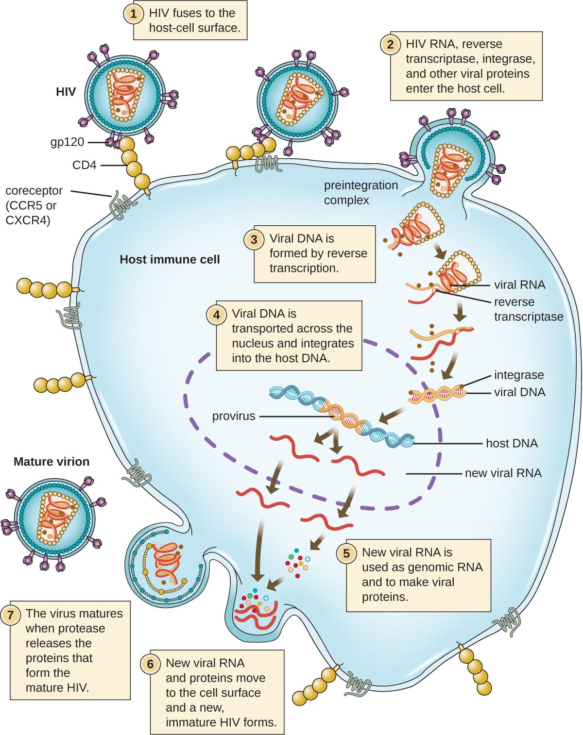
When defenses are too weak
If hypersensitivity and autoimmunity are the immune system doing too much, immunodeficiency is it doing too little. A part of the immune system is missing or not working, so the patient cannot clear infections that a healthy person would shrug off — and they become prey to opportunistic organisms, microbes that rarely trouble the immunocompetent. Immunodeficiencies split into two groups by origin.
Primary immunodeficiencies are congenital — the person is born with a genetic defect in some component of immunity. They range from mild (selective IgA deficiency, often silent) to catastrophic. The most severe is severe combined immunodeficiency (SCID), in which both B cells and T cells are absent or nonfunctional; affected infants have almost no defense and, without treatment such as a bone-marrow transplant, do not survive infancy — the origin of the "bubble" isolation once used to protect them.
Secondary (acquired) immunodeficiencies are far more common and develop after birth from some external cause: malnutrition (the leading cause worldwide), certain cancers (especially leukemias and lymphomas), chemotherapy and other immunosuppressive drugs, radiation, aging, and viral infection — most importantly HIV. Many of your patients are secondarily immunodeficient by treatment: anyone on chemotherapy, high-dose steroids, or anti-rejection drugs.
| Feature | Primary immunodeficiency | Secondary immunodeficiency |
|---|---|---|
| Origin | Congenital (genetic) | Acquired after birth |
| Onset | Usually infancy/childhood | Any age |
| Examples | SCID, selective IgA deficiency, DiGeorge syndrome | HIV/AIDS, malnutrition, chemotherapy, leukemia, steroids |
| Frequency | Rare | Common — many hospitalized patients |
HIV and AIDS
The human immunodeficiency virus (HIV) is a retrovirus that targets the very cell that coordinates the whole immune response: the CD4 helper T cell. Over years, HIV steadily infects and destroys these cells. As the CD4 count falls, the immune system loses its conductor, and the patient becomes progressively unable to fight off infection. HIV is transmitted through blood, sexual contact, and from mother to child in pregnancy, birth, or breastfeeding — not through casual contact.
AIDS (acquired immunodeficiency syndrome) is the advanced stage of HIV infection, defined by a CD4 count that falls below 200 cells/µL (normal is roughly 500–1,500) or by the appearance of an AIDS-defining opportunistic illness — infections and cancers that a healthy immune system holds at bay, such as Pneumocystis jirovecii pneumonia, Candida of the esophagus, disseminated tuberculosis, cytomegalovirus, and Kaposi sarcoma. The lower the CD4 count, the wider the range of opportunists that can take hold.
The story is far more hopeful than it once was. Antiretroviral therapy (ART) — combinations of drugs that block different steps of the viral life cycle — can suppress HIV to undetectable levels, preserve the CD4 count, prevent progression to AIDS, and render the virus untransmissible sexually. HIV has become, for many, a chronic manageable condition. Nursing priorities include supporting adherence to a lifelong regimen, watching for opportunistic infections, and applying standard precautions consistently — the same barrier practices that protect against all bloodborne pathogens.
- Immunodeficiency = too little immune function, opening the door to opportunistic infections.
- Primary = congenital (e.g., SCID); secondary = acquired (HIV, malnutrition, chemotherapy, steroids) and far more common.
- HIV destroys CD4 helper T cells; AIDS = CD4 < 200 cells/µL or an AIDS-defining opportunistic illness.
- ART suppresses HIV and preserves immunity; nurses support adherence and use standard precautions.
Section 5Transfusion and transplant reactions
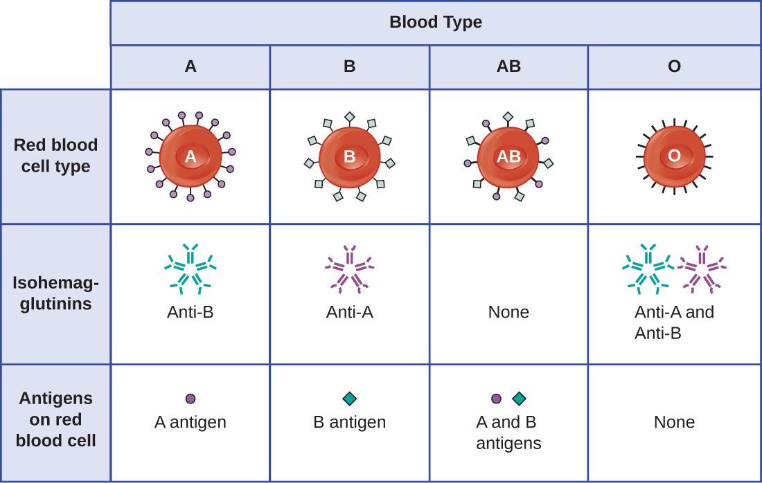
ABO and Rh: the blood you can safely give
Red blood cells carry surface molecules that the immune system reads as antigens. The most important set is the ABO system: type A cells carry the A antigen, type B carry B, type AB carry both, and type O carry neither. Crucially, each person naturally makes antibodies against the ABO antigen they lack — a type A person has anti-B antibodies, a type O person has both anti-A and anti-B. This is why blood must be matched: transfusing the wrong ABO type sends antibody against donor red cells, and they are destroyed.
A second system, Rh, adds "positive" or "negative": Rh-positive cells carry the D antigen, Rh-negative cells do not. Unlike ABO, anti-Rh antibodies are not present from birth — an Rh-negative person makes them only after being exposed to Rh-positive blood. Because type O-negative blood carries none of these antigens, it is the emergency universal donor for red cells; type AB-positive, carrying no anti-A/anti-B antibodies, is the universal recipient.
| Blood type | Antigens on red cells | Antibodies in plasma | Can receive red cells from |
|---|---|---|---|
| A | A | anti-B | A, O |
| B | B | anti-A | B, O |
| AB | A and B | none | A, B, AB, O (universal recipient) |
| O | none | anti-A and anti-B | O only (universal donor) |
When a transfusion goes wrong
An acute hemolytic transfusion reaction is a type II hypersensitivity and a medical emergency, almost always caused by an ABO mismatch — usually a clerical or identification error at the bedside. The recipient's antibodies bind the transfused donor cells and destroy (hemolyze) them, triggering fever, chills, flank or back pain, dark urine, hypotension, and potentially kidney failure and death. The nurse's response is immediate: stop the transfusion, keep the IV line open with saline, and notify the provider and blood bank. Because these reactions come from misidentification, the two-person bedside verification of patient and unit is not paperwork — it is the actual safeguard against a fatal error.
A related problem crosses the placenta. Hemolytic disease of the newborn (HDN) occurs when an Rh-negative mother, sensitized to Rh-positive blood in an earlier pregnancy, makes anti-Rh antibodies that cross into a subsequent Rh-positive fetus and destroy its red cells. It is largely preventable: giving Rh immune globulin (RhoGAM) to Rh-negative mothers stops them from becoming sensitized in the first place.
Transplants and rejection
Transplanting tissue raises the same problem in a different form: the recipient's immune system recognizes donor cells as foreign (chiefly by their MHC/HLA molecules) and attacks the graft. How much it attacks depends on how closely donor and recipient are matched. An autograft (self to self, like a skin graft) and an isograft (between identical twins) are not rejected. An allograft (between two genetically different people — the usual organ transplant) is rejected unless the immune response is suppressed, and a xenograft (between species) faces the strongest rejection of all.
Rejection comes in three tempos: hyperacute (within minutes, from preexisting antibodies), acute (days to weeks, T-cell mediated), and chronic (months to years). To keep an allograft alive, recipients take immunosuppressant drugs for life — which, as ever, trades rejection risk for infection risk. One further danger runs the opposite direction: in a bone-marrow or stem-cell transplant, the graft's immune cells can attack the recipient's body — graft-versus-host disease (GVHD).
- ABO: you make antibodies against the antigen you lack; O-negative is the universal red-cell donor, AB-positive the universal recipient.
- An acute hemolytic transfusion reaction is a type II reaction from ABO mismatch — stop the transfusion at once.
- HDN comes from Rh incompatibility and is prevented with Rh immune globulin.
- Grafts: autograft/isograft not rejected, allograft needs immunosuppression; GVHD is the graft attacking the host.
The chapter in one breath
- Hypersensitivity is an immune response that harms the host; the four types differ by mediator and timing (I–III antibody, IV T-cell).
- Type I allergy runs from sensitization and IgE to mast-cell degranulation; systemic release is anaphylaxis, treated with IM epinephrine immediately.
- Autoimmunity is lost self-tolerance — organ-specific (Graves, type 1 diabetes, myasthenia gravis) or systemic (RA, SLE).
- Immunodeficiency is too little immunity: primary (congenital, e.g., SCID) or secondary (acquired — HIV, chemotherapy, malnutrition).
- HIV destroys CD4 T cells; AIDS is CD4 < 200/µL or an opportunistic illness; ART controls it.
- ABO/Rh matching prevents the type II hemolytic transfusion reaction; identification errors are the cause, so the bedside check is the safeguard.
- Grafts are rejected by degree of mismatch; allografts need lifelong immunosuppression, and GVHD is the graft attacking the host.
ReferenceWords worth knowing
A quick reference for the terms in this chapter — skim it now, or circle back whenever a word trips you up.
- Allergen
- An otherwise harmless antigen that provokes a type I (allergic) hypersensitivity response.
- Allograft
- A transplant between two genetically different individuals of the same species; the usual organ transplant, requiring immunosuppression.
- Anaphylaxis
- A severe, systemic type I reaction with airway swelling and distributive shock; a life-threatening emergency treated with epinephrine.
- Autoantibody
- An antibody directed against the body's own tissues.
- Autoimmunity
- A failure of self-tolerance in which the immune system attacks the body's own cells and tissues.
- CD4 helper T cell
- The coordinating lymphocyte of the immune response and the primary target of HIV.
- Graft-versus-host disease (GVHD)
- A reaction in which transplanted (graft) immune cells attack the recipient's tissues, seen with bone-marrow/stem-cell transplants.
- Hemolytic disease of the newborn (HDN)
- Destruction of a fetus's red cells by maternal anti-Rh antibodies; prevented with Rh immune globulin.
- Hemolytic transfusion reaction
- A type II reaction from an ABO-mismatched transfusion; the recipient's antibodies destroy donor red cells.
- Hypersensitivity
- An exaggerated or misdirected immune response that damages the host's own tissues.
- IgE
- The antibody class that arms mast cells and basophils and mediates type I hypersensitivity.
- Immunodeficiency
- A deficient immune response; primary (congenital) or secondary (acquired).
- Mast cell
- A tissue cell coated with IgE that releases histamine and other mediators when it degranulates.
- Molecular mimicry
- A microbial antigen resembling a self-protein, so anti-microbe antibodies also attack the body (e.g., rheumatic fever).
- Opportunistic infection
- An infection by a normally harmless microbe that takes hold when host defenses are weakened.
- Primary immunodeficiency
- A congenital, genetically based defect in immune function, such as SCID.
- Rh factor
- The D red-cell antigen; Rh-positive cells carry it, Rh-negative do not.
- Secondary immunodeficiency
- An acquired loss of immune function from an external cause (HIV, malnutrition, drugs, cancer).
- Self-tolerance
- The immune system's normal ability to recognize and not attack the body's own molecules.
- Sensitization
- The silent first exposure in which the immune system builds antibodies or primed cells against an antigen, setting up a later reaction.
- Severe combined immunodeficiency (SCID)
- A severe primary immunodeficiency lacking functional B cells and T cells.
Check yourself
Infectious Diseases by Body System
Microbes do not respect the neat chapter boundaries of a textbook — they land somewhere on a real patient and cause trouble that a nurse has to recognize, report, and help treat. This chapter reorganizes what you already know about pathogens around the place they strike: the skin, the lungs, the gut, the urinary and reproductive tracts, and the bloodstream and brain. For each region we will name the usual offenders, describe what the infection looks like at the bedside, and connect it to the assessments and precautions that keep one patient's infection from becoming the whole unit's problem. Think of it as a field guide — organized the way you actually meet infections on the floor.
By the end of this chapter you can
- Recognize the common skin and wound infections — impetigo, cellulitis, and MRSA — and the assessment findings that flag them.
- Distinguish the major respiratory infections (pneumonia, tuberculosis, influenza, strep throat) and the isolation precautions each requires.
- Explain how gastrointestinal pathogens cause illness through infection versus toxin, and why C. difficile and norovirus demand soap-and-water hand hygiene.
- Describe the pathogens and presentations of urinary tract infections and common sexually transmitted infections.
- Recognize the warning signs of bacteremia, sepsis, and meningitis and the urgency each carries.
- Match each body-system infection to its typical causative organism and route of transmission.
- Connect bedside nursing actions — hand hygiene, device care, early recognition — to the prevention of healthcare-associated infection.
Section 1Skin & wound infections
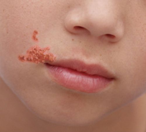
The skin is a wall, until it isn't
Intact skin is one of the body's best defenses: a dry, slightly acidic, constantly shedding barrier that most microbes simply cannot cross. Skin and soft-tissue infections almost always begin where that wall is breached — a scratch, an insect bite, a surgical incision, a pressure injury, an IV site, or the cracked skin of eczema. Once organisms get past the surface, the two players you will meet again and again are Staphylococcus aureus and Streptococcus pyogenes (group A strep). Their reservoirs differ, and the difference matters: S. aureus is a true resident, colonizing the anterior nares of about 30% of adults and moist skin folds, which is why nasal decolonization with mupirocin is used before some surgery. S. pyogenes is not normal skin or nasal flora — it is carried asymptomatically in the pharynx (roughly 5–15% of children) and only transiently on skin.
What the infection looks like depends mostly on how deep it goes. Impetigo is a superficial infection of the outermost skin, common in children, producing fragile blisters that rupture into a golden, honey-colored crust. Cellulitis is deeper, involving the dermis and the fat beneath it: a spreading area of warmth, redness, swelling, and tenderness with poorly defined edges, often on a leg. Erysipelas is a brighter, sharply raised, well-demarcated cellulitis of the upper dermis, classically caused by group A strep. The deepest and most dangerous, necrotizing fasciitis, races along the tissue planes and produces pain out of proportion to what the skin shows — a surgical emergency, not an antibiotic-only problem.
MRSA and the infected wound
MRSA — methicillin-resistant Staphylococcus aureus — is ordinary S. aureus that has picked up the mecA gene, which builds an altered penicillin-binding protein (PBP2a) that beta-lactam drugs cannot grip. The name understates the problem: MRSA resists all the beta-lactams — penicillins, the anti-staphylococcal penicillins (nafcillin, oxacillin), cephalosporins such as cefazolin and ceftriaxone, and the carbapenems — with only the newer anti-MRSA cephalosporins (ceftaroline) as an exception. Treatment falls to vancomycin, linezolid, or daptomycin, or, for uncomplicated skin infection, TMP-SMX, doxycycline, or clindamycin. It is a leading cause of skin abscesses (patients often report a "spider bite" they never actually saw a spider make) and a notorious cause of surgical-site and device-related infections. MRSA is spread by contact — hands, shared equipment, contaminated surfaces — which is why colonized or infected patients are placed on contact precautions (gown and gloves) and why hand hygiene between patients is non-negotiable.
A wound infection announces itself with the classic signs of local inflammation — increasing redness, warmth, swelling, pain, and purulent (pus) drainage — sometimes with a spreading red border, foul odor, or fever. The single most useful nursing skill here is the ability to tell colonization from infection: nearly every open wound grows bacteria on a swab, but only a wound with the signs of tissue invasion needs antibiotics. A clean-looking chronic wound with a positive culture and no inflammation is usually colonized, and treating it does more harm than good.
| Infection | Depth | Usual cause | Hallmark finding |
|---|---|---|---|
| Impetigo | Superficial epidermis | S. aureus, group A strep | Honey-colored crust |
| Erysipelas | Upper dermis | Group A strep | Bright, raised, sharply bordered |
| Cellulitis | Dermis & subcutaneous fat | S. aureus, strep | Warm, red, tender, ill-defined edges |
| Abscess / MRSA | Localized pus pocket | MRSA, S. aureus | Fluctuant lump; may need drainage |
| Necrotizing fasciitis | Fascia (surgical emergency) | Group A strep, mixed flora | Pain out of proportion, rapid spread |
- Skin infections start where the barrier is broken; S. aureus and group A strep are the usual causes.
- Depth drives the picture: impetigo (superficial) → cellulitis (dermis) → necrotizing fasciitis (surgical emergency).
- MRSA resists the penicillin family, spreads by contact, and warrants contact precautions.
- A wound with spreading redness, warmth, pain, and pus is infected; a positive swab without those signs is usually just colonized.
Section 2Respiratory infections
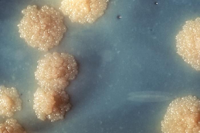
Upper tract, lower tract
The respiratory system is split into an upper tract (nose, sinuses, throat, larynx) and a lower tract (trachea, bronchi, and the alveoli of the lungs), and the division matters because lower-tract infections are generally more serious. Upper-tract infections — the common cold, most sore throats, sinusitis — are usually viral and self-limited. The important exception is strep throat (streptococcal pharyngitis), caused by group A strep, which produces a sudden painful sore throat, fever, and swollen tonsils without the cough and runny nose of a cold. It matters because untreated it can lead to rheumatic fever, so it is one of the few sore throats worth culturing and treating with antibiotics.
The lungs are supposed to be nearly sterile, defended by the mucus blanket, the sweeping cilia, and the cough reflex. When those defenses fail — because a patient is intubated, sedated, weak, or aspirating — organisms reach the alveoli and cause pneumonia. Nurses classify pneumonia by where it was acquired: community-acquired pneumonia (CAP), most often from Streptococcus pneumoniae; hospital-acquired and ventilator-associated pneumonia (VAP), which involve tougher, more resistant organisms; and aspiration pneumonia, from inhaled mouth or stomach contents in patients with impaired swallowing or a depressed gag reflex.
Tuberculosis and influenza
Tuberculosis (TB), caused by the slow-growing acid-fast bacillus Mycobacterium tuberculosis, is the respiratory infection whose precautions you must never get wrong. TB spreads by tiny airborne droplet nuclei that hang in the air, so a suspected case goes into a negative-pressure airborne isolation room, and staff wear a fitted N95 respirator — not a surgical mask. Classic active TB brings a chronic productive cough (sometimes with blood), night sweats, fever, and weight loss. Treatment is long — months of multiple drugs — and the nurse's central role is supporting adherence, because stopping early is exactly how drug-resistant TB is bred.
Influenza is a viral lower-and-upper respiratory infection that arrives abruptly with fever, body aches, headache, and a dry cough. It spreads by droplets, so influenza patients are placed on droplet precautions (a surgical mask within a few feet), and because it is viral, antibiotics do nothing — antiviral drugs help only if started early. The most important nursing contribution against influenza happens before flu season: vaccination of patients and staff alike.
| Infection | Typical cause | Transmission | Precaution |
|---|---|---|---|
| Strep throat | Group A strep (bacterial) | Droplet/contact | Droplet for infants and young children until 24 h of effective antibiotics; standard thereafter and in adults |
| Community pneumonia | Streptococcus pneumoniae | Droplet | Standard/droplet |
| Influenza | Influenza virus | Droplet | Droplet (surgical mask) |
| Tuberculosis | Mycobacterium tuberculosis | Airborne | Airborne (N95 + negative pressure) |
- Upper-tract infections are mostly viral; strep throat is the bacterial exception worth treating.
- Pneumonia is classified by where it was acquired — community, hospital/ventilator, or aspiration — which predicts the likely organism.
- TB is airborne: negative-pressure room and N95; its long therapy makes adherence a nursing priority.
- Influenza is viral and droplet-spread; vaccination and droplet precautions matter more than antibiotics.
Section 3Gastrointestinal infections
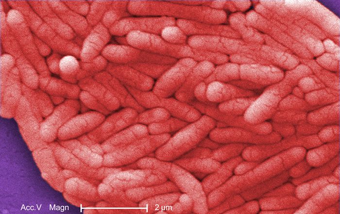
Two ways the gut gets sick
Gastrointestinal (GI) pathogens almost all share one route of entry — the fecal-oral route, meaning microbes (or their toxins) from stool reach a new mouth by way of contaminated food, water, or hands. But they make you sick in two very different ways. In a true infection, the organism is swallowed, colonizes the gut, and invades or irritates the lining; symptoms take a day or more to appear because the microbe has to multiply first. In an intoxication, the microbe has already grown in the food and produced a toxin, and it is the pre-formed toxin you swallow — so symptoms hit fast, often within hours (the vomiting of Staphylococcus aureus food poisoning is the textbook example).
Foodborne illness is the umbrella term for all of this, and the usual suspects — Salmonella, Campylobacter, Shiga-toxin-producing E. coli, Norovirus — produce overlapping pictures of nausea, vomiting, crampy abdominal pain, and diarrhea that may be watery or bloody. For most healthy adults the illness is self-limited and the treatment is rehydration, not antibiotics. The nurse's clinical eye is on the vulnerable: infants, older adults, and immunocompromised patients, who can dehydrate dangerously, and on the warning signs of bloody diarrhea, which can signal the Shiga-toxin-producing E. coli strain that damages the kidneys. That organism deserves a specific caution: it never invades the bloodstream — it sticks to the colon lining and releases a toxin that is absorbed — and antibiotics and antimotility drugs are contraindicated in suspected cases, because both increase toxin release and the risk of hemolytic uremic syndrome. Watch urine output, creatinine, platelets, and hemoglobin.
The two that spread on the unit
Clostridioides difficile (C. diff) is the infection you help cause if you are not careful with antibiotics. Broad-spectrum antibiotics flatten the normal gut flora, and this spore-forming organism blooms into severe, foul-smelling watery diarrhea and colitis. Two facts drive nursing practice: C. diff forms spores that alcohol hand rub does not kill, so you must wash with soap and water after caring for these patients; and the room needs a bleach-based (sporicidal) cleaner. Patients go on contact precautions, and new-onset diarrhea in anyone on antibiotics should raise the question immediately.
Norovirus is the explosive, highly contagious cause of "stomach flu" that shuts down cruise ships, long-term-care units, and hospital wards. It takes only a handful of viral particles to infect, it spreads by the fecal-oral route and by aerosolized vomit, and — like C. diff spores — it resists alcohol gel, so soap-and-water hand hygiene and bleach cleaning are again the rule. Sudden onset of vomiting and watery diarrhea sweeping through a group of people points straight at norovirus.
| Cause | Mechanism | Onset | Key nursing point |
|---|---|---|---|
| Salmonella/Campylobacter | Infection (invasion) | 1–3 days | Rehydrate; usually no antibiotics |
| S. aureus food poisoning | Pre-formed toxin | Hours | Fast onset, short course |
| Shiga-toxin E. coli | Non-invasive adherence + Shiga toxin | 2–4 days | Bloody diarrhea; do not give antibiotics or antimotility drugs — they raise the risk of hemolytic uremic syndrome; monitor urine output, creatinine, platelets, hemoglobin |
| C. difficile | Overgrowth after antibiotics | Days–weeks | Soap & water; contact precautions |
| Norovirus | Infection (very low dose) | 12–48 hours | Soap & water; extremely contagious |
- GI pathogens spread by the fecal-oral route and cause illness by infection (slow onset) or pre-formed toxin (fast onset).
- Most foodborne illness is self-limited; the mainstay is rehydration, and the risk is dehydration in the very young, old, and immunocompromised.
- C. difficile follows antibiotic use, forms spores, and demands soap-and-water hand hygiene plus contact precautions.
- Norovirus is intensely contagious, resists alcohol gel, and calls for the same soap-and-water and bleach-cleaning response.
Section 4Urogenital infections & STIs
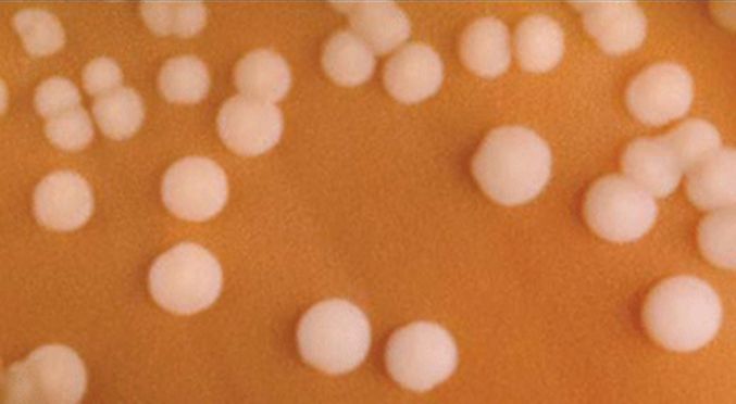
Urinary tract infections
The urinary tract infection (UTI) is one of the most common infections nurses manage, and it is usually an inside job: the culprit is most often the patient's own Escherichia coli, harmless in the colon but troublesome once it climbs the short distance up the urethra into the bladder. Anatomy explains why women get far more UTIs than men — a shorter urethra sits closer to the anus. A lower UTI, or cystitis (bladder infection), brings dysuria (burning with urination), urinary frequency and urgency, and cloudy or foul-smelling urine. If the infection ascends to the kidneys — pyelonephritis — the patient turns systemically ill: fever, chills, and flank (back) pain over the kidney.
The version that belongs to nursing above all others is the catheter-associated UTI (CAUTI). An indwelling urinary catheter is a superhighway that lets bacteria bypass every normal defense and travel straight to the bladder, and the risk climbs with every day the catheter stays in. The single most effective prevention is simple and squarely a nursing responsibility: do not place a catheter unless it is truly indicated, and remove it the moment it is no longer needed. A subtle but crucial point — a positive urine culture in a patient with no symptoms (asymptomatic bacteriuria) is usually not treated, because treating it breeds resistance without helping the patient.
Sexually transmitted infections
Sexually transmitted infections (STIs) are spread through sexual contact and range from bacterial infections that antibiotics cure to viral infections that we manage but cannot eradicate. Two features make STIs a distinct nursing challenge: many are asymptomatic, so people transmit them unknowingly, and all of them carry stigma, which means your ability to take a nonjudgmental history and protect confidentiality is part of the clinical care. Chlamydia (from Chlamydia trachomatis) is the most commonly reported bacterial STI and is frequently silent, especially in women, where untreated infection can scar the fallopian tubes and cause infertility. Gonorrhea (from Neisseria gonorrhoeae) often travels with it. Syphilis, caused by the spirochete Treponema pallidum, progresses in stages if untreated, beginning with a painless sore.
The viral STIs behave differently from the curable bacterial ones, though not all in the same way. Genital herpes (herpes simplex virus) is a lifelong infection that causes recurring outbreaks of painful blisters. Human papillomavirus (HPV) causes genital warts and, in its high-risk types, cervical and other cancers — which is exactly why the HPV vaccine is a cancer-prevention tool. HPV is not lifelong for most people: roughly 90% of infections are cleared or suppressed within about two years, and it is persistent infection with a high-risk type (16 and 18 above all) that carries the cancer risk — an important reassurance to give after an abnormal Pap or HPV result. HIV attacks the immune system itself. Across all of them the nursing themes are consistent: encourage screening (because many are silent), support partner treatment to stop the ping-pong of reinfection, and teach prevention without shame.
| Infection | Organism | Type | Nursing note |
|---|---|---|---|
| Chlamydia | Chlamydia trachomatis | Bacterial (curable) | Often silent; screen & treat partners |
| Gonorrhea | Neisseria gonorrhoeae | Bacterial (curable) | Rising drug resistance |
| Syphilis | Treponema pallidum | Bacterial (curable) | Painless sore; stages if untreated |
| Genital herpes | Herpes simplex virus | Viral (lifelong) | Recurrent painful blisters |
| HPV | Human papillomavirus | Viral (usually clears in 1–2 years) | Warts; persistent high-risk types drive cancer; vaccine prevents |
- Most UTIs are caused by the patient's own E. coli; cystitis = burning and frequency, pyelonephritis = fever and flank pain.
- CAUTI risk rises with catheter days; the top prevention is avoiding and promptly removing catheters.
- Asymptomatic bacteriuria is usually not treated — treating it breeds resistance without benefit.
- Many STIs are silent; nursing care centers on nonjudgmental screening, partner treatment, and prevention (including the HPV vaccine).
Section 5Bloodstream & CNS infections
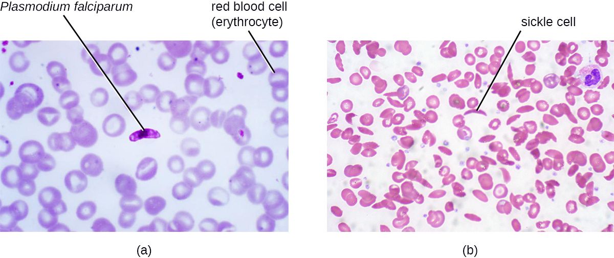
From bacteremia to sepsis
The bloodstream is supposed to be sterile, so finding microbes there is always significant. Bacteremia simply means bacteria are present in the blood — sometimes transiently and harmlessly (brushing your teeth can do it), but sometimes as the launching point for disaster. The danger is sepsis: not the infection itself but the body's dysregulated, overwhelming response to it, in which widespread inflammation causes blood vessels to leak and dilate, blood pressure to fall, and organs to lose their perfusion. Left unchecked it progresses to septic shock — dangerously low blood pressure that does not correct with fluids — and to multi-organ failure. Common sources include pneumonia, UTIs, abdominal infections, and, critically for hospitalized patients, infected central lines (a CLABSI, or central-line-associated bloodstream infection).
Recognizing sepsis early is one of the highest-value skills a nurse has, because survival is measured in hours. The bedside picture is a patient who looks and acts wrong on top of an infection: fever or an abnormally low temperature, a fast heart rate, fast breathing, and — often the first and most sensitive sign — new confusion or altered mental status. Falling blood pressure, decreasing urine output, and mottled skin signal that shock is setting in. When sepsis is recognized, care moves fast along a bundle: draw blood cultures, give broad-spectrum antibiotics quickly, and infuse fluids to restore perfusion.
Meningitis
Meningitis is inflammation of the meninges, the membranes wrapping the brain and spinal cord, and bacterial meningitis is one of the true medical emergencies in this book — it can kill a previously healthy young person within a day. The classic triad is fever, severe headache, and a stiff neck (nuchal rigidity), often with photophobia (light hurts), vomiting, and a rapidly declining level of consciousness. A spreading purple, non-blanching rash points to the especially feared meningococcus, Neisseria meningitidis. Because that organism spreads by respiratory droplets and can move through close-contact groups like dorms and barracks, a suspected case goes on droplet precautions, and close contacts may need preventive antibiotics.
Diagnosis rests on a lumbar puncture to sample the cerebrospinal fluid, and the nursing imperative is speed: antibiotics should not wait. Viral (aseptic) meningitis, by contrast, is far more common and usually much milder, resolving on its own. The nurse's job is to keep bacterial meningitis at the front of the mind whenever fever meets a stiff neck and a changed mental status — because the cost of moving slowly is measured in lives and lifelong disability.
| Condition | What it is | Red-flag signs | Urgency |
|---|---|---|---|
| Bacteremia | Bacteria in the blood | May be silent or feverish | Investigate the source |
| Sepsis | Dysregulated response to infection | Confusion, fast HR/RR, low BP | Hours — cultures, antibiotics, fluids |
| Septic shock | Sepsis + fluid-resistant low BP | Mottled skin, low urine output | Critical |
| Bacterial meningitis | Infected meninges | Fever, stiff neck, headache, confusion | Emergency — droplet precautions |
- Bacteremia is bacteria in the blood; sepsis is the body's overwhelming, harmful response to infection.
- Septic shock is sepsis with fluid-resistant low blood pressure; early recognition and the sepsis bundle save lives.
- New confusion, fast heart and breathing rates, and falling blood pressure are the sepsis warning signs to escalate.
- Bacterial meningitis (fever + stiff neck + headache + confusion) is an emergency; meningococcal cases need droplet precautions.
The chapter in one breath
- Skin infections begin at a break in the barrier; depth predicts severity, from impetigo to necrotizing fasciitis, and MRSA drives contact precautions.
- Respiratory infections range from viral colds to pneumonia; the precaution level is everything, and TB is airborne (N95, negative pressure) while flu is droplet.
- GI pathogens spread fecal-orally by infection or pre-formed toxin; C. difficile and norovirus resist alcohol gel and demand soap-and-water hygiene.
- UTIs usually come from the patient's own E. coli; CAUTI is prevented by removing catheters early, and asymptomatic bacteriuria is left untreated.
- STIs are often silent; nursing centers on nonjudgmental screening, partner treatment, and vaccination (HPV).
- Sepsis is the body's dangerous response to infection — new confusion, fast vitals, and low blood pressure demand rapid cultures, antibiotics, and fluids.
- Bacterial meningitis — fever, stiff neck, headache, confusion — is a true emergency requiring immediate treatment and droplet precautions.
ReferenceWords worth knowing
A quick reference for the terms in this chapter — skim it now, or circle back whenever a word trips you up.
- Bacteremia
- The presence of bacteria in the bloodstream, which is normally sterile.
- Catheter-associated urinary tract infection (CAUTI)
- A UTI that develops because an indwelling urinary catheter lets bacteria bypass normal defenses; risk rises with each catheter day.
- Cellulitis
- A spreading bacterial infection of the dermis and subcutaneous fat, marked by warmth, redness, swelling, and tenderness with ill-defined edges.
- Clostridioides difficile (C. diff)
- A spore-forming gut bacterium that overgrows after antibiotics wipe out normal flora, causing severe diarrhea and colitis.
- Cystitis
- Infection of the bladder (a lower UTI), causing dysuria, urgency, and frequency.
- Dysuria
- Pain or burning with urination, a hallmark symptom of a lower UTI.
- Empiric therapy
- Treatment started against the most likely organisms before culture results are back, based on the clinical picture.
- Enterotoxin
- A toxin that acts on the intestinal lining; when pre-formed in food it causes rapid-onset food poisoning.
- Impetigo
- A superficial skin infection, common in children, producing blisters that crust over with a honey-colored scab.
- Meningitis
- Inflammation of the meninges around the brain and spinal cord; the bacterial form is a medical emergency.
- Methicillin-resistant Staphylococcus aureus (MRSA)
- S. aureus carrying mecA/PBP2a, which makes it resistant to all beta-lactam antibiotics — penicillins, cephalosporins, and carbapenems — except the anti-MRSA cephalosporins; spread by contact and a common cause of skin and wound infections.
- Norovirus
- A highly contagious virus causing explosive vomiting and diarrhea; resists alcohol gel, so soap-and-water hygiene is required.
- Pneumonia
- Infection of the lung alveoli; classified as community-acquired, hospital/ventilator-associated, or aspiration.
- Pyelonephritis
- Infection of the kidney (an upper UTI), marked by fever, chills, and flank pain.
- Sepsis
- A dysregulated, life-threatening body-wide response to infection that can lead to organ dysfunction.
- Septic shock
- Sepsis with dangerously low blood pressure that does not correct with fluids alone.
- Sexually transmitted infection (STI)
- An infection spread through sexual contact; often asymptomatic, ranging from curable bacterial to lifelong viral illness.
- Tuberculosis (TB)
- A chronic lung infection by Mycobacterium tuberculosis, spread by airborne nuclei and requiring airborne isolation.
- Urinary tract infection (UTI)
- Infection of the urinary tract, most often by E. coli ascending the urethra into the bladder.
- Wound infection
- Invasion of a break in the skin by microbes, signaled by spreading redness, warmth, pain, and purulent drainage.