Neurology
The nervous system is the body’s command center. When it fails, the stakes are immediate. Minutes decide the outcome of a stroke. Rising pressure inside the skull can be deadly. This chapter walks through the major neurologic emergencies and diseases. You will cover stroke, raised intracranial pressure, head and spinal trauma, seizures, and the slow diseases that wear down the brain and nerves.
By the end of this chapter you can
- Tell an ischemic stroke from a hemorrhagic one, and use FAST.
- Explain raised intracranial pressure, the Monro-Kellie doctrine, and Cushing’s triad.
- Compare an epidural and a subdural hematoma, and know where a spinal cord injury does the most harm.
- Describe the seizure types, status epilepticus, and how to keep a person safe.
- Contrast Alzheimer, Parkinson, MS, myasthenia gravis, and Guillain-Barre.
Section 1Stroke
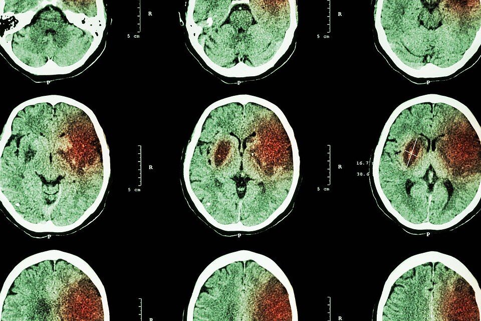
Your brain is the hungriest organ you have. It is only about 2 percent of your body weight. Yet it uses roughly 20 percent of your oxygen and glucose. It also has almost no way to store fuel of its own. So brain tissue needs a constant supply of oxygen-rich blood, second by second. That blood arrives through the carotid and vertebral arteries. When the supply stops, neurons cannot switch to a backup plan the way muscle can. Within minutes they start to fail. Soon after, they start to die. That is just what a stroke is. Blood flow to part of the brain stops suddenly, so that region loses its function. Another name for it is a cerebrovascular accident, or CVA.
Two ways the flow fails: ischemic vs hemorrhagic
Two very different problems can starve brain tissue. Telling them apart drives everything you do next. Explore the figure below to see them side by side.
An ischemic stroke is a plumbing blockage. A vessel gets clogged, and the tissue past the clog is cut off. This is by far the most common type. It makes up about 87 percent of strokes. Hemorrhagic strokes make up the other 13 percent or so. The clog comes in two forms. In a thrombotic stroke, a clot forms right there in a brain artery, on top of atherosclerotic plaque. Plaque is a fatty buildup in the vessel wall. In an embolic stroke, the clot forms somewhere else and travels. It is often a piece thrown from the heart during atrial fibrillation. It rides the bloodstream and lodges in a smaller brain vessel.
A hemorrhagic stroke works the other way. It is a leak. A weak or overstretched vessel ruptures and bleeds into or around the brain. Now two injuries happen at once. The tissue past the rupture loses its blood flow. And the escaping blood forms a mass. That mass raises intracranial pressure and irritates nearby tissue. Long-standing hypertension and ruptured aneurysms are common causes. Hemorrhagic strokes are less common, but they are often more deadly.
| Feature | Ischemic (~87%) | Hemorrhagic |
|---|---|---|
| Core problem | A blockage; a clot cuts off flow | A bleed; the vessel ruptures |
| Common causes | Atherosclerosis (thrombotic), A-fib emboli (embolic) | Hypertension, ruptured aneurysm |
| Clot-buster tPA | Can help if given in time | Never give it; absolutely contraindicated |
| Typical headache | Often mild, or none at all | Often sudden and severe, the "worst of my life" |
The penumbra, and why time is brain
When an artery blocks, the tissue at the center of the blocked zone dies fast. Around that dead core sits a rim of tissue. It is called the ischemic penumbra. These neurons are starving and silent, so they have stopped working. But they are not yet dead. Trickles of blood from nearby vessels keep them barely alive. This rim is the whole reason we rush. Restore the flow fast and those neurons can wake back up. The deficit can then shrink. Wait too long and the rim you could have saved turns into dead tissue for good.
Experts figure the brain loses about 1.9 million neurons every minute that an ischemic stroke goes untreated. That rate is why we say time is brain. Every minute of delay is brain tissue you cannot get back.
Recognizing it fast: FAST
Because minutes matter, there is a quick screen called FAST:
- F - Face: Ask the person to smile. Does one side droop?
- A - Arm: Ask them to raise both arms. Does one drift downward?
- S - Speech: Is speech slurred, absent, or strange? Can they repeat a simple sentence?
- T - Time: If you see any of these, note the time the symptoms began. Then call for emergency help right away.
That last letter is easy to skip. It may be the most important one. Whether a patient can get treatment rests on knowing when the patient was last known well.
Why the CT scan comes before the clot-buster
The main clot-dissolving drug is tPA (tissue plasminogen activator, also called alteplase). It works beautifully on a clot. But it is a clot-dissolving (thrombolytic) drug. If the stroke is really a bleed, tPA pours fuel on the fire. It would make the hemorrhage much worse. Here is the catch. Ischemic and hemorrhagic strokes can look the same. You cannot tell them apart by symptoms alone.
That is why an emergency non-contrast CT scan of the head comes first, every time. Its main job is to rule out a bleed. Only after blood is ruled out can tPA be considered. Even then the window is strict. For patients who qualify, it is usually within 3 to 4.5 hours of symptom onset. Some patients also qualify for a mechanical thrombectomy. There the clot is pulled out by hand, and the window is longer.
Which side, which deficit
Motor nerve tracts cross over in the lower medulla. That crossing is called the pyramidal decussation. So a stroke on one side of the brain causes weakness on the opposite (contralateral) side of the body. A right-brain stroke causes left-sided weakness. A left-brain stroke causes right-sided weakness. Language adds another twist. In most people the language centers sit in the left hemisphere. So a left-brain stroke often causes aphasia, which is trouble making or understanding language. It comes along with right-sided weakness. A right-brain stroke more often spares speech. But it may cause left-sided neglect, where the patient ignores their affected side completely.
- Ischemic strokes (~87%) are blockages, either thrombotic or embolic. Hemorrhagic strokes are bleeds. The treatment for one would harm the other.
- The ischemic penumbra is tissue you can still save around the dead core. Fast blood flow rescues it. That is why time is brain.
- Screen quickly with FAST: Face droop, Arm drift, Speech trouble, Time to call for help and note onset.
- A non-contrast CT must rule out a bleed before tPA, because the clot-buster would make a hemorrhage worse. tPA is usually given within 3 to 4.5 hours of onset.
- Deficits are contralateral, which means on the opposite side. A left-brain stroke often causes aphasia plus right-sided weakness.
Section 2Raised intracranial pressure
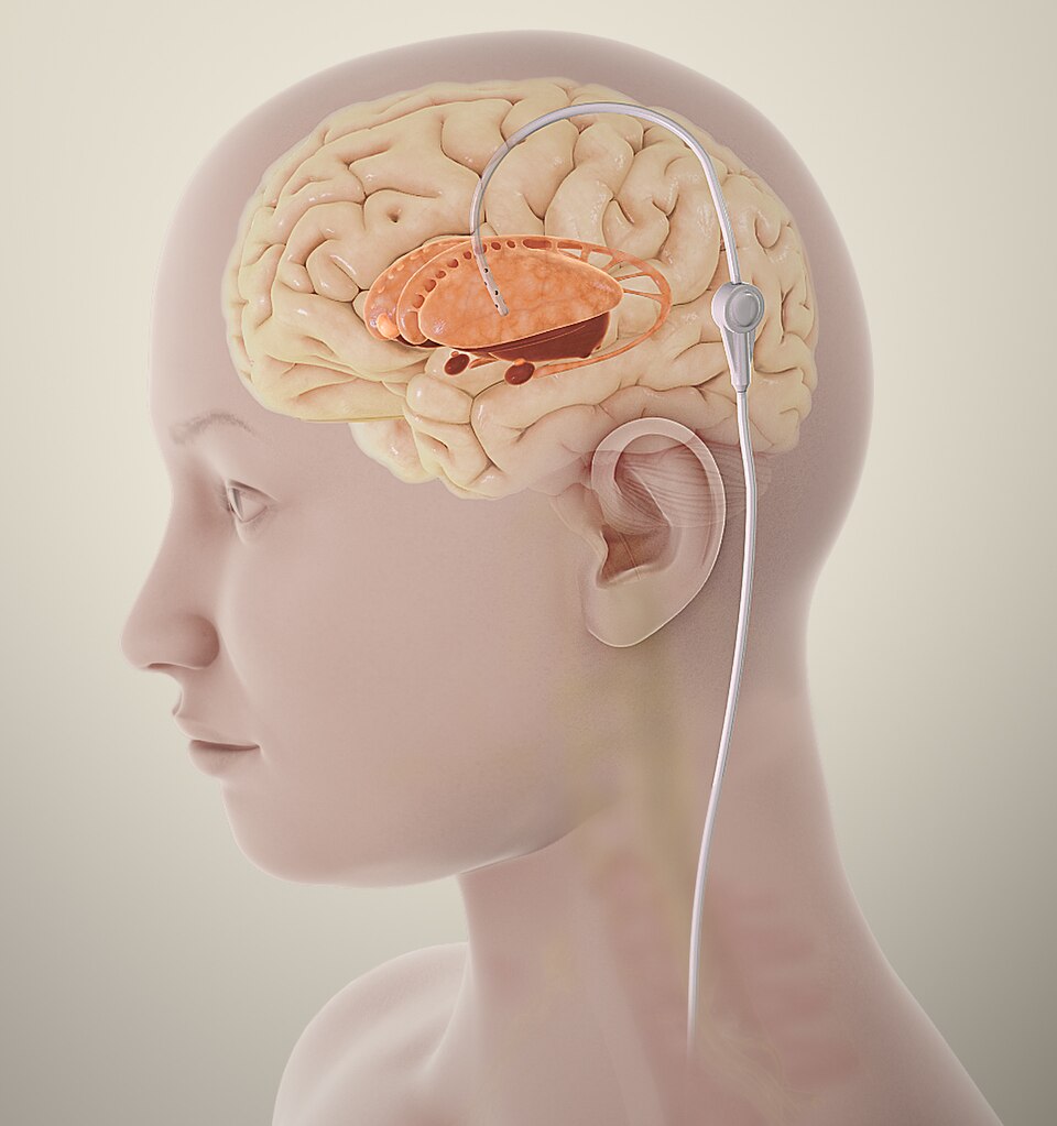
Picture the brain as a delicate organ inside a hard, sealed box. That box is the skull. In a healthy person the box holds three things, and they share the space in a careful balance. There is the brain tissue itself, about 80% of the volume. There is the blood in its vessels, about 10%. And there is the clear cerebrospinal fluid (CSF) that cushions and bathes it, also about 10%. The skull cannot stretch, so the total volume inside is fixed. The gentle pressure these three create is the intracranial pressure (ICP). Normally it sits around 5 to 15 mmHg.
The Monro-Kellie doctrine: a box that cannot expand
One rule governs all of this. It is the Monro-Kellie doctrine. The skull is a rigid, fixed container. So the three volumes inside must always add up to the same total. If one grows, another has to shrink to make room. If neither shrinks, the pressure climbs. At first the brain is clever about this. It buys time through compensation. It shunts CSF down into the spinal canal. It also squeezes some venous blood out of the skull. That is why a slow-growing problem can stay quiet for a while. But this wiggle room runs out. Once those buffers are used up, even a tiny added volume sends the pressure up steeply. The patient falls off a compensation cliff. Now small changes cause big, dangerous swings in ICP.
What raises the pressure
Anything that adds volume to that sealed box can raise ICP. The usual causes line up with the three normal contents, plus a few new intruders:
- Bleeding (hemorrhage) - an epidural, subdural, subarachnoid, or intracerebral bleed adds a pool of blood that takes up space.
- Tumor (mass lesion) - a growing mass literally crowds out brain, blood, and CSF.
- Cerebral edema - swelling of the brain tissue itself. Fluid leaks into or around the cells. It often follows trauma, a stroke, or an infection.
- Hydrocephalus - a backup of CSF. The body makes too much, the flow is blocked, or it is not taken back up. Fluid then builds up in the ventricles.
The earliest warning: a change in consciousness
Here is the single most important point. The earliest and most sensitive sign of rising ICP is a change in level of consciousness (LOC). The brain's alertness centers react fast to pressure and to falling oxygen. So before anything dramatic happens, the patient just seems different. They may be restless, confused, drowsy, hard to wake, slow to answer, or newly agitated. Families often notice it first ("she's just not herself"). Trust that. A falling Glasgow Coma Scale (GCS) score is your red flag. It comes long before the classic textbook signs. Do not wait for the pupils to change.
Cushing's triad: the late, ominous sign
The pressure inside the skull can rise high enough to threaten blood flow to the brainstem. Then the body tries one last rescue. To keep pushing blood past that crushing pressure, it drives the systolic blood pressure up. Baroreceptors sense the surge. They reflexively slow the heart, which gives bradycardia. At the same time, the pressure squeezes the brainstem's respiratory centers. Breathing turns irregular, as in Cheyne-Stokes breathing. Those three signs together make up Cushing's triad. It is a late and grave finding. It tells you the brainstem is in trouble.
| Feature | Early rising ICP | Cushing's triad (late) |
|---|---|---|
| Consciousness | Mild confusion, restlessness, drowsiness, a falling GCS | Deeply obtunded, meaning barely rousable, or in a coma |
| Blood pressure | Often normal | Rising systolic with widening pulse pressure |
| Heart rate | Often normal | Bradycardia |
| Breathing | Usually normal | Irregular (e.g., Cheyne-Stokes) |
| Meaning | Act now; it can still be turned around | The brainstem is in trouble; herniation is near |
Herniation: the feared end result
If the pressure is not relieved, the brain gets forced across its normal walls. It may push under the falx, across the tentorial notch, or down through the foramen magnum at the base of the skull. This shift of brain tissue past its normal borders is called herniation. As tissue is pushed down, it crushes the brainstem, which runs breathing, heart rate, and consciousness. The results are catastrophic. You see a fixed, dilated pupil on the affected side, from pressure on cranial nerve III. Protective reflexes are lost. Breathing stops, and the patient dies. Herniation is where untreated intracranial hypertension ends up. Everything above is about catching the problem long before this point. Explore the figure below to see how a growing volume drives the pressure up and pushes the brain toward herniation.
- The Monro-Kellie doctrine: the skull is a fixed box holding brain, blood, and CSF. When one volume grows, another must shrink, or ICP rises. Compensation buys time, then fails sharply.
- Common causes are a bleed, tumor, cerebral edema, or hydrocephalus - anything that adds volume to the sealed skull.
- The earliest sign is a change in level of consciousness (restlessness, confusion, drowsiness, a falling GCS). Act on it early.
- Cushing's triad - rising systolic BP with a widening pulse pressure, bradycardia, and irregular breathing - is a late, ominous sign that the brainstem is in trouble.
- Herniation, brain tissue forced through the foramen magnum onto the brainstem, is the feared and often fatal end result.
Section 3Brain & spinal cord injury
Your brain is soft, delicate tissue. It floats inside a hard, unforgiving skull, cushioned by a thin layer of cerebrospinal fluid. That fluid normally works like a shock absorber. But in trauma the same skull that protects your brain becomes the problem. The brain can slam against the bone. And swelling has nowhere to go. That is why head injuries are so dangerous. The box is sealed, so any extra blood or swelling presses on the brain itself.
Concussion vs contusion, and the coup-contrecoup pattern
A concussion is a functional injury. The brain gets shaken hard enough to upset how neurons signal. But a scan usually shows no bleeding and no bruising. Patients often have a brief loss of consciousness, confusion, headache, and amnesia around the event. A contusion is a step worse. It is a real bruise of brain tissue, with capillary bleeding and swelling you can often see on CT. Contusions can grow over hours, so repeat neuro checks matter.
A coup-contrecoup injury often causes both. Picture a moving head striking a dashboard. The brain first bruises right under the point of impact. That is the coup. Then it rebounds and slams into the other side of the skull. That is the contrecoup. One blow, two injured areas.
Bleeding between the layers: epidural vs subdural hematoma
Three membranes wrap the brain. The tough outer one is the dura mater. The middle one is the arachnoid. The delicate pia mater lies right against the brain. Two things change how fast the patient crashes. One is where the blood collects. The other is which vessel is torn.
An epidural hematoma is bleeding above the dura. The classic source is a torn middle meningeal artery after a temporal skull fracture. It is arterial, so it fills fast under high pressure. The textbook picture is a lucid interval. The patient is knocked out. Then they wake up and seem fine. Then they go downhill fast, as the clot grows and presses on the brain. This is a neurosurgical emergency.
A subdural hematoma is bleeding below the dura, from torn bridging veins. It is venous, so it oozes slowly. Symptoms can take hours, days, or even weeks to show up. It is common in older adults and in people with chronic alcohol use. In both, brain atrophy stretches those bridging veins and leaves them fragile.
| Feature | Epidural hematoma | Subdural hematoma |
|---|---|---|
| Vessel | Artery (middle meningeal) | Bridging veins |
| Speed | Fast, high pressure | Slow ooze |
| Classic clue | A lucid interval, then a decline | Slow symptoms that come and go |
| Typical patient | Temporal skull fracture | Older adults, chronic alcohol use |
Spinal cord injury by level
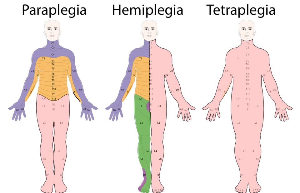
The spinal cord is the main data cable between the brain and the body. Where it is damaged sets what is lost below that point, because signals can no longer travel past the injury. As a rule, the higher the injury, the more of the body is affected.
- Lumbar/sacral injuries may affect the legs, bladder, and bowel.
- Thoracic injuries can cause paraplegia, loss of function in the legs and lower trunk.
- Cervical injuries can cause tetraplegia (quadriplegia), affecting all four limbs.
The scariest zone is the high cervical cord. The line to memorize is "C3, C4, C5 keep the diaphragm alive." The phrenic nerve drives the diaphragm, and it comes off those levels. An injury at or above C3-C5 can paralyze the diaphragm. Then the patient cannot breathe on their own and needs a ventilator right away. That is why airway and breathing come first in any suspected cervical injury.
Two shock states to keep straight
Spinal shock comes right after trauma. All reflexes, movement, and feeling below the injury drop out for a time. Think of the cord as stunned. It can last days to weeks. Neurogenic shock is a circulatory problem. A high injury wipes out sympathetic tone, so vessels cannot hold their squeeze. That gives the classic triad of hypotension, bradycardia, and warm dry skin. The circulatory instability usually runs one to three weeks. Autonomic dysreflexia shows up later. It is a dangerous, overblown reflex in injuries at or above T6. A full bladder or bowel usually sets it off. Blood pressure spikes suddenly, and that is a true emergency.
Explore the figure below to see how these head injuries take shape.
- Concussion is functional, with no bleeding. Contusion is a true bruise. Coup-contrecoup hurts both the impact site and the opposite side.
- Epidural is arterial and fast, with the classic lucid interval. Subdural is venous and slower. It is common in older adults and in people with chronic alcohol use.
- Higher spinal injuries affect more of the body. A C3-C5 injury threatens the phrenic nerve and the diaphragm, so breathing is the priority.
- Spinal shock is a short-term loss of reflexes. Neurogenic shock is hypotension with bradycardia. Autonomic dysreflexia is a dangerous BP spike at or above T6.
Section 4Seizures & epilepsy
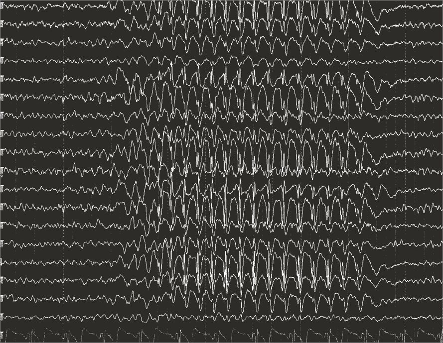
To understand a seizure, start with the normal brain. Your neurons talk to each other all day with tiny electrical signals. In health that chatter is coordinated but not synchronized. Different regions fire at different times for different jobs. Think of a huge orchestra, where each section plays its own part. A tug-of-war keeps that balance. On one side are excitatory signals, mostly the neurotransmitter glutamate, which says "fire." On the other side are inhibitory signals, mostly GABA, which says "settle down." As long as the balance holds, the brain hums along smoothly.
A seizure happens when that balance breaks. A group of neurons suddenly fires all at once. It is a burst of abnormal, hypersynchronous electrical activity. Hypersynchronous means they all fire in lockstep. Instead of the orchestra playing its parts, every instrument blasts the same note. The outward signs depend on where this starts and how far it spreads. Keep one difference clear. A seizure is a single event. Epilepsy is a chronic condition. It is a tendency toward recurrent, unprovoked seizures. It is usually diagnosed after two or more, or after one seizure plus a high risk of another.
Focal vs. generalized: where the storm begins
Seizures are grouped by where the abnormal firing starts. A focal seizure begins in one area of one hemisphere. The older term for it was partial. Only part of the brain is involved, so the signs match that region. You may see a twitching hand, an odd smell, a rising feeling in the stomach, or a spell of staring and confusion. The person may stay aware (focal aware). Or their awareness may change (focal impaired awareness). A focal seizure can also spread and become generalized.
A generalized seizure involves both hemispheres from the start. So consciousness is lost right away. The two you will see most on exams are tonic-clonic and absence.
| Feature | Tonic-clonic (generalized) | Absence |
|---|---|---|
| Classic look | Stiffening then rhythmic jerking | Brief blank staring spell |
| Phases | Tonic (muscles rigid) then clonic (rhythmic jerks) | No motor phases; the eyelids may flutter |
| Duration | Usually 1 to 3 minutes | Often 5 to 10 seconds |
| Awareness | Lost, and the person falls | Lost for a moment, with no fall |
| After the event | Postictal confusion, fatigue, sore muscles | Goes right back to the task, often unaware |
| Typical patient | Any age | Often school-age children |
In a tonic-clonic seizure you may also see cyanosis, a blue tint from pauses in breathing during the tonic phase. Tongue-biting and loss of bladder control are common too. Whole-brain firing overrides normal muscle and autonomic control, which explains all of it. Absence seizures are so short that people mistake them for daydreaming. That is why a child who seems "not to be paying attention" sometimes turns out to be seizing dozens of times a day.
What sets a seizure off: common triggers
Two kinds of things lower the seizure threshold. One is anything that pushes the brain toward excitation. The other is losing something that was calming it:
- Fever in young children - rapid temperature spikes cause febrile seizures, usually between 6 months and 5 years.
- Low sodium (hyponatremia) or low glucose (hypoglycemia) - with a low sodium the blood outside the cells is dilute, so water moves into brain cells by osmosis and the brain swells. That swelling is what sets the seizure off. A low glucose is different. It starves neurons of the fuel they need to hold a steady membrane.
- Missed antiseizure medication - the drug level that was calming the brain falls too low. This is the most common reason a person with epilepsy breaks through.
- Alcohol withdrawal - alcohol holds the brain down. Stop it suddenly and the brain rebounds into a hyperexcitable state.
- Others: sleep deprivation, flashing lights, and infections such as meningitis.
Status epilepticus: the emergency
Status epilepticus is a seizure that lasts 5 minutes or longer. It also counts when seizures repeat without full recovery of consciousness in between. This is a true neurologic emergency. Nonstop firing burns through oxygen and glucose faster than the brain can supply them. The steady muscle activity drives the temperature up and can injure neurons. The result is hypoxia, acidosis, and lasting damage. Treatment is fast IV benzodiazepines, such as lorazepam, plus airway support.
Priorities during a seizure
Your job is protection, not stopping the seizure itself. You cannot switch off the electrical activity by hand. So focus on preventing injury:
- Protect the airway and head - turn the person on their side, the recovery position. That lets secretions drain and lowers the risk of aspiration. Cushion the head and clear away nearby hazards.
- Do not restrain - holding the limbs down can cause fractures or dislocations. Let the movements run their course.
- Nothing in the mouth - never put in objects or fingers. You risk broken teeth, bites, and a blocked airway.
- Time it - note the start time. If it reaches 5 minutes, treat it as status epilepticus and get help.
- Stay and observe - loosen tight clothing and give the person privacy. Write down what you saw for the provider: the movements, how long it lasted, and any incontinence.
Tip the balance yourself in the figure below, then watch the trace and the clock.
- A seizure is a burst of abnormal, hypersynchronous electrical firing. Epilepsy is a tendency toward recurrent, unprovoked seizures.
- Focal seizures start in one area, so the signs match that region. Generalized seizures involve both hemispheres. Tonic-clonic means stiffen then jerk, with postictal confusion. Absence means a brief staring spell, common in children.
- Common triggers lower the seizure threshold: fever in children, low sodium or glucose, missed medication, and alcohol withdrawal. Correct the cause you can fix.
- Status epilepticus is a seizure of 5 minutes or longer, or back-to-back seizures with no recovery. It is an emergency. It needs IV benzodiazepines and airway support.
- During a seizure: turn side-lying to protect the airway, cushion the head, do NOT restrain or put anything in the mouth, and time it.
Section 5Neurodegenerative disease
Your brain runs on about 86 billion neurons. Here is the humbling part. You were born with nearly all of them, and adult brains make very few new ones. Neurons are not like skin or blood cells, which replace themselves all the time. Neurons are built to last a lifetime. That is exactly why the diseases in this section are so devastating. When these cells die, the body usually cannot grow replacements. The losses stack up, so the damage is progressive and irreversible. Picture a city slowly losing its power lines, one by one, with no crew coming to fix them.
Healthy neurons keep themselves tidy. They fold their proteins the right way. They clear out cell garbage. And they pass chemical messages, called neurotransmitters, across tiny gaps called synapses. Each neurodegenerative disease is a different way this housekeeping and signaling breaks down. Each one hits a different neighborhood of the nervous system.
Alzheimer disease: the memory system clogs first
Alzheimer disease is the most common cause of dementia. The trouble is misfolded protein. Two abnormal proteins build up. Beta-amyloid plaques collect in the spaces between neurons. Tau tangles, also called neurofibrillary tangles, twist up inside them. These deposits are toxic. They jam the machinery neurons use to talk and to stay alive, so the cells wither and die. The damage often starts in the hippocampus, the brain's memory-forming hub. So the first and loudest symptom is usually short-term memory loss. The person forgets recent conversations, while old memories stay clear much longer. As it spreads across the cortex, it takes language and judgment. In the end it takes the ability to recognize loved ones and to handle basic self-care.
Parkinson disease: a dopamine shortage in the movement circuit
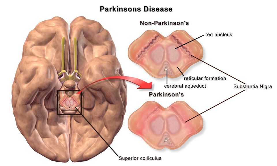
Parkinson disease targets one spot deep in the brain. It is called the substantia nigra, a cluster of neurons that make dopamine. Dopamine is the chemical that starts and smooths your movements. As these neurons die off, dopamine drops. The movement circuit, called the basal ganglia, loses its balance. The classic picture goes by the word TRAP:
- Tremor at rest, often a "pill-rolling" motion of the fingers that eases with purposeful movement
- Rigidity, stiff, cogwheel-like resistance in the limbs
- Akinesia/Bradykinesia, slow and shrinking movements, such as small handwriting, a masked face, and a shuffling gait
- Postural instability, poor balance and a tendency to fall, usually a later sign
ALS: the motor neurons fail while the mind stays largely intact
Amyotrophic lateral sclerosis (ALS) is also called Lou Gehrig disease. It attacks the motor neurons, the wiring that carries "move" commands from the brain and spinal cord to the muscles. Both upper and lower motor neurons break down. So patients develop steady muscle weakness, atrophy, and fasciculations. Atrophy means wasting, and fasciculations are muscle twitches. It often starts in one hand or foot and spreads from there. Here is the cruel twist. In most patients, cognition, senses, and awareness stay largely intact. The person stays alert inside a body that is losing the power to move, to swallow, and in time to breathe. The brain is not untouched, though. About 15 percent go on to develop frontotemporal dementia, which changes behavior and language. Failure of the breathing muscles is usually what limits life.
| Feature | Alzheimer | Parkinson | ALS |
|---|---|---|---|
| Cells lost | Cortical and hippocampal neurons | Dopamine neurons (substantia nigra) | Upper & lower motor neurons |
| Hallmark pathology | Amyloid plaques + tau tangles | Dopamine loss (Lewy bodies) | Motor neuron degeneration |
| First symptom | Short-term memory loss | Resting tremor, slowness | Weakness in one spot |
| Cognition | Lost step by step | Often kept early on | Usually kept |
One common thread ties all three together, and it is worth memorizing. A specific group of neurons is slowly and permanently lost. So the symptoms only get worse over time. Current treatments slow progression or ease symptoms. They do not regrow the lost cells, and they do not cure the disease. That is why care centers on safety, function, dignity, and family support. Explore the figure below to see where each disease strikes in the nervous system.
- Neurodegenerative diseases share one theme: progressive, irreversible loss of specific neurons that the body cannot replace.
- Alzheimer = beta-amyloid plaques + tau tangles, starting in the hippocampus, so short-term memory loss comes first.
- Parkinson = dopamine-neuron loss in the substantia nigra, causing resting tremor, rigidity, bradykinesia, and postural instability (TRAP).
- ALS = upper and lower motor-neuron death causing spreading weakness and atrophy, while cognition and senses usually stay intact.
- Treatments slow or ease symptoms but do not reverse the neuron loss, so care emphasizes safety, function, and support.
Section 6Demyelinating & neuromuscular disorders
Every time you lift a coffee cup, wiggle your toes, or take a breath, a tiny chemical talk happens between a nerve and a muscle. It takes place at the neuromuscular junction (NMJ). That is the special synapse where a motor nerve meets a muscle fiber. Learn this handshake and you will see why the disorders in this section cause weakness in such different patterns.
The neuromuscular junction: how the signal crosses
An electrical impulse races down a motor neuron. That impulse is an action potential. When it reaches the nerve ending, it triggers the release of a neurotransmitter called acetylcholine (ACh). The ACh floats across the tiny gap, called the synaptic cleft. It binds to nicotinic ACh receptors on the muscle membrane. That binding opens ion channels. The muscle membrane depolarizes, and the muscle contracts. Then an enzyme called acetylcholinesterase chews up the ACh. The signal switches off cleanly, so the muscle can relax. Three things have to be in place. Enough ACh must be released. Enough working receptors must be there to catch it. And a healthy nerve must carry the impulse in the first place. Break any one link and you get weakness. But the pattern of that weakness tells you which link failed.
Multiple sclerosis: the insulation frays
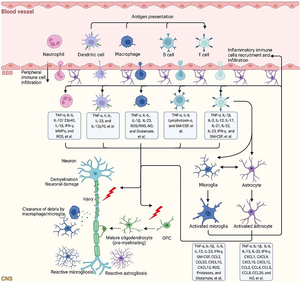
Nerve fibers in the central nervous system are wrapped in myelin. Myelin is a fatty insulation. It lets impulses jump quickly from gap to gap. In multiple sclerosis (MS), the body's own immune cells attack that myelin and strip it off CNS neurons. The CNS means the brain, the spinal cord, and the optic nerves. This is autoimmune demyelination. Where myelin is lost, a plaque, or patch of sclerosis, forms. Signals then slow, scatter, or stop for good. Myelin can be damaged almost anywhere in the CNS. So the symptoms are famously scattered. They include blurred or double vision from optic neuritis, numbness and tingling, weakness, spasticity, fatigue, and bladder problems. The classic course is relapsing-remitting. Flares appear, then partly fade as the inflammation settles and some remyelination happens. Even so, scarring builds up over the years. A handy phrase is lesions separated in space and time.
Myasthenia gravis: the receptors get blocked
In myasthenia gravis (MG), the nerve and its ACh are fine. The muscle's receptors are the ones under attack. The immune system makes anti-ACh receptor antibodies. These block and destroy nicotinic receptors at the NMJ. With fewer receptors to catch ACh, each signal lands weakly. The hallmark is fatigable weakness. Muscles work at first, then tire fast with repeated use, and they recover with rest. That is why symptoms are usually worse at the end of the day. It classically strikes muscles that work all the time. So you see ptosis (drooping eyelids), diplopia (double vision), and trouble chewing, swallowing, or holding the head up. The feared complication is myasthenic crisis. There the respiratory muscles weaken and breathing fails.
Guillain-Barre syndrome: the nerves lose their coating from the outside in
In Guillain-Barre syndrome (GBS), the immune system attacks the myelin of peripheral nerves. It often starts a week or two after an infection, usually a gut or airway illness. The commonest germ identified is Campylobacter jejuni, a bacterium that causes diarrhea. The reason is molecular mimicry. Antibodies aimed at the bug also react with nerve myelin, because the two look alike. The signature is ascending paralysis. Weakness and reduced reflexes begin in the feet and climb upward. They can reach the diaphragm, and then the patient needs a ventilator. Unlike MG, the weakness is usually symmetric and steadily progressive, not fatigable. It tends to peak, then slowly recover.
| Feature | Myasthenia gravis | Guillain-Barre |
|---|---|---|
| Target | ACh receptors at NMJ | Peripheral nerve myelin |
| Pattern | Fatigable, often eyes first | Ascending, feet upward |
| Reflexes | Usually normal | Reduced or gone |
| Trigger | Autoimmune, linked to the thymus | Often after an infection |
| Feared crisis | Myasthenic crisis (breathing) | Respiratory failure |
Explore the neuromuscular junction in the figure below to see where ACh crosses and where these diseases strike.
- At the neuromuscular junction, acetylcholine crosses from nerve to muscle and binds receptors to trigger contraction, then acetylcholinesterase clears it.
- Multiple sclerosis is autoimmune demyelination of the CNS, often relapsing-remitting, with scattered symptoms separated in space and time.
- Myasthenia gravis is caused by antibodies against ACh receptors, producing fatigable weakness that worsens with use and improves with rest.
- Guillain-Barre syndrome is peripheral demyelination causing ascending paralysis, often after an infection, with lost reflexes.
- MG and GBS can both weaken the diaphragm, so respiratory monitoring is a top priority.
The chapter in one breath
- Ischemic stroke (about 87%) is a clot blocking flow. Hemorrhagic stroke is a burst vessel. The FAST screen catches both fast, because time is brain. FAST is Face droop, Arm drift, Speech slurred, Time to call 911.
- Only ischemic strokes get tPA, within roughly 3 to 4.5 hours. A stat CT must rule out bleeding first. Giving a clot-buster into a hemorrhage is deadly.
- Raised intracranial pressure shows up late as Cushing's triad: rising BP with a widening pulse pressure, bradycardia, and irregular breathing. A sluggish, blown pupil goes with it. All of it warns that the brain is being squeezed toward herniation.
- Traumatic brain injury is tracked with the Glasgow Coma Scale, which scores eyes, verbal, and motor. A score of 8 or less usually means secure the airway. Watch for the lucid-interval pattern of an epidural hematoma.
- Spinal cord injury above T6 risks neurogenic shock early. That looks like warm, dry skin with hypotension and bradycardia. Later it risks autonomic dysreflexia: a pounding headache with dangerously high BP, often from a full bladder.
- Seizures call for protecting the patient and timing them. Turn them side-lying, put nothing in the mouth, and never restrain. A seizure lasting over 5 minutes is status epilepticus, a true emergency.
- Neurodegenerative diseases differ by mechanism. Parkinson's comes from dopamine loss, with resting tremor, rigidity, and bradykinesia. Alzheimer's comes from plaques and tangles, with steady memory loss. Multiple sclerosis comes from demyelination, with relapsing-remitting deficits.
- Myasthenia gravis causes fatigable weakness that worsens with use, such as ptosis and diplopia. Antibodies against acetylcholine receptors are the cause. Guillain-Barre syndrome is an ascending paralysis that threatens the diaphragm. In both, watch respiratory function closely.
ReferenceWords worth knowing
A quick reference for the terms in this chapter. Skim it now, or come back whenever a word trips you up.
- Bradykinesia
- Slowness in starting and carrying out movement. It is a core motor sign of Parkinson disease. Dopamine-producing neurons in the substantia nigra break down, so the basal ganglia can no longer start voluntary movement smoothly.
- Cushing's triad
- A late and ominous set of three vital-sign changes: hypertension with a widening pulse pressure, bradycardia, and irregular (often slow) respirations. It signals dangerously high intracranial pressure, with brainstem herniation close behind.
- Demyelination
- The loss or breakdown of the fatty myelin sheath that insulates axons. Nerve signals then slow down or stop. That is what produces the neurologic deficits seen in diseases like multiple sclerosis.
- Epidural hematoma
- A pool of blood between the skull and the outer dura mater. It usually comes from a torn middle meningeal artery after temporal trauma. The classic pattern is a brief lucid interval, then a fast decline.
- Hemorrhagic stroke
- A stroke caused by a ruptured blood vessel that bleeds into or around the brain. It starves tissue of blood. It also raises intracranial pressure, as the growing pool of blood presses on nearby neurons.
- Herniation
- The life-threatening shift of brain tissue across or through rigid structures inside the skull, when pressure builds. It may pass under the falx, or through the tentorium or foramen magnum. It often presses on the brainstem and its vital centers.
- Intracranial pressure
- The pressure inside the skull from the brain, blood, and cerebrospinal fluid together. It is normally about 5–15 mmHg. It turns dangerous when a mass, a bleed, or swelling pushes it higher.
- Ischemic stroke
- The most common type of stroke. A clot or narrowing blocks an artery and cuts off blood flow. The brain tissue it fed is starved of the oxygen and glucose it needs to survive.
- Monro-Kellie doctrine
- The rule that the skull is a fixed, rigid box. It holds three contents: brain, blood, and cerebrospinal fluid. If one of them grows, another must shrink to match. If not, intracranial pressure rises.
- Multiple sclerosis
- A chronic autoimmune disease. The body attacks myelin in the central nervous system. Scattered plaques form, and symptoms follow a relapsing-remitting pattern. They include vision changes, weakness, and numbness.
- Myasthenia gravis
- An autoimmune disorder. Antibodies block or destroy acetylcholine receptors at the neuromuscular junction. Muscle weakness then comes and goes. It worsens with use and improves with rest.
- Penumbra
- The ring of at-risk but still salvageable brain tissue around the dead core of an ischemic stroke. Restore blood flow quickly and it can be saved. That is why we say "time is brain."
- Seizure
- A sudden burst of abnormal, excessive electrical activity in the brain's neurons. It can change movement, sensation, behavior, or consciousness.
- Status epilepticus
- A neurologic emergency. It is a seizure lasting 5 minutes or longer, or repeated seizures with no full recovery in between. It risks permanent brain injury and needs treatment right away.
- Subdural hematoma
- A pool of blood between the dura and the arachnoid, usually from torn bridging veins. It often bleeds slowly, so it may show up days to weeks later. Older adults and people on anticoagulants are most at risk.
- tPA
- Tissue plasminogen activator, a clot-dissolving (thrombolytic) drug. It is given within a strict time window for ischemic stroke, to break down the blocking clot and restore blood flow. It is never used in hemorrhagic stroke.
DrillFlashcards for this unit
These are the Week 5 cards. It is the same deck as the hub, pulled in here so you can review the moment you finish reading. Tap the card to flip it. Hit Shuffle to mix them up. To grade yourself and track mastery across the whole deck, open the full flashcards page.
Check yourself
| Feature | Epidural | Subdural |
|---|---|---|
| Bleed source | Artery (fast) | Vein / bridging veins (slower) |
| Location | Above the dura | Below the dura |
| Onset | Fast, often with a lucid interval | Minutes to weeks; often chronic in older adults |
| CT shape | Lens / convex (lemon) | Crescent (banana) |
| Feature | Alzheimer | Parkinson |
|---|---|---|
| Core deficit | Memory / cognition | Movement |
| Pathology | Amyloid plaques & tau tangles | Dopamine loss in substantia nigra |
| Hallmark signs | Short-term memory loss, disorientation | Resting tremor, rigidity, bradykinesia, shuffling gait |