The Respiratory System
Every cell in your body burns oxygen to make energy, and every one of them produces carbon dioxide as waste — so twelve to sixteen times a minute, without a single conscious command, your body trades one gas for the other. This chapter follows a breath from the nostril to the microscopic air sac where the exchange actually happens, then explains the physics that pulls air in, the diffusion that swaps the gases, the clever chemistry that ferries them through the blood, and the volumes a clinician measures to tell a healthy lung from a sick one. By the end you will understand not just how you breathe but why the whole elegant machine works the way it does.
By the end of this chapter you can
- Trace the path of air through the conducting and respiratory zones and name each structure it passes.
- Describe how the airway warms, humidifies, and filters incoming air, and explain the mucociliary escalator.
- Diagram the respiratory membrane and explain why its thinness and huge surface area make it ideal for gas exchange.
- Explain the job of surfactant and predict what happens when it is missing.
- Use Boyle’s law to explain inspiration and expiration, and name the muscles and pressures involved in each.
- Apply partial-pressure gradients to external and internal respiration, saying which way each gas moves and why.
- Describe how oxygen and carbon dioxide are carried in the blood, including hemoglobin and the bicarbonate system.
- Define the lung volumes and capacities and interpret how spirometry separates obstructive from restrictive disease.
Section 1The respiratory tract
Two zones: a highway and a destination
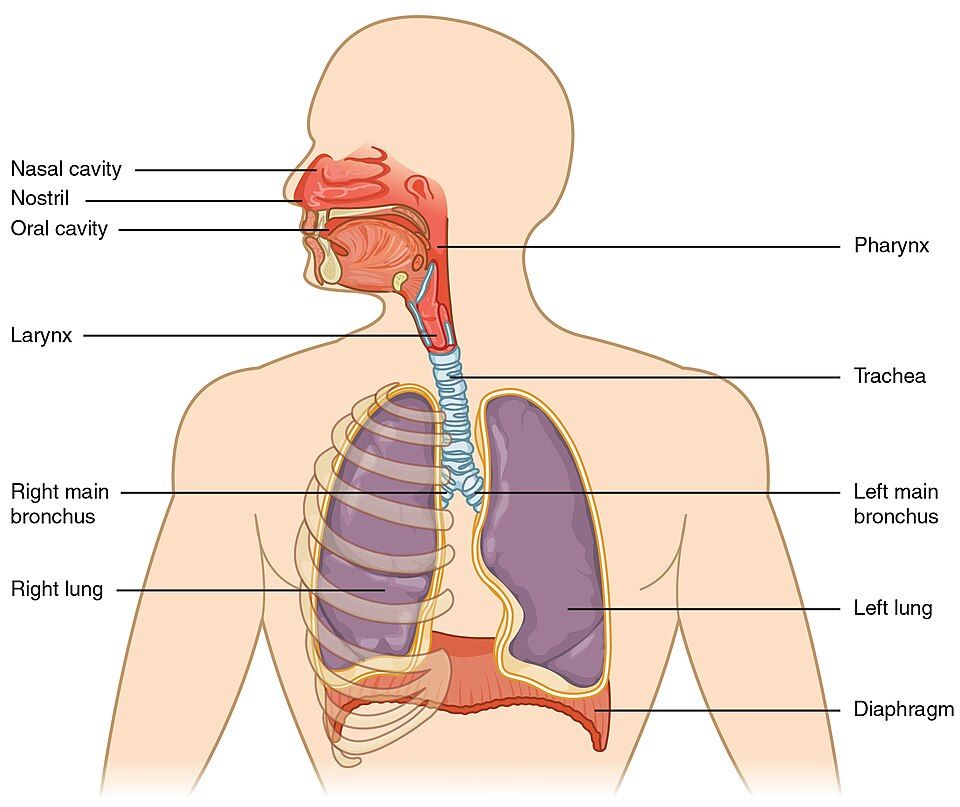
The respiratory system does one headline job — move oxygen in and carbon dioxide out — but it also warms and humidifies air, filters out debris, gives you the sense of smell, produces your voice, and helps regulate blood pH. To do all this, the airway is split into two functional zones. The conducting zone is plumbing: the nose, pharynx, larynx, trachea, bronchi, and bronchioles that carry air but never exchange gas. The respiratory zone — the respiratory bronchioles, alveolar ducts, and alveoli — is where the actual trade of oxygen for carbon dioxide takes place. Think of the conducting zone as the highway and the respiratory zone as the destination; air only does its real work once it arrives.
Clinicians also divide the tract by location. The upper respiratory tract is everything above the larynx (nose, nasal cavity, sinuses, pharynx), and the lower respiratory tract is the larynx on down (trachea, bronchi, lungs). This is why a "cold" (an upper-tract infection) feels so different from bronchitis or pneumonia (lower-tract problems).
The journey of a breath
Follow one inhalation. Air enters the nostrils, swirls through the nasal cavity, and passes into the three-part pharynx (throat). It slips through the larynx — the voice box, guarded by the epiglottis that flips down during swallowing so food goes to the esophagus instead of the lungs — and enters the trachea (windpipe), held permanently open by C-shaped rings of cartilage. The trachea forks into the right and left primary bronchi, which branch again and again like an upside-down tree into ever-smaller bronchi and then bronchioles, until the tubes end in clusters of alveoli. The whole branching pattern is called the bronchial tree.
| Structure | Zone | Key feature |
|---|---|---|
| Nasal cavity | Conducting | Warms, humidifies, filters; lined by mucous membrane |
| Pharynx | Conducting | Shared passage for air and food (naso-, oro-, laryngo-) |
| Larynx | Conducting | Voice box; epiglottis guards the airway |
| Trachea | Conducting | C-shaped cartilage rings keep it open |
| Bronchi | Conducting | Branch into each lung, then lobes and segments |
| Bronchioles | Conducting | No cartilage; smooth muscle sets their diameter |
| Alveoli | Respiratory | Thin-walled air sacs where gas exchange occurs |
The air that arrives is conditioned
By the time air reaches the alveoli it has been transformed: warmed to body temperature, saturated with water vapor, and scrubbed nearly clean. The nasal cavity does most of this conditioning, its rich blood supply radiating heat and its mucous lining trapping dust and microbes. Beneath that mucus sits a carpet of microscopic cilia beating in waves toward the throat — the mucociliary escalator — which sweeps trapped debris up and out to be swallowed or coughed away. It is a tireless housekeeping system that protects the delicate air sacs from the world.
Walk the whole tract, structure by structure, in the interactive below.
- The conducting zone (nose → bronchioles) moves air; the respiratory zone (respiratory bronchioles → alveoli) exchanges gas.
- Air path: nose → pharynx → larynx → trachea → bronchi → bronchioles → alveoli.
- Incoming air is warmed, humidified, and filtered; the mucociliary escalator sweeps debris up and out.
Section 2The alveolus & respiratory membrane
A surface the size of a room
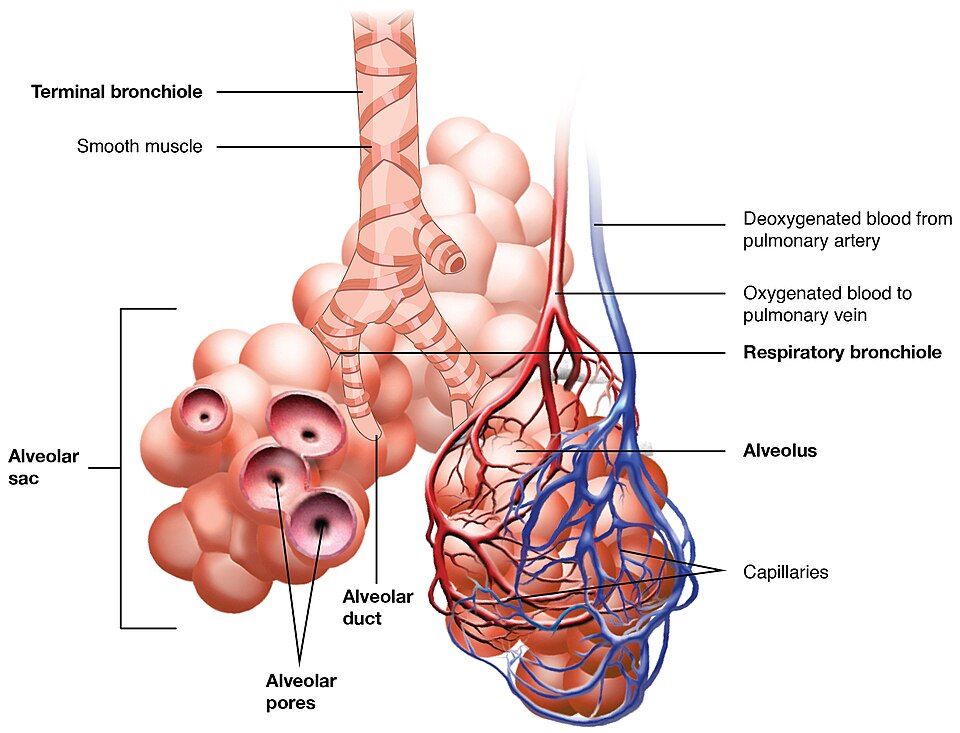
The lungs hold roughly 300 million alveoli, each a hollow, bubble-like sac wrapped in a mesh of capillaries. Individually they are tiny, but together their walls unfold into an astonishing 70 square meters of surface — about the floor area of a small apartment — packed inside your chest. This enormous area is the whole point: the more membrane you spread out, the more gas can cross it at once. Structure fits function, and here the function is to make oxygen and carbon dioxide diffusion as fast and abundant as physically possible.
The respiratory membrane: three thin layers
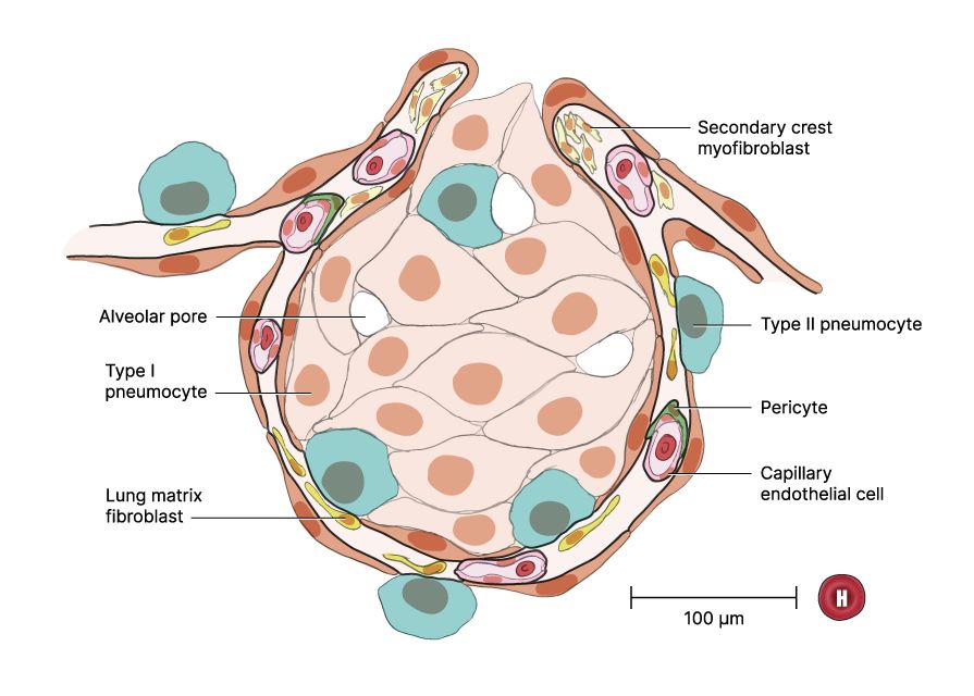
Gas exchange happens across the respiratory membrane, the barrier separating alveolar air from capillary blood. Despite the important-sounding name, it is almost nothing at all — about 0.5 micrometers thick, thinner than a soap bubble. It has just three layers: the alveolar wall (a single flat layer of type I cells), the capillary wall (a single flat layer of endothelial cells), and their fused basement membranes glued between them. Oxygen crosses all three and enters the blood in a fraction of a second; carbon dioxide crosses the other way just as fast.
| Cell / layer | What it is | Job |
|---|---|---|
| Type I alveolar cell | Simple squamous (flat) cell | Forms most of the wall; thin enough for gases to cross |
| Type II alveolar cell | Rounded cell scattered in the wall | Secretes surfactant |
| Alveolar macrophage | Roaming immune cell ("dust cell") | Engulfs debris and microbes that reach the sac |
| Capillary endothelium | Flat cells lining the capillary | The blood side of the respiratory membrane |
Why does the thinness matter so much? Because diffusion slows sharply as the barrier thickens. In diseases that thicken this membrane — pulmonary edema floods it with fluid, pulmonary fibrosis stiffens it with scar — oxygen suddenly has farther to travel, and the patient becomes short of breath even though the air and the blood are both right there.
Surfactant: keeping the sacs from collapsing
A wet-walled bubble wants to collapse, because water molecules lining it pull toward one another — a force called surface tension. Left unchecked, that tension would snap the smallest alveoli shut with every exhalation. To prevent this, the type II alveolar cells secrete surfactant, a soapy mix of lipids and proteins that coats the inner surface and breaks up the water’s pull, lowering surface tension and keeping the sacs open and easy to inflate.
Zoom into a single sac and its capillary in the interactive below.
- ~300 million alveoli give ~70 m² of surface for gas exchange.
- The respiratory membrane is ~0.5 µm thick and has three layers: alveolar wall, fused basement membranes, and capillary wall.
- Type II cells make surfactant, which lowers surface tension and keeps alveoli from collapsing.
Section 3Breathing mechanics
You make room, and air follows (Boyle’s law)
Here is the surprise most students never expect: you do not actively suck air into your lungs. You make the chest bigger, and air rushes in on its own to fill the extra space. The physics behind this is Boyle’s law — at a fixed temperature, the pressure of a gas rises as its volume falls, and falls as its volume rises. Squeeze a container and the pressure inside climbs; enlarge it and the pressure drops. Air always flows from higher pressure to lower pressure, so all your muscles have to do is change the volume of the chest, and the pressure difference moves the air for you.
Inspiration: an active pull
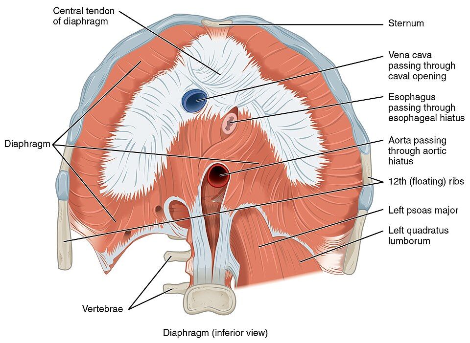
Breathing in is active — it takes muscle work. The diaphragm, a dome of muscle under the lungs, contracts and flattens downward, while the external intercostal muscles lift the ribs up and out. Both actions enlarge the thoracic cavity. By Boyle’s law, that larger volume drops the pressure inside the lungs (the intrapulmonary pressure) just below atmospheric pressure, and air flows in until the two pressures equalize. The lungs themselves are not muscular; they follow the chest wall because a thin film of fluid and a permanently negative intrapleural pressure keep them stuck to it, so when the chest expands, the lungs expand with it.
Expiration: mostly letting go
Breathing out at rest is the opposite — and easier, because it is usually passive. The diaphragm and external intercostals simply relax, the stretched lungs and chest wall recoil like a released rubber band, thoracic volume shrinks, intrapulmonary pressure rises above atmospheric, and air flows out. No muscle contraction is required for a quiet exhale. Only when you need to force air out — blowing out candles, coughing, exercising hard — do the internal intercostals and abdominal muscles pitch in to actively squeeze the chest.
| Inspiration | Expiration (quiet) | |
|---|---|---|
| Effort | Active (muscles contract) | Passive (muscles relax) |
| Main muscles | Diaphragm, external intercostals | None — elastic recoil |
| Thoracic volume | Increases | Decreases |
| Intrapulmonary pressure | Falls below atmospheric | Rises above atmospheric |
| Air movement | In | Out |
Watch the diaphragm change the chest’s volume and pressure in the interactive below.
- Boyle’s law: enlarging the chest lowers lung pressure so air flows in; shrinking it raises pressure so air flows out.
- Inspiration is active (diaphragm + external intercostals); quiet expiration is passive (elastic recoil).
- A negative intrapleural pressure keeps the lungs adhered to the chest wall; break the seal and the lung collapses (pneumothorax).
Section 4Gas exchange
Partial pressure: each gas pushes on its own
Air is a mixture, and in a mixture every gas exerts its own share of the total pressure — its partial pressure — as if the others were not there. Oxygen makes up about 21% of air, so at sea level its partial pressure (written PO₂) is roughly 160 mmHg in the atmosphere and about 104 mmHg once diluted inside the alveoli. This matters because gases, like everything else in diffusion, move from where their partial pressure is high to where it is low, and they keep moving until the two sides even out. Every gas exchange in the body is just this one rule applied over and over.
External respiration: loading O₂, dumping CO₂
External respiration is the swap that happens in the lungs, between alveolar air and the blood. Blood arriving from the body is oxygen-poor (PO₂ ≈ 40 mmHg) and carbon-dioxide-rich (PCO₂ ≈ 45 mmHg). The alveolar air is the reverse — high in oxygen (≈ 104 mmHg) and low in carbon dioxide (≈ 40 mmHg). So oxygen diffuses down its gradient into the blood, and carbon dioxide diffuses the other way out into the alveoli to be exhaled. Blood leaves the lungs fully loaded with oxygen and having given up a small share of its carbon dioxide — just enough to bring PCO₂ from about 45 back down to about 40 mmHg. Most of the blood’s carbon dioxide stays with it, parked in the large bicarbonate pool you will meet in Section 5; each pass through the lungs trims that pool rather than emptying it.
Internal respiration: the reverse at the tissues
Internal respiration is the same physics run backward, out in the body’s tissues. Cells constantly burn oxygen and pour out carbon dioxide, so the tissue PO₂ is low (≈ 40 mmHg) and its PCO₂ is high (≈ 45 mmHg). Blood arriving in the tissue capillaries is the opposite, so oxygen diffuses out of the blood into the cells and carbon dioxide diffuses into the blood to be hauled back to the lungs. Same rule, opposite direction — and the cycle repeats with every heartbeat.
| Gas | Alveoli (air) | Deoxygenated blood | Tissues |
|---|---|---|---|
| PO₂ | ~104 mmHg | ~40 mmHg | ~40 mmHg |
| PCO₂ | ~40 mmHg | ~45 mmHg | ~45 mmHg |
| Net movement | O₂ in, CO₂ out (external) | — | O₂ out, CO₂ in (internal) |
Switch between the lungs and the tissues in the interactive below.
- Each gas diffuses from high to low partial pressure and stops when the pressures equalize.
- External respiration (lungs): O₂ into blood, CO₂ out to be exhaled.
- Internal respiration (tissues): O₂ out to cells, CO₂ into blood — the same rule reversed.
Section 5Gas transport in the blood
Oxygen rides on hemoglobin
Oxygen barely dissolves in water, so blood plasma alone could never carry enough of it to keep you alive — only about 1.5% of the oxygen in blood is dissolved. The other 98.5% is bound to hemoglobin, the iron-containing protein packed inside red blood cells. Each hemoglobin molecule grips up to four oxygen molecules on its iron sites, forming oxyhemoglobin. This is the whole reason red blood cells exist: hemoglobin lets blood carry roughly seventy times more oxygen than it otherwise could.
The oxygen–hemoglobin dissociation curve
Hemoglobin has a wonderful property: it grabs oxygen tightly where oxygen is plentiful (the lungs) and releases it where oxygen is scarce (the tissues). Plot how saturated hemoglobin is against the surrounding PO₂ and you get the S-shaped oxygen–hemoglobin dissociation curve. Better still, hemoglobin reads the tissue’s needs: in an active, hard-working tissue — warmer, more acidic, richer in carbon dioxide — hemoglobin loosens its grip and hands off more oxygen right where it is needed most. This shift, driven by low pH and high CO₂, is the Bohr effect. It is homeostasis in miniature: the busiest cells automatically get the biggest oxygen delivery.
Carbon dioxide travels three ways
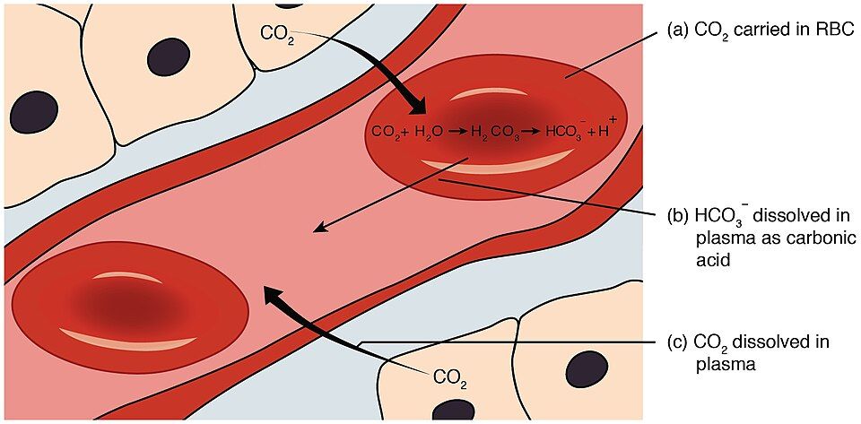
Carbon dioxide is more sociable than oxygen and hitches three different rides back to the lungs. A small share (~7%) simply dissolves in plasma; about 23% binds to hemoglobin (as carbaminohemoglobin, on a different site than oxygen); and the majority — about 70% — is converted inside red blood cells into bicarbonate. The enzyme carbonic anhydrase combines CO₂ with water into carbonic acid, which splits into a hydrogen ion and a bicarbonate ion (HCO₃⁻) that slips out into the plasma. At the lungs the whole reaction runs in reverse, releasing CO₂ to be breathed out.
| Gas | Form in blood | Share |
|---|---|---|
| Oxygen | Bound to hemoglobin (oxyhemoglobin) | ~98.5% |
| Oxygen | Dissolved in plasma | ~1.5% |
| Carbon dioxide | As bicarbonate (HCO₃⁻) | ~70% |
| Carbon dioxide | Bound to hemoglobin (carbaminohemoglobin) | ~23% |
| Carbon dioxide | Dissolved in plasma | ~7% |
Switch between oxygen and carbon dioxide transport in the interactive below.
- Oxygen is carried mostly on hemoglobin (~98.5%); each molecule binds four O₂.
- The Bohr effect — low pH plus high CO₂ — makes hemoglobin release more oxygen in hard-working tissues; the warmth of those tissues shifts the curve the same way by a separate route.
- Carbon dioxide travels as bicarbonate (~70%), on hemoglobin (~23%), and dissolved (~7%); this links breathing to blood pH.
Section 6Lung volumes & capacities
Four volumes
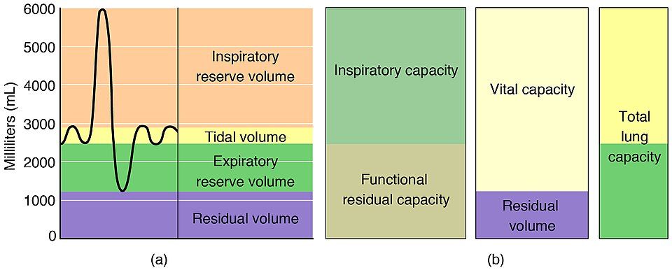
A spirometer records how much air you move as you breathe, and its trace is divided into four basic volumes. Tidal volume (TV) is the modest amount — about 500 mL — you move in a normal quiet breath. On top of that, inspiratory reserve volume (IRV, ~3000 mL) is the extra you can pull in with a maximal inhale, and expiratory reserve volume (ERV, ~1100 mL) is the extra you can force out after a normal exhale. Finally, residual volume (RV, ~1200 mL) is the air that always stays behind — you can never fully empty your lungs, and that leftover air keeps the alveoli from collapsing between breaths.
Four capacities (volumes added together)
A capacity is simply two or more volumes added together, and each one answers a practical question. Vital capacity (VC) — the total you can move from a maximal inhale to a maximal exhale (TV + IRV + ERV, ~4600 mL) — is the number clinicians care about most. Total lung capacity (TLC) adds the residual volume back in for everything the lungs can hold (~5800 mL). Inspiratory capacity (IC = TV + IRV) is all you can breathe in from rest, and functional residual capacity (FRC = ERV + RV) is what remains after a normal exhale.
| Measure | Definition | Typical value |
|---|---|---|
| Tidal volume (TV) | Air per normal quiet breath | ~500 mL |
| Inspiratory reserve (IRV) | Extra air forcibly inhaled | ~3000 mL |
| Expiratory reserve (ERV) | Extra air forcibly exhaled | ~1100 mL |
| Residual volume (RV) | Air that cannot be exhaled | ~1200 mL |
| Vital capacity (VC) | TV + IRV + ERV | ~4600 mL |
| Total lung capacity (TLC) | VC + RV (everything) | ~5800 mL |
Dead space & what spirometry reveals
Not every milliliter you inhale reaches the alveoli. About 150 mL stays in the conducting airways — the anatomical dead space — where no gas exchange happens, so of a 500 mL breath only ~350 mL is fresh air that actually does work. Spirometry becomes truly powerful when timed: the FEV₁/FVC ratio (how much of your vital capacity you can blow out in the first second) separates the two great families of lung disease. In obstructive disease (asthma, COPD) the airways are narrowed, so air comes out slowly and the ratio drops. In restrictive disease (fibrosis) the lungs are stiff and small, so volumes shrink but the air that is there comes out briskly, keeping the ratio normal or high.
Trace each volume on a spirometer in the interactive below.
- Four volumes: tidal, inspiratory reserve, expiratory reserve, and residual; capacities are volumes summed (VC, TLC, IC, FRC).
- Residual volume always stays behind and keeps alveoli open; it cannot be measured by simple spirometry.
- A low FEV₁/FVC ratio signals obstructive disease; shrunken volumes with a normal ratio signal restrictive disease.
The chapter in one breath
- The airway has a conducting zone that moves and conditions air and a respiratory zone where gas is actually exchanged; air travels nose → pharynx → larynx → trachea → bronchi → bronchioles → alveoli.
- Gas exchange occurs across the paper-thin respiratory membrane of ~300 million alveoli; surfactant from type II cells keeps the sacs from collapsing.
- Breathing obeys Boyle’s law: inspiration is active (diaphragm + external intercostals enlarge the chest), while quiet expiration is passive elastic recoil.
- Gases diffuse from high to low partial pressure — O₂ in and CO₂ out at the lungs (external respiration), and the reverse at the tissues (internal respiration).
- Oxygen is carried mostly on hemoglobin; carbon dioxide travels mostly as bicarbonate, tying breathing to blood pH.
- The lung’s volumes and capacities quantify how much air you move; a low FEV₁/FVC ratio flags obstructive disease and shrunken volumes flag restrictive disease.
ReferenceWords worth knowing
A quick reference for the terms in this chapter — skim it now, or circle back whenever a word trips you up.
- Alveolus
- A microscopic, thin-walled air sac (plural: alveoli) where oxygen and carbon dioxide are exchanged with the blood.
- Bicarbonate (HCO₃⁻)
- The ion into which most carbon dioxide is converted for transport in the blood; also a key player in acid–base balance.
- Bohr effect
- The fall in hemoglobin’s oxygen affinity caused by rising H⁺ (lower pH) and rising carbon dioxide, so hemoglobin releases more oxygen in active tissue. Warmth shifts the curve the same direction but by a separate mechanism, not by the Bohr effect.
- Boyle’s law
- At constant temperature, the pressure of a gas is inversely related to its volume; the basis of how the chest moves air.
- Bronchiole
- A small airway of the conducting zone with no cartilage, whose diameter is set by smooth muscle.
- Conducting zone
- The airways (nose through bronchioles) that carry, warm, humidify, and filter air but do not exchange gas.
- Dead space (anatomical)
- The ~150 mL of inhaled air that stays in the conducting airways and never reaches the alveoli for exchange.
- Diaphragm
- The dome-shaped muscle beneath the lungs; its contraction is the main driver of inspiration.
- Expiration
- Breathing out; passive at rest (elastic recoil) and active when forced.
- External respiration
- Gas exchange in the lungs between alveolar air and blood — oxygen in, carbon dioxide out.
- Hemoglobin
- The iron-containing protein in red blood cells that binds and carries up to four oxygen molecules.
- Inspiration
- Breathing in; an active process in which the diaphragm and external intercostals enlarge the chest.
- Internal respiration
- Gas exchange in the tissues between blood and cells — oxygen out to cells, carbon dioxide into blood.
- Intrapleural pressure
- The pressure in the fluid-filled space between the lung and chest wall; kept negative so the lung stays inflated against the wall.
- Partial pressure
- The share of total gas pressure contributed by one gas in a mixture; gases diffuse from high to low partial pressure.
- Residual volume
- The air (~1200 mL) that remains in the lungs after the most forceful exhale and cannot be voluntarily expelled.
- Respiratory membrane
- The ~0.5 µm barrier — alveolar wall, fused basement membranes, and capillary wall — across which gases diffuse.
- Spirometry
- A test that measures the volumes and flow rates of air a person breathes, used to diagnose lung disease.
- Surfactant
- A lipid-and-protein mixture from type II alveolar cells that lowers surface tension and keeps alveoli from collapsing.
- Tidal volume
- The volume of air (~500 mL) moved in a single normal, quiet breath.
- Vital capacity
- The maximum air a person can exhale after a maximal inhale (tidal + inspiratory reserve + expiratory reserve volumes).