The Digestive System
Every meal you have ever eaten was, in a sense, still outside your body until this system took it apart. The digestive system is a long muscular tube — roughly nine meters from mouth to anus — that ingests food, physically and chemically breaks it into molecules small enough to cross a cell membrane, absorbs those molecules into the blood, and packages the leftovers for disposal. In this chapter we will walk that tube from one end to the other, watch the muscle in its walls push and mix, step inside the acid bath of the stomach and the velvet lining of the small intestine, meet the accessory glands that pour in their juices, and finally follow a bite of food down to the individual building blocks your cells can burn. By the end you should be able to trace a sandwich from lips to bloodstream and name what happens at every stop.
By the end of this chapter you can
- Distinguish the alimentary canal from the accessory organs and list the organs in each group.
- Name the six essential digestive processes and the four layers of the gut wall.
- Contrast peristalsis and segmentation and explain what each accomplishes.
- Describe the regions of the stomach and the gastric gland cells along with what each secretes.
- Explain how the small intestine multiplies its surface area and how a single villus absorbs nutrients.
- State what the salivary glands, liver, gallbladder, and pancreas contribute and how their secretions reach the gut.
- Follow the chemical digestion of carbohydrates, proteins, fats, and nucleic acids to their absorbable end products.
- Connect common disorders — ulcers, celiac disease, gallstones, lactose intolerance — to the anatomy that explains them.
Section 1The digestive tract
One long tube, two kinds of organs
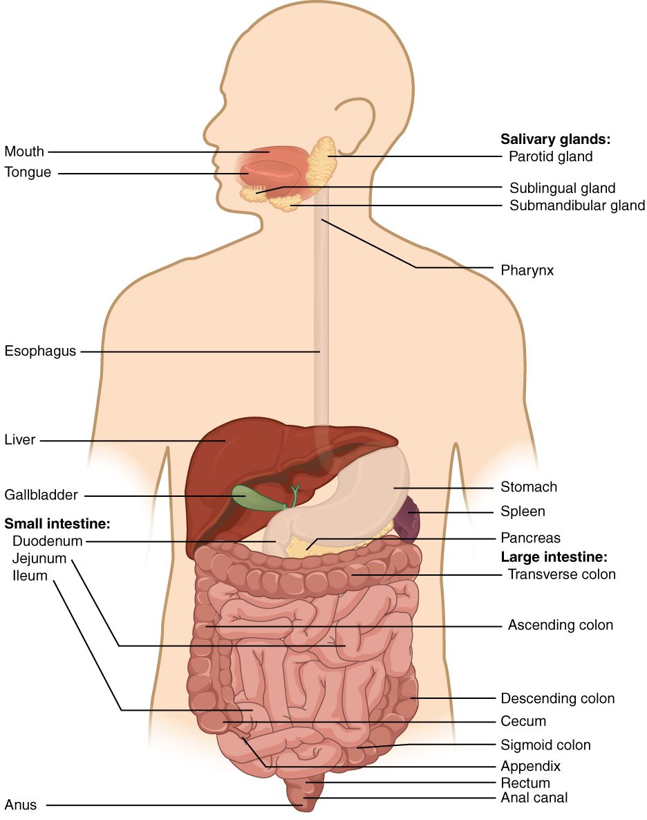
The digestive system has two divisions that are easy to keep straight once you see the logic. The alimentary canal — also called the gastrointestinal (GI) tract — is the continuous muscular tube that food actually passes through: the mouth, pharynx, esophagus, stomach, small intestine, large intestine, and finally the anus. Laid out end to end it runs about nine meters in a cadaver, though muscle tone shortens it considerably in life. The second division is the accessory organs — the teeth, tongue, salivary glands, liver, gallbladder, and pancreas. These never see the food pass through them; instead they either help mechanically (teeth, tongue) or pour secretions into the canal through ducts. Keeping "tube" and "helpers" separate is the first organizing idea of the whole chapter.
Six jobs, start to finish
Everything the system does can be sorted into six essential processes, and it helps to name them in order. Ingestion is taking food in through the mouth. Propulsion moves it along, first by voluntary swallowing and then by involuntary peristalsis. Mechanical digestion physically breaks food apart — chewing, the churning of the stomach, and segmentation in the intestine — increasing the surface area exposed to enzymes. Chemical digestion uses those enzymes to split large molecules into small ones. Absorption moves the small molecules across the gut wall into blood or lymph. And defecation eliminates whatever cannot be absorbed. A seventh word, secretion, threads through all of them: water, acid, enzymes, bile, and mucus are released along the way to make digestion possible.
The wall has four layers
From the esophagus to the anus the tube is built from the same four concentric layers, and knowing them lets you read any part of the tract. Innermost is the mucosa, the moist lining that secretes and absorbs and faces the hollow center, or lumen. Around it sits the submucosa, a layer of connective tissue packed with blood vessels, lymphatics, and a nerve network. Outside that is the muscularis externa, usually two sheets of smooth muscle — an inner circular layer and an outer longitudinal layer — whose contractions do the propelling and mixing. The outermost layer is the serosa (the visceral peritoneum) where the organ hangs free in the abdomen, or an adventitia of fibrous tissue where it is anchored to the body wall, as in the esophagus.
| Layer | What it is | Main job |
|---|---|---|
| Mucosa | Epithelium + thin connective tissue + a wisp of muscle, facing the lumen | Secretion, absorption, and defense at the surface |
| Submucosa | Loose connective tissue with vessels, lymphatics, glands, nerves | Supplies the mucosa; houses the submucosal nerve plexus |
| Muscularis externa | Inner circular and outer longitudinal smooth muscle | Peristalsis and segmentation; forms sphincters |
| Serosa / Adventitia | Outer visceral peritoneum, or fibrous anchoring layer | Wraps and suspends, or tethers, the organ |
Tap each organ in the interactive below to see where it sits and what job it does.
- The alimentary canal is the tube food passes through; the accessory organs (teeth, tongue, salivary glands, liver, gallbladder, pancreas) help from the side.
- The six processes are ingestion → propulsion → mechanical digestion → chemical digestion → absorption → defecation, with secretion throughout.
- The gut wall has four layers: mucosa, submucosa, muscularis externa, serosa/adventitia.
- The lumen is topologically outside the body; nutrients only truly enter once absorbed across the wall.
Section 2Moving and mixing
Two muscle layers, two kinds of motion
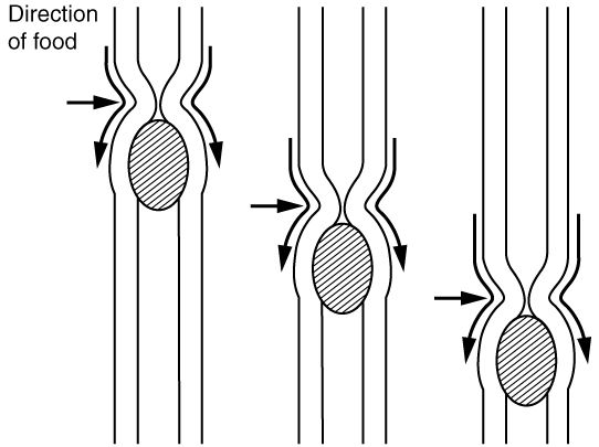
The smooth muscle of the muscularis externa produces two very different movements, and they solve two different problems. When the muscle works to move food forward, that is propulsion; when it works to mix food in place, that is mechanical digestion. The inner circular layer narrows the tube when it contracts; the outer longitudinal layer shortens it. By timing these two sheets against each other, the gut can either march a bolus along or churn it like a washing machine. Almost all of this happens automatically, without a thought, orchestrated by the gut's own nerve network with only background input from the brain.
Peristalsis: the wave that pushes
Peristalsis is the traveling wave of contraction that propels material along the tract. Just behind the bolus the circular muscle contracts to squeeze, while just ahead of it the muscle relaxes to receive — so the food is milked forward, the way you might squeeze the last of the toothpaste up the tube. Peristalsis is what carries a swallowed bite down the esophagus in a few seconds, and it is powerful enough to work against gravity: you can swallow while lying down, or even upside down, and the bolus still arrives at the stomach because the muscle, not gravity, is doing the work.
Segmentation: mixing in place
Segmentation is a nonmoving rhythm seen mainly in the small intestine. Rings of circular muscle contract at intervals, chopping the contents into segments, then relax as neighboring rings contract — sloshing the chyme back and forth without sending it anywhere in particular. This does two useful things at once: it blends food thoroughly with digestive enzymes, and it repeatedly presses the mixture against the absorptive wall so nutrients can be taken up. Only as absorption nears completion does segmentation give way to peristalsis, which finally moves the residue onward. The whole tempo is set by pacemaker cells — the interstitial cells of Cajal — that fire slow electrical waves the muscle follows.
| Feature | Peristalsis | Segmentation |
|---|---|---|
| Net effect | Moves food forward along the tract | Mixes food in place, little net movement |
| Muscle action | Contract behind, relax ahead — a traveling wave | Alternating stationary rings of contraction |
| Main location | Whole tract — esophagus, stomach, intestines | Small intestine (and colon) |
| Main purpose | Propulsion | Mechanical mixing and absorption contact |
Switch between the two motions in the interactive and watch how each one moves the contents.
- Peristalsis propels: contract behind the bolus, relax ahead, so it travels forward — even against gravity.
- Segmentation mixes chyme in place and presses it against the wall for absorption; it dominates the small intestine.
- Interstitial cells of Cajal set the rhythm both motions follow.
- Thickened circular muscle forms sphincters that act as one-way valves between regions.
Section 3The stomach
A muscular bag with regions and folds
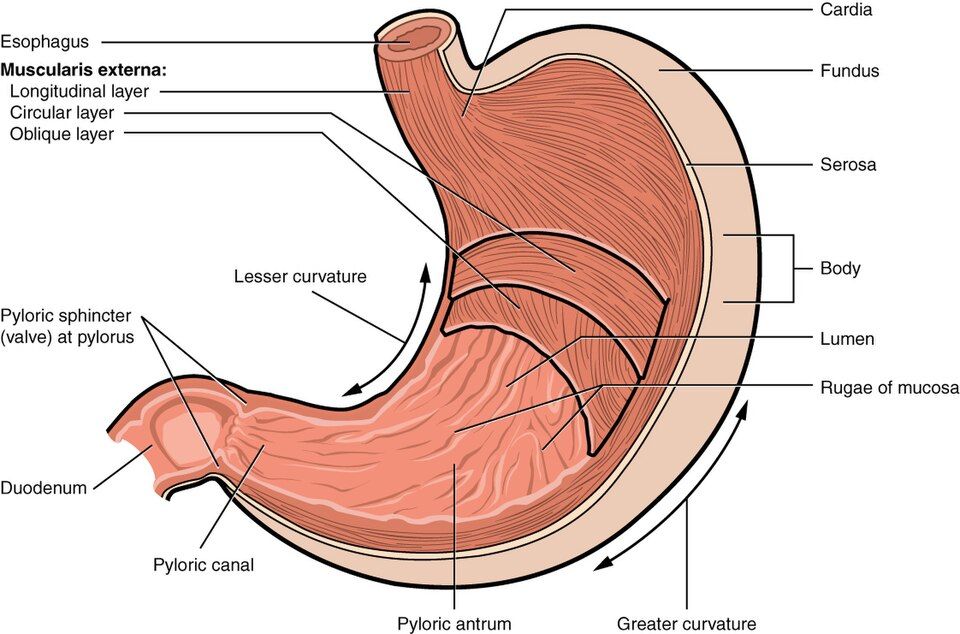
The stomach is a J-shaped expansion of the tract that acts as a storage tank, a mixing vat, and a chemical reactor all at once. It has four regions: the cardia, where the esophagus enters; the domed fundus that bulges up beside it; the large central body; and the funnel-shaped pylorus that narrows to the pyloric sphincter guarding the exit into the small intestine. When empty its lining is thrown into deep folds called rugae that flatten out as the stomach fills, letting it stretch to hold a liter or more. The stomach also has a third muscle layer the rest of the tract lacks — an inner oblique layer beneath the circular and longitudinal sheets — which lets it churn food in several directions at once, grinding a meal into the creamy, acidic soup called chyme.
The gastric glands and their cells
The stomach lining is dotted with millions of gastric pits that lead down into gastric glands, and the different cells lining those glands each make one signature product. Surface mucous cells, the columnar cells lining the lumen and the pits themselves, secrete the thick, bicarbonate-rich alkaline mucus that shields the lining from acid. Deeper in, in the neck of each gland, mucous neck cells secrete a different, thinner and slightly acidic mucus whose job is not that protective coat. Parietal cells are the workhorses that pump out hydrochloric acid (HCl) and, just as importantly, intrinsic factor. Chief cells release pepsinogen, the inactive precursor of the protein-splitting enzyme pepsin, plus a little gastric lipase. Scattered among them are enteroendocrine cells such as the G cells that release the hormone gastrin into the blood, which in turn drives the other cells to secrete. Together these cells produce two to three liters of gastric juice a day.
| Cell | Secretes | What it does |
|---|---|---|
| Surface mucous cell | Thick, bicarbonate-rich alkaline mucus | Coats and protects the lining from acid and pepsin |
| Mucous neck cell | Thin, acidic mucus | Lubricates within the neck of the gland; not the protective coat |
| Parietal cell | HCl and intrinsic factor | Acidifies chyme, activates pepsin, kills microbes; intrinsic factor is needed to absorb vitamin B₁₂ |
| Chief cell | Pepsinogen, gastric lipase | Pepsinogen becomes pepsin in acid and digests protein |
| Enteroendocrine (G) cell | Gastrin (a hormone) | Stimulates gastric secretion and motility |
Making acid — and surviving it
Parietal cells drive the stomach's pH down to about 1.5 to 2, acidic enough to unravel proteins and to convert pepsinogen into active pepsin. Secretion is switched on in three overlapping phases: the cephalic phase (the sight, smell, and thought of food, via the brain), the gastric phase (stretch and protein in the stomach itself, the largest phase), and the intestinal phase that first briefly encourages and then dampens secretion as chyme moves on. The obvious question is how the stomach avoids digesting itself. Its mucosal barrier answers this three ways: a thick coat of bicarbonate-rich mucus, tight junctions sealing the epithelial cells to one another, and rapid replacement of surface cells every few days, so any damaged patch is quickly resurfaced.
Switch between the stomach's regions and its secreting cells in the interactive.
- Regions: cardia, fundus, body, pylorus; rugae let it stretch; a third oblique muscle layer churns food into chyme.
- Gland cells: parietal (HCl + intrinsic factor), chief (pepsinogen), mucous neck (mucus), G cells (gastrin).
- Acid pH ~1.5–2 activates pepsin; secretion runs in cephalic, gastric, and intestinal phases.
- The mucosal barrier (bicarbonate mucus, tight junctions, fast cell turnover) keeps the stomach from digesting itself.
Section 4The small intestine
Three segments, one enormous surface
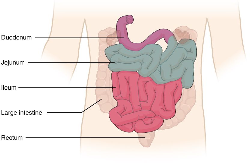
The small intestine is where nearly all chemical digestion finishes and nearly all absorption happens, so almost everything about it is built to maximize the surface exposed to chyme. It has three segments in sequence: the short, curved duodenum that receives chyme and the bile and pancreatic juices; the long jejunum, the main site of absorption; and the ileum, which finishes the job and empties into the large intestine at the ileocecal valve. Although it is only about an inch wide, it is by far the longest part of the tract, and it packs three levels of folding to turn that length into a surface roughly the area of a tennis court — on the order of 200 square meters.
Three amplifiers of surface area
The magnification comes in three nested tiers, each one folding the layer above it. First, the wall itself is thrown into permanent circular folds (plicae circulares) that force chyme to spiral slowly along, giving it more time and contact. Second, the surface of every fold is carpeted with millions of fingerlike villi, each a projection of mucosa about a millimeter tall. Third, the apical membrane of every absorptive cell on a villus is fringed with thousands of tiny microvilli, so densely packed they look furry under the microscope — the brush border. Together these three amplifiers multiply the flat tube's area more than 500-fold.
| Amplifier | What it is | Rough effect |
|---|---|---|
| Circular folds | Permanent ridges of the whole wall | Slow and spiral the chyme; ~3× area |
| Villi | Fingerlike mucosal projections, ~1 mm | Carpet every fold; ~10× area |
| Microvilli (brush border) | Membrane fringe on each cell's apex | Vast final surface; ~20× area; hold digestive enzymes |
Inside a single villus
Zoom into one villus and you find the whole absorption machine in miniature. Its outer layer is a sheet of absorptive epithelial cells (enterocytes) carrying the brush border, whose membrane enzymes finish digesting sugars and peptides right at the point of uptake. Inside the villus sits a network of capillaries that carry off absorbed sugars and amino acids to the liver, and a single blind lymphatic vessel called a lacteal that absorbs digested fats. Between the villi, deep pits called intestinal crypts continually manufacture fresh cells to replace the ones worn off the tips, which is why the lining renews itself every few days.
Tap each level of folding, then explore the parts of a single villus, in the interactive.
- Segments in order: duodenum → jejunum → ileum; this is where most digestion and absorption happen.
- Three surface amplifiers: circular folds → villi → microvilli (brush border), giving ~200 m².
- A villus contains a capillary network (sugars, amino acids) and a lacteal (fats), fed by cells born in the crypts.
- Destroying the villi — as in celiac disease — wrecks absorption.
Section 5The accessory organs
Glands that pour in from the side
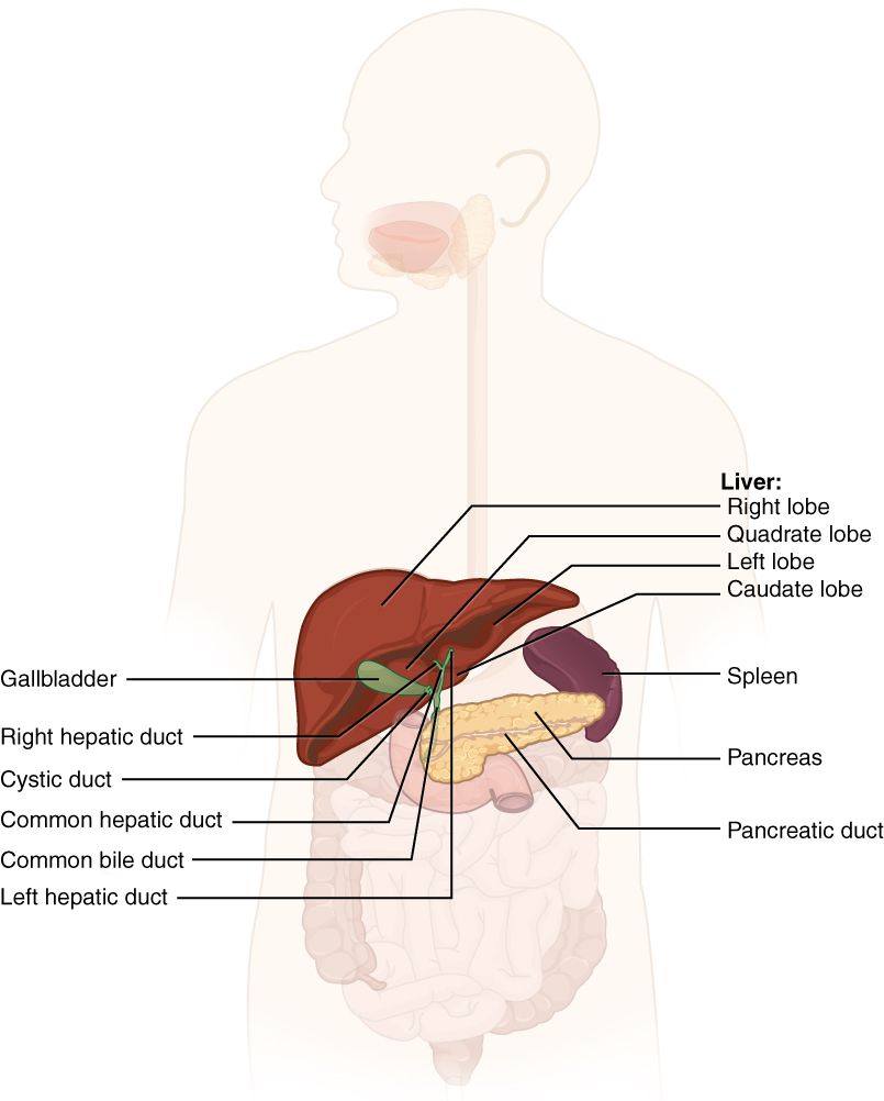
The accessory glands never let food pass through them; instead they manufacture secretions and deliver them into the canal through ducts, usually into the mouth or the duodenum. Three sets matter most. The salivary glands — the parotid in front of each ear, the submandibular under the jaw, and the sublingual under the tongue — release up to a liter and a half of saliva a day. Saliva moistens food into a swallowable bolus, begins starch digestion with salivary amylase, and defends the mouth with the antibacterial enzyme lysozyme and antibodies.
The liver and gallbladder
The liver is the body's largest gland and a chemical factory with dozens of jobs, but its one digestive contribution is bile. Bile is a greenish fluid containing bile salts that act on fat the way dish soap acts on grease: they emulsify it, breaking large fat globules into a fine suspension of tiny droplets so that fat-digesting enzymes have vastly more surface to attack. Between meals bile is diverted into the gallbladder, a small muscular pouch that stores and concentrates it. When a fatty meal reaches the duodenum, the hormone cholecystokinin (CCK) signals the gallbladder to squeeze, sending bile down the common bile duct into the small intestine.
The pancreas: two glands in one
The pancreas lies behind the stomach and does double duty. Its exocrine tissue — clusters of acinar cells — produces pancreatic juice, the most complete digestive cocktail in the body: enzymes for every food group (pancreatic amylase, lipase, the protein-splitters trypsin and chymotrypsin, and nucleases) plus a flood of bicarbonate that neutralizes the acidic chyme arriving from the stomach so the intestinal enzymes can work near neutral pH. Its endocrine tissue — the pancreatic islets — instead secretes insulin and glucagon into the blood to regulate blood sugar. The pancreatic duct joins the common bile duct at the hepatopancreatic ampulla, and their combined flow enters the duodenum through the sphincter of Oddi.
| Organ | Secretion | Digestive contribution |
|---|---|---|
| Salivary glands | Saliva (amylase, lysozyme, mucus) | Moisten food; start starch digestion; defend the mouth |
| Liver | Bile (bile salts) | Emulsifies fats into small droplets |
| Gallbladder | — (stores & concentrates bile) | Releases bile on demand via CCK |
| Pancreas (exocrine) | Pancreatic juice (enzymes + bicarbonate) | Digests all food groups; neutralizes acid |
Tap each accessory organ in the interactive to trace where its secretion goes.
- Salivary glands (parotid, submandibular, sublingual) moisten food and start starch digestion with amylase.
- The liver makes bile, which emulsifies fat; the gallbladder stores it and releases it via CCK.
- The pancreas supplies enzymes for every food group plus bicarbonate to neutralize acid, and separately makes insulin/glucagon.
- Pancreatic and bile ducts merge at the hepatopancreatic ampulla to enter the duodenum.
Section 6Chemical digestion
Why big molecules must be cut down
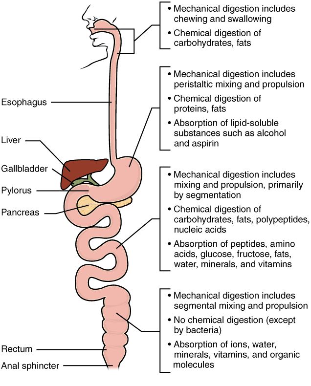
The nutrients in food arrive as large polymers — starch, proteins, fats, nucleic acids — and they are simply too big to cross the cell membranes of the gut lining. Chemical digestion solves this by hydrolysis: enzymes split the bonds holding each polymer together, adding a molecule of water at every cut, until only the small absorbable building blocks remain. Each food group has its own team of enzymes and its own set of end products, and it is worth learning them side by side because the exam and the clinic both ask you to trace a nutrient from plate to bloodstream.
Carbohydrates, proteins, and nucleic acids
Carbohydrate digestion starts in the mouth with salivary amylase and resumes in the small intestine with pancreatic amylase, both breaking starch into the disaccharide maltose and related fragments. Brush-border enzymes — maltase, sucrase, and lactase — then split those disaccharides into the monosaccharides glucose, fructose, and galactose, which are absorbed. Protein digestion begins in the stomach with pepsin and continues in the intestine with the pancreatic proteases trypsin and chymotrypsin, finishing with brush-border peptidases that release single amino acids. Nucleic acids are cut by pancreatic nucleases into nucleotides, and the brush-border nucleosidases and phosphatases then take those nucleotides apart into nitrogenous bases, pentose sugars, and phosphate ions — and it is those pieces, not whole nucleotides, that are absorbed.
Fats take a different route
Fat digestion is the special case. Because fat will not dissolve in the watery chyme, bile salts must first emulsify it into tiny droplets; only then can pancreatic lipase clip each triglyceride into monoglycerides and free fatty acids. These products are ferried to the villus surface in little bile-salt packages called micelles, absorbed into the enterocyte, and there reassembled into triglycerides, coated in protein, and shipped out as droplets called chylomicrons. Too big for capillaries, chylomicrons enter the lacteal instead — which is why dietary fat travels first through the lymph, not straight to the liver like sugars and amino acids.
| Nutrient | Key enzymes | Absorbed as |
|---|---|---|
| Carbohydrate | Salivary & pancreatic amylase; brush-border maltase, sucrase, lactase | Monosaccharides (glucose, fructose, galactose) |
| Protein | Pepsin (stomach); trypsin, chymotrypsin; brush-border peptidases | Amino acids |
| Fat | Bile salts (emulsify) + pancreatic lipase | Monoglycerides & fatty acids → chylomicrons |
| Nucleic acid | Pancreatic nucleases (to nucleotides); brush-border nucleosidases & phosphatases | Nitrogenous bases, pentose sugars, phosphate ions |
Tap a nutrient in the interactive to follow it down to its absorbable building blocks.
- Chemical digestion is hydrolysis — enzymes cut polymers into absorbable monomers.
- Carbs → monosaccharides (amylase then brush-border maltase/sucrase/lactase); proteins → amino acids (pepsin, trypsin, chymotrypsin, peptidases); nucleic acids → nucleotides → bases, pentose sugars, and phosphates.
- Fats need bile emulsification before lipase, then travel as chylomicrons into the lacteal.
- A missing brush-border enzyme (lactase) explains lactose intolerance.
The chapter in one breath
- The digestive system is an alimentary canal (mouth → anus) plus accessory organs, carrying out ingestion, propulsion, mechanical and chemical digestion, absorption, and defecation.
- The gut wall's four layers — mucosa, submucosa, muscularis externa, serosa/adventitia — repeat along the whole tube, and the lumen is topologically outside the body.
- Smooth muscle produces peristalsis (propels food forward) and segmentation (mixes it for absorption), with sphincters as one-way gates.
- The stomach churns food into chyme and secretes acid, pepsinogen, and intrinsic factor from parietal, chief, mucous, and G cells, protected by its mucosal barrier.
- The small intestine absorbs almost everything, using circular folds, villi, and microvilli to build a huge surface; each villus holds capillaries and a lacteal.
- The salivary glands, liver, gallbladder, and pancreas pour saliva, bile, and enzyme-rich, bicarbonate-buffered pancreatic juice into the canal.
- Chemical digestion hydrolyzes carbs to monosaccharides, proteins to amino acids, fats (after bile emulsification) to fatty acids and chylomicrons, and nucleic acids to nucleotides and then to bases, sugars, and phosphates.
ReferenceWords worth knowing
A quick reference for the terms in this chapter — skim it now, or circle back whenever a word trips you up.
- Absorption
- The movement of digested nutrients across the gut wall into the blood or lymph.
- Accessory organs
- Digestive structures — teeth, tongue, salivary glands, liver, gallbladder, pancreas — that aid digestion without food passing through them.
- Alimentary canal
- The continuous muscular tube (mouth, pharynx, esophagus, stomach, small and large intestine, anus) that food travels through; the GI tract.
- Bile
- A liver secretion containing bile salts that emulsifies fats; stored and concentrated in the gallbladder.
- Chylomicron
- A protein-coated droplet of reassembled fat that carries absorbed lipids from the intestine into the lacteal and lymph.
- Chyme
- The soupy, acidic mixture of partly digested food and gastric juice that leaves the stomach.
- Emulsification
- The physical breakup of large fat globules into tiny droplets by bile salts, increasing the surface available to lipase.
- Hydrolysis
- The enzyme-driven splitting of a large molecule by adding water; the chemistry of chemical digestion.
- Intrinsic factor
- A parietal-cell protein required for absorption of vitamin B₁₂ in the ileum; its loss causes pernicious anemia.
- Lacteal
- The blind lymphatic vessel in the center of a villus that absorbs digested fats (chylomicrons).
- Mucosa
- The innermost gut layer, facing the lumen, that secretes and absorbs.
- Parietal cell
- A gastric gland cell that secretes hydrochloric acid and intrinsic factor.
- Pepsin
- The stomach's protein-digesting enzyme, formed when acid activates pepsinogen from chief cells.
- Peristalsis
- A traveling wave of smooth-muscle contraction that propels material forward along the tract.
- Rugae
- Deep folds of the empty stomach lining that flatten as it fills, allowing it to stretch.
- Segmentation
- Stationary rings of contraction that mix chyme in place and press it against the absorptive wall.
- Serosa
- The outer visceral-peritoneum layer of a gut organ that hangs free in the abdomen.
- Sphincter
- A thickened ring of circular muscle that acts as a valve between regions of the tract.
- Villus
- A fingerlike mucosal projection of the small intestine, carpeted with microvilli, that absorbs nutrients.