The Appendicular Skeleton
Think of the axial skeleton as the central mast of your body. The appendicular skeleton is everything that hangs off that mast and lets you act on the world. It includes the arms that reach and grip and the legs that carry you and spring. It also includes two bony girdles, which are frames that bolt the limbs to the trunk. This part of the skeleton has 126 bones, and almost every one of them is a lever built for movement. In this chapter, we work outward from each girdle to the fingertips and toes. Along the way, you will see one theme again and again. The shoulder is built for freedom, and the hip is built for strength. The whole design is a set of trade-offs between the two. Learn the bones in this order, and the hand and foot will make sense. With 27 and 26 bones each, they stop being a jumble and become a clear map.
By the end of this chapter you can
- Define the appendicular skeleton, list its four divisions, and account for all 126 bones.
- Name the two bones of the pectoral girdle. Explain why the sternoclavicular joint is the only bony link between the arm and the axial skeleton.
- Name the bones of the upper limb (humerus, radius, and ulna), and place the radius on the thumb side.
- Sort the 27 bones of the hand and wrist into carpals, metacarpals, and phalanges, and name the eight carpals.
- Describe how three bones fuse to form each hip bone. Explain why the hip is steadier than the shoulder, and why the shoulder moves more freely.
- Compare the male and female pelvis, and explain how the female pelvis is shaped for childbirth.
- Name the bones of the lower limb, and tell the weight-bearing tibia from the fibula, which carries almost no weight.
- Sort the 26 bones of the foot and ankle into tarsals, metatarsals, and phalanges. Explain how the foot's arches absorb shock.
Section 1The pectoral girdle
Two bones, one clever compromise
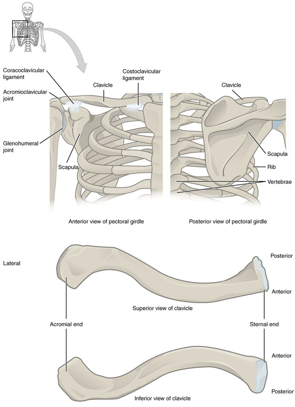
The pectoral girdle is your shoulder girdle. It is the frame that connects each arm to your trunk. It has only two bones on each side. The clavicle (collarbone) is in front, and the scapula (shoulder blade) is in back.
What is surprising is how loosely the whole arm is attached to the rest of the skeleton. The entire arm hangs from the axial skeleton at one small joint. This is the sternoclavicular joint, where the inner end of the clavicle meets the top of the breastbone (the manubrium of the sternum). The scapula is not joined by bone to the ribs or spine at all. It floats on the back of the rib cage, and only muscles hold it in place.
This loose setup is a trade. The shoulder gives up steadiness to gain a huge range of motion, which means how far a joint can move. You can swing your arm around in almost a full circle in every direction. The price is that the shoulder is one of the easiest joints in the body to dislocate, or pop out of place.
The clavicle: a slender strut
The clavicle is an S-shaped bone that works like a brace. It holds the shoulder out and away from the chest, so the arm can swing freely. Its rounded inner end is the sternal (medial) end, and it forms a joint with the sternum. Its flat outer end is the acromial (lateral) end. It meets a part of the scapula called the acromion at the acromioclavicular (AC) joint.
The clavicle is thin, and it sits right under the skin with nothing to pad it. So it is the most frequently fractured bone in the body. It usually breaks in its middle third. This often happens when someone falls onto an outstretched arm or onto the tip of the shoulder.
The scapula: a triangle of landmarks
The scapula is a flat, triangle-shaped bone. It is covered with bumps and ridges where muscles attach. A ridge called the spine runs across its back. On the outer side, the spine ends in the acromion, which is the bony tip of your shoulder that you can feel. A hook called the coracoid process curves forward like a bent finger. It anchors muscles and ligaments.
The most important feature is the glenoid cavity, also called the glenoid fossa. This is a shallow socket on the outer corner of the scapula. The rounded head of the upper arm bone (humerus) fits into it to form the shoulder joint. That shallow socket is the key to the whole design. It lets the arm bone swing widely, but it also lets the head slip out too easily. Picture a ball resting on a saucer instead of sitting in a cup.
| Bone / feature | What it is | Why it matters |
|---|---|---|
| Clavicle | S-shaped collarbone at the front of the girdle | Holds the shoulder out. It is the bone broken most often. |
| Scapula | Triangle-shaped shoulder blade at the back | Held on only by muscles. It carries the socket for the arm. |
| Glenoid cavity | Shallow socket on the scapula | Holds the head of the humerus. Shallow means it moves a lot but is not steady. |
| Acromion | Outer tip of the spine of the scapula | Forms the point of the shoulder. It joins the clavicle. |
| Sternoclavicular joint | Where the clavicle meets the sternum | The only bony joint that links the arm to the axial skeleton |
Explore both bones and their landmarks in the interactive below.
- The pectoral girdle has just two bones on each side, the clavicle and the scapula.
- The arm's only bony link to the axial skeleton is the sternoclavicular joint. The scapula floats on muscle.
- The glenoid cavity is shallow. It gives up steadiness for a huge range of motion.
- The clavicle is the most commonly fractured bone in the body.
Section 2Bones of the upper limb
Three long bones between shoulder and wrist
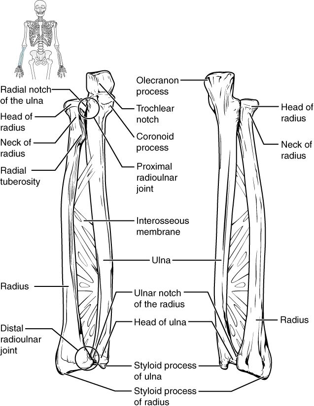
Each upper limb has 30 bones, but three big long bones do most of the work of holding it up. The upper arm is called the brachium, and it has one bone, the humerus. The forearm is called the antebrachium, and it has two bones side by side, the radius and the ulna.
To tell them apart, use the anatomical position. That means standing with your palms facing forward. In this position, the radius is on the thumb (lateral) side. The ulna is on the little-finger (medial) side. Here is a phrase that sticks: the radius rotates around the ulna, and the radius is on the same side as the thumb.
The humerus and the elbow
The humerus is the long bone of the upper arm. Its rounded head fits into the glenoid cavity at the shoulder. Just below the head is the surgical neck. This is a common place for breaks, and it sits very close to an important nerve (the axillary nerve). Farther down the shaft is the deltoid tuberosity, a rough bump where the deltoid muscle attaches.
At the elbow end, two rounded surfaces form the joint. The capitulum meets the radius. The trochlea is shaped like a pulley, and it grips the ulna. On either side of them are two bumps, the medial and lateral epicondyles. The ulnar nerve runs behind the inner (medial) one, in a shallow groove. There it lies just under the skin, with no muscle to cover it. So when you bump that spot, you press right on the nerve. A jolt shoots down your forearm. This is your "funny bone."
Radius and ulna: a rotating pair
The two forearm bones are joined along their length by a sheet of tough tissue, the interosseous membrane. They also meet at small joints near the top and bottom. This setup lets the forearm do a neat trick. It can turn the palm up, which is supination, or turn it down, which is pronation. In pronation, the radius turns and crosses over the ulna.
At the elbow, the ulna is the main partner. Its olecranon forms the bony point of your elbow. Its trochlear notch wraps around the trochlea of the humerus, like a wrench on a bolt. At the wrist, the radius takes over. It is the forearm bone that forms most of the joint with the wrist bones (carpals). Both bones end in a pointed tip called a styloid process.
| Bone | Where it is | Main job |
|---|---|---|
| Humerus | Upper arm (brachium) | Its head fits the glenoid cavity. Its lower end forms the elbow. |
| Radius | Forearm, thumb (lateral) side | Turns during pronation and supination. Forms the wrist joint. |
| Ulna | Forearm, pinky (medial) side | Its olecranon forms the point of the elbow. Keeps the forearm steady. |
Tap each of the three bones. Then turn the forearm from supination to pronation.
- The upper limb has three long bones: the humerus in the upper arm, and the radius and ulna in the forearm.
- With palms facing forward, the radius is lateral (thumb side) and the ulna is medial (pinky side).
- The ulna is the main bone at the elbow (the olecranon). The radius is the main bone at the wrist, and it does the turning.
Section 3The hand & wrist
Twenty-seven bones in three groups
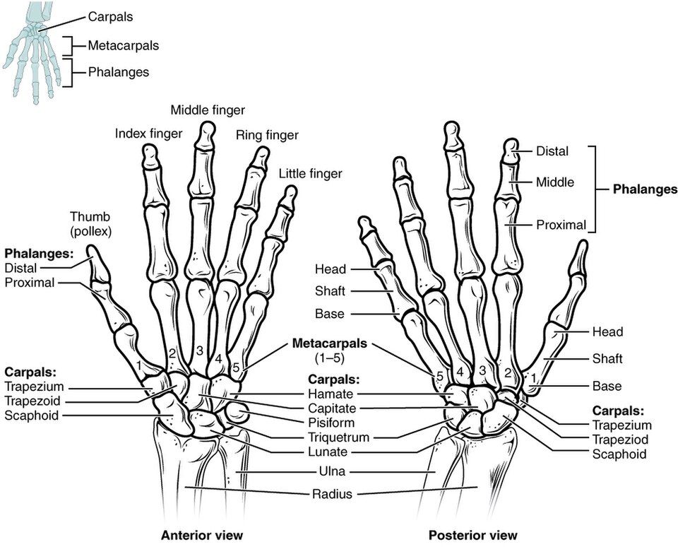
Your hand is the most skillful part of your body. That skill comes from packing 27 bones, and all the joints between them, into a small space. They sort neatly into three groups, from the wrist outward. There are 8 carpals in the wrist, 5 metacarpals in the palm, and 14 phalanges in the fingers. Learn those three numbers (8, 5, 14) in order, and you know the whole hand.
The carpals: eight little stones
The carpals are eight small wrist bones set in two rows of four. Ligaments bind them tightly, so the wrist bends as one unit. Start on the thumb side of the row nearest the forearm, then move to the row nearest the fingers. In that order, they are the scaphoid, lunate, triquetrum, pisiform, then the trapezium, trapezoid, capitate, hamate. A classic memory trick keeps the order: "Sally Left The Party To Take Cathy Home."
The scaphoid needs special attention. It is the carpal bone that breaks most often, usually in a fall onto an outstretched hand. Its blood supply enters from only one end. So a break can cut off blood to part of the bone, and that part can die.
Metacarpals and phalanges
The metacarpals are the five bones of the palm. They are numbered 1 to 5, starting at the thumb. Their rounded ends near the fingers are your knuckles. The thumb's metacarpal meets the trapezium at a saddle joint, shaped like a rider sitting in a saddle. This special joint lets the thumb swing across the palm to touch the other fingers. This is the opposable grip that makes the human hand so useful.
The phalanges are the finger bones. Each finger has three: proximal (nearest the palm), middle, and distal (at the tip). But the thumb, or pollex, has only two. That makes 14 per hand. (One finger bone is called a phalanx.)
| Group | Count | Where it is & notes |
|---|---|---|
| Carpals | 8 | Wrist, in two rows of four. The scaphoid is the one broken most often. |
| Metacarpals | 5 | Palm, numbered 1–5 from the thumb. Their heads are the knuckles. |
| Phalanges | 14 | Each finger has 3. The thumb has only 2. |
Tap each of the three groups below: the wrist, the palm, and the fingers.
- The hand has 27 bones: 8 carpals + 5 metacarpals + 14 phalanges.
- In order, the eight carpals are the scaphoid, lunate, triquetrum, pisiform, trapezium, trapezoid, capitate, and hamate.
- Each finger has 3 phalanges, and the thumb has 2. The thumb's saddle joint lets it touch the other fingers.
Section 4The pelvic girdle
Built for strength, not freedom
The shoulder is built to move freely. The pelvic girdle, or hip girdle, is built for the opposite. Its jobs are strength and weight-bearing. It must pass the whole weight of your upper body down into your legs. It must also take the pounding of every step.
So the pelvic girdle does not float on muscle. Instead, it attaches firmly to the axial skeleton at the sacroiliac joints. These are strong joints that barely move. The girdle itself is the pair of hip bones, also called coxal bones or os coxae. Together with the sacrum and coccyx, they form the bony pelvis, a bowl of bone at the base of your trunk.
Three bones fused into one
Each hip bone starts out as three separate bones. They fuse into one bone during adolescence, as a child grows into an adult. They meet at a deep, cup-shaped socket called the acetabulum. The ilium is the large, fan-shaped upper part. Its rim, the iliac crest, is the "hip" where you rest your hands. The ischium is the lower back part. Its rough ischial tuberosity is the bone you sit on. The pubis is the front part. The two pubic bones meet in front at the pubic symphysis, a joint made of tough cartilage (fibrocartilage).
Notice how different this is from the shoulder. The acetabulum is a deep socket that grips the head of the thigh bone (femur) firmly. So the hip almost never dislocates. It is the mirror image of the shallow glenoid cavity, which moves freely.
Male and female pelvis: shaped by childbirth
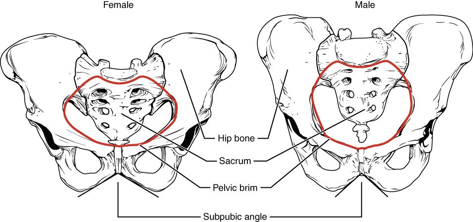
The pelvis is the part of the skeleton that differs most clearly between males and females. That is because the female pelvis is shaped to let a baby pass through. Compared with the male pelvis, the female pelvis is wider and shallower. Its opening at the top, the pelvic inlet, is larger and more rounded. The angle under the pubic bones, called the subpubic angle, is also wider. In a female it is greater than 90°.
The male pelvis is narrower and heavier. Its inlet is heart-shaped, and its subpubic angle is less than 90°. These differences are reliable. So anatomists and forensic scientists use the pelvis to tell whether a skeleton is male or female.
| Feature | Female pelvis | Male pelvis |
|---|---|---|
| Overall shape | Wider, shallower, lighter | Narrower, deeper, heavier |
| Pelvic inlet | Rounded, oval, and larger | Heart-shaped and smaller |
| Subpubic angle | Wide, more than 90° | Narrow, less than 90° |
| Reason for the difference | Roomy birth canal for childbirth | — |
Tap the ilium, ischium, and pubis. Then switch between the male and female pelvis.
- The pelvic girdle is two hip bones. Each is made of the ilium + ischium + pubis, fused where they meet at the acetabulum.
- It attaches firmly to the axial skeleton at the sacroiliac joints. It is built to bear weight, not to move a lot.
- The female pelvis is wider, with a subpubic angle greater than 90°. This shape helps with childbirth.
Section 5Bones of the lower limb
The strongest bones in the body
The lower limb carries your whole body weight and powers every step. So its bones are the largest and strongest you have. Like the arm, each lower limb has 30 bones. Four of them are the big bones that hold it up. The thigh has one bone, the femur. The patella, or kneecap, guards the knee. The lower leg has two bones, the tibia and fibula. They match the radius and ulna of the forearm, but with one key difference, which we will get to soon.
Femur and patella
The femur is the longest, heaviest, and strongest bone in the body. Its ball-shaped head sits deep in the acetabulum. Below the head is the angled neck. This is where a "hip fracture" usually happens in older adults with thin bones. Two bumps, the greater and lesser trochanters, anchor strong hip muscles. A ridge down the back, the linea aspera, gives muscles more places to attach. At the knee, the femur's rounded ends, the condyles, roll on top of the tibia.
The patella is the largest sesamoid bone in the body. A sesamoid bone is a bone that forms inside a tendon. The patella sits in the tendon of the quadriceps, the big muscle on the front of the thigh. It works like a pulley. It gives the thigh muscles better leverage when you straighten your knee. It also shields the knee from a direct hit.
Tibia and fibula: one carries, one steadies
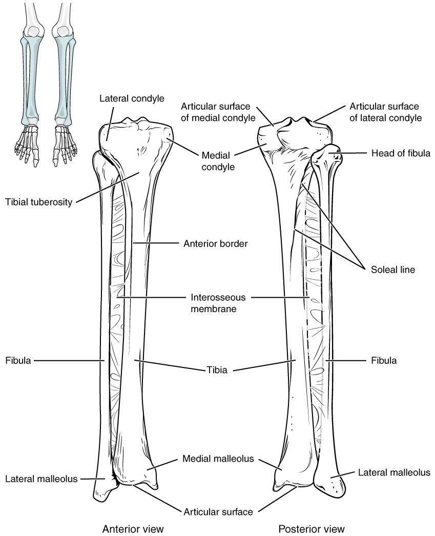
Here is how the leg differs from the forearm. In the leg, the two bones do not share the load. The tibia, or shinbone, is the thick inner (medial) bone. It carries almost all the weight. Its wide, flat top, the tibial plateau, supports the femur. Its lower end forms the medial malleolus, the bump on the inside of your ankle.
The fibula is the thin outer (lateral) bone. It carries almost no weight. Its jobs are to stabilize the ankle, or keep it steady, and to give muscles a place to attach. Its lower end forms the lateral malleolus, the bump on the outside of your ankle. In fact, the fibula does not even reach the knee. Only the tibia meets the femur there.
| Bone | Where it is | Job |
|---|---|---|
| Femur | Thigh | Longest, strongest bone. Its head fits the acetabulum. |
| Patella | Front of the knee | Largest sesamoid bone. Gives the quadriceps better leverage. |
| Tibia | Lower leg, medial | Weight-bearing shinbone. Forms the medial malleolus. |
| Fibula | Lower leg, lateral | Carries almost no weight. Steadies the ankle and forms the lateral malleolus. |
Tap each of the four bones. Then find the two malleoli at the ankle.
- The four big bones of the lower limb are the femur, patella, tibia, and fibula.
- The femur is the longest and strongest bone in the body. The patella is its largest sesamoid bone.
- The tibia (medial) bears the weight and forms the knee joint with the femur. The fibula (lateral) mainly steadies the ankle.
Section 6The foot & ankle
Twenty-six bones, built to bear weight
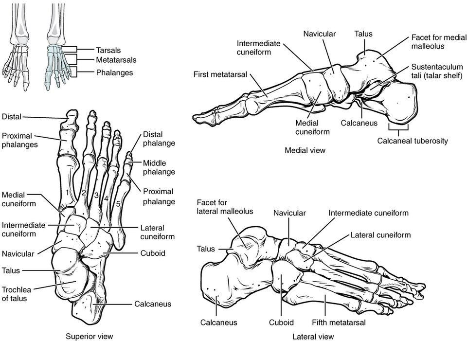
The foot is a lot like the hand, with three groups of bones. But it is tuned for a very different job. Instead of grabbing, it must bear weight and spring you forward. It has 26 bones. There are 7 tarsals in the ankle and heel, 5 metatarsals in the sole, and 14 phalanges in the toes. It is the same three-group plan as the hand, with slightly different numbers to remember (7, 5, 14).
The tarsals and the weight it takes
The tarsals are seven bones that form the ankle and the back of the foot. Two of them do the hard work. The talus sits on top, and the tibia and fibula grip it. It takes the body's weight from the leg and passes it down. The calcaneus, or heel bone, is the largest tarsal. It forms the heel and takes the weight when your foot hits the ground. The other five tarsals link the ankle to the sole. They are the navicular, the cuboid, and three cuneiforms.
The metatarsals are numbered 1–5, starting at the big toe. They form the sole and the ball of the foot. The phalanges are the toe bones. Each toe has three, but the big toe, or hallux, has only two. This is exactly the same pattern as the hand.
The arches: the foot's suspension
What really sets the foot apart from the hand is its arches. Ligaments and tendons hold the tarsals and metatarsals in three curves. The medial longitudinal arch is the high one along the inside of the foot. The lateral longitudinal arch runs along the outside. The transverse arch runs across the foot.
These arches make the foot a natural spring and shock absorber, like the springs in a car. They flatten a little to soak up the impact of each step. Then they spring back to push you off. When the arches drop, the result is flat feet (pes planus). This can make the feet and legs tired, because the shock absorber is gone.
| Group | Count | Where it is & notes |
|---|---|---|
| Tarsals | 7 | Ankle and heel. The talus takes weight from the leg. The calcaneus is the heel. |
| Metatarsals | 5 | Sole, numbered 1–5 from the big toe. Their heads form the ball of the foot. |
| Phalanges | 14 | Each toe has 3. The big toe (hallux) has 2. |
Tap each of the three groups (ankle, sole, and toes) below, and see the arches.
- The foot has 26 bones: 7 tarsals + 5 metatarsals + 14 phalanges.
- The talus takes weight from the leg. The calcaneus is the heel and the largest tarsal.
- Three arches make the foot a spring and shock absorber. When they fall, the result is flat feet.
The chapter in one breath
- The appendicular skeleton has 126 bones. These are the two limb girdles plus the arms and legs that hang from them.
- The pectoral girdle (clavicle + scapula) links the arm to the trunk by just one bony joint, the sternoclavicular joint. It gains freedom of movement but loses steadiness.
- The upper limb has three long bones: the humerus, the radius (thumb side), and the ulna (pinky side). The ulna leads at the elbow, and the radius leads at the wrist.
- The hand has 27 bones: 8 carpals, 5 metacarpals, and 14 phalanges. Its thumb can touch the other fingers, which makes the hand skillful.
- The pelvic girdle is two hip bones. Each is the ilium + ischium + pubis, fused at the acetabulum. It is built for strength and bearing weight. The female pelvis is shaped for childbirth.
- The lower limb has the strongest bones in the body: the femur, the patella, the weight-bearing tibia, and the steadying fibula.
- The foot has 26 bones: 7 tarsals, 5 metatarsals, and 14 phalanges. Its arches make it a spring and shock absorber.
- The whole design is a trade-off. The shoulder chooses freedom, and the hip chooses strength.
ReferenceWords worth knowing
Here is a quick list of the terms in this chapter. Skim it now, or come back when a word trips you up.
- Acetabulum
- The deep, cup-shaped socket on the hip bone. The head of the femur fits into it to form the hip joint.
- Appendicular skeleton
- The 126 bones of the limbs and the two girdles (pectoral and pelvic). The girdles attach the limbs to the axial skeleton.
- Calcaneus
- The heel bone. It is the largest tarsal, and it bears weight when your foot hits the ground.
- Carpals
- The eight small bones of the wrist, set in two rows of four.
- Clavicle
- The S-shaped collarbone that holds the shoulder out. It is the bone broken most often in the body.
- Femur
- The thigh bone. It is the longest, heaviest, and strongest bone in the body.
- Fibula
- The thin outer (lateral) bone of the lower leg. It carries almost no weight. It steadies the ankle and forms the lateral malleolus.
- Glenoid cavity
- The shallow socket on the scapula. The head of the humerus fits into it at the shoulder joint.
- Hip bone (os coxae)
- Also called the coxal bone. Each one is the ilium, ischium, and pubis fused together, and the pair forms the pelvic girdle.
- Humerus
- The single long bone of the upper arm. It runs from the shoulder to the elbow.
- Metacarpals
- The five bones of the palm, numbered 1 to 5 from the thumb. Their heads form the knuckles.
- Metatarsals
- The five bones of the sole of the foot, numbered 1 to 5 from the big toe.
- Patella
- The kneecap. It is the largest sesamoid bone, and it forms inside the quadriceps tendon to improve leverage.
- Pectoral girdle
- The shoulder girdle, made of the clavicle and scapula. It attaches each arm to the axial skeleton.
- Pelvic girdle
- The two hip bones. They attach the legs to the axial skeleton and bear the body's weight.
- Phalanges
- The bones of the fingers and toes. Each digit has three, except the thumb and big toe, which have two. One bone is a phalanx.
- Radius
- The outer (thumb-side) bone of the forearm. It turns during pronation and forms most of the wrist joint.
- Scapula
- The triangle-shaped shoulder blade. Only muscles hold it to the trunk, and it carries the glenoid cavity.
- Talus
- The tarsal bone that the tibia and fibula grip. It takes the body's weight from the leg.
- Tarsals
- The seven bones of the ankle and the back of the foot. They include the talus and calcaneus.
- Tibia
- The inner (medial) shinbone. It is the weight-bearing bone of the lower leg. It forms the knee joint with the femur and forms the medial malleolus.
- Ulna
- The inner (pinky-side) bone of the forearm. Its olecranon forms the point of the elbow.