Assessing the Thorax and Lungs
The chest is the one region where a nurse can detect a patient deteriorating before any monitor announces it. Respiratory rate is the earliest vital sign to change in sepsis, in hemorrhage, in opioid oversedation and in heart failure — and it is the vital sign most often invented rather than counted. This chapter teaches the thoracic exam the way it is actually performed at the bedside: the bony landmarks you find with your fingertips, the lobes lying underneath them, and then the four steps in their fixed order — inspection, palpation, percussion, auscultation. You will learn what resonance sounds like against dullness, why bronchial breath sounds in the periphery mean consolidation, why a wheeze that disappears is worse than a wheeze that does not, and how to write a lung note precise enough that the next nurse can tell whether the patient improved. Every finding here has an escalation threshold attached, because a chest exam that ends in your head instead of in an SBAR call helps no one.
By the end of this chapter you can
- Locate the suprasternal notch, sternal angle (angle of Louis), vertebra prominens, costal angle and the standard thoracic reference lines, and name the lobe lying beneath any point you touch.
- Perform the thoracic exam in the correct order — inspection, palpation, percussion, auscultation — and explain why the abdomen is the single exception to that sequence.
- Count and describe respiratory rate, depth and pattern, and state the expected ranges for the newborn, infant, toddler, preschooler, school-age child, adolescent, adult and older adult.
- Assess chest expansion and tactile fremitus, and interpret increased and decreased fremitus.
- Distinguish the five percussion tones and measure diaphragmatic excursion (expected 3–5 cm).
- Differentiate vesicular, bronchovesicular and bronchial breath sounds and identify crackles, wheezes, rhonchi, stridor and pleural friction rub.
- Elicit and interpret bronchophony, egophony and whispered pectoriloquy.
- Recognize respiratory red flags requiring immediate escalation and communicate them using SBAR.
- Document a lung assessment in precise, defensible language and apply the five rights of delegation to respiratory tasks.
Section 1Landmarks, Lobes and the Respiratory Baseline
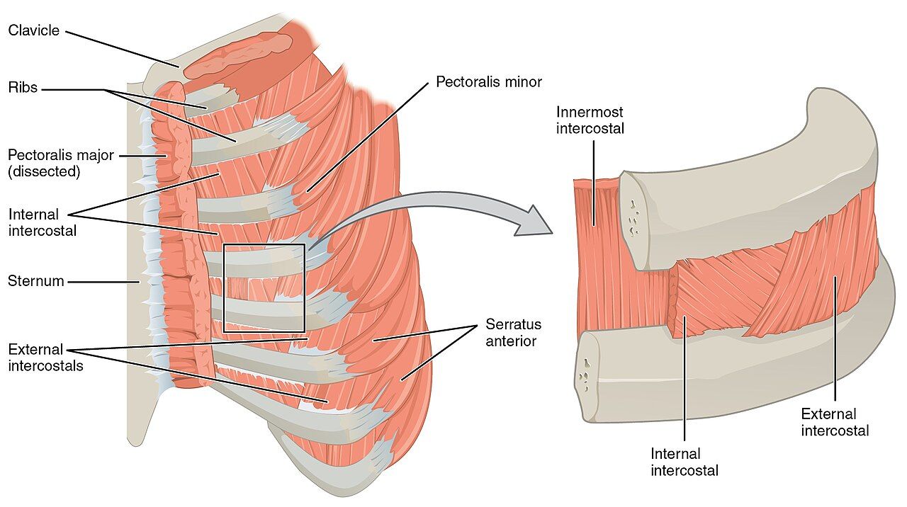
Reference lines and the bony landmarks you can find in the dark
A lung finding without a location is nearly useless. “Crackles in the chest” tells the next nurse nothing; “fine end-inspiratory crackles, bilateral posterior bases, below the level of the scapular tips” tells them exactly where to listen at 0300 to see whether the patient got better. Naming a location requires two coordinates: a horizontal one from the ribs and intercostal spaces, and a vertical one from a reference line. Learn to find both by touch, because you will often be assessing in a dim room with a patient who does not want to sit forward.
Start at the suprasternal notch, the hollow at the top of the sternum. Walk your fingers down about 5 cm until you feel a firm ridge — the sternal angle, or angle of Louis, where the manubrium meets the body of the sternum. The second rib articulates here, so the space just below your finger is the second intercostal space. From that single landmark you can count every rib on the anterior chest. Count in the intercostal spaces, not on the ribs, and remember that the costal cartilages of ribs eight through ten fuse rather than attach to the sternum, so anterior counting becomes unreliable below about the seventh rib. The sternal angle also marks the level where the trachea bifurcates into the right and left mainstem bronchi (roughly T4 to T5) — useful, because that is exactly where normal breath sounds are loudest and most tubular.
Posteriorly, ask the patient to flex the neck forward and find the vertebra prominens, the prominent spinous process of C7. Below it lies T1. The inferior angle of the scapula sits at approximately the seventh rib or seventh intercostal space and is the practical marker for “the bases.” The vertical reference lines are: midsternal, midclavicular and anterior axillary anteriorly; anterior, mid- and posterior axillary laterally; and vertebral (midspinal) and scapular posteriorly. The costal angle, formed where the two costal margins meet at the xiphoid, is normally less than 90 degrees and widens in chronic air trapping.
What lies beneath: lobes, fissures and lung borders
The right lung has three lobes, the left has two plus the lingula, which is the left lung’s anatomic answer to the right middle lobe. The clinically important fact is where those lobes sit relative to the skin. The oblique (major) fissures run from about the T3 spinous process posteriorly, around the chest, to the sixth rib at the midclavicular line. Everything you auscultate on the posterior chest below the scapular tips is lower lobe. The right horizontal (minor) fissure runs from the fourth rib at the sternum to the fifth rib at the midaxillary line, so the right middle lobe is heard on the anterior and right lateral chest only — never on the back. This is why nurses who auscultate only the posterior chest miss right middle lobe pneumonia and aspiration, and why the lateral chest wall at the midaxillary line is a mandatory part of the exam, not an optional one.
The lung apices extend 2 to 4 cm above the inner third of the clavicles, so a finding “above the clavicle” is still lung. The lower borders sit at roughly the sixth rib at the midclavicular line, the eighth rib at the midaxillary line, and the level of T10 posteriorly, descending to about T12 on deep inspiration. Below those borders you are percussing liver on the right and stomach or spleen on the left, which is why a dull note at the right base near the sixth rib is normal liver and not consolidation.
The respiratory baseline and the order of the exam
Count respirations for a full 30 seconds and multiply by two when the pattern is regular, and for a full 60 seconds when it is irregular, when the patient is an infant, or when the rate is at either extreme. Count without announcing it — keep your fingers on the radial pulse and watch or feel the chest, because a patient who knows you are counting breathes differently. Document rate, depth (shallow, normal, deep) and rhythm (regular, irregular), plus effort and oxygen saturation with the delivery device and flow rate. “RR 18” alone is an incomplete vital sign.
| Age group | Expected respiratory rate (breaths/min) | Bedside notes |
|---|---|---|
| Newborn (birth to 1 month) | 30–60 | Irregular periodic breathing with pauses under 20 seconds is normal; a pause of 20 seconds or more, or any pause with color or heart-rate change, is apnea |
| Infant (1–12 months) | 30–53 | Nose breathers in the early months; a blocked nose alone can cause distress |
| Toddler (1–2 years) | 22–37 | Breathing remains abdominal/diaphragmatic |
| Preschooler (3–5 years) | 20–28 | Thoracic breathing emerges around age 6 to 7 |
| School-age (6–11 years) | 18–25 | Chest wall still thin; sounds transmit widely |
| Adolescent (12–18 years) | 12–20 | Adult pattern established |
| Adult | 12–20 | Over 20 is tachypnea; under 12 is bradypnea; both are reportable trends |
| Older adult | 12–20 | May sit at the upper end; a sustained rate over 24 in an older adult is a sepsis and pneumonia screen, not normal aging |
| Pregnancy (any trimester) | 12–20 (usually unchanged) | Tidal volume rises 30% to 40%; rate itself should not rise much — tachypnea in pregnancy is abnormal |
Oxygen saturation by pulse oximetry is normally 95% to 100% on room air. A saturation of 90% corresponds roughly to a PaO2 of 60 mmHg — the shoulder of the oxyhemoglobin dissociation curve, below which small further drops in saturation mean large drops in PaO2. For most patients with chronic carbon dioxide retention the prescribed target is 88% to 92%, because excess oxygen worsens ventilation-perfusion matching and can blunt ventilatory drive; titrate only within the ordered range. That target is never a reason to withhold oxygen — a patient who is acutely and severely hypoxemic is given oxygen first, and the target range is re-established once they are safe. Always chart saturation with its context: “SpO2 94% on 2 L/min nasal cannula” means something entirely different from “SpO2 94% on room air.”
The thoracic exam runs inspection, palpation, percussion, auscultation — in that order, every time. The abdomen is the one exception: there you inspect, then auscultate, then percuss, then palpate, because pressing on the abdomen first changes bowel sounds and can produce falsely absent or hyperactive findings. Learn the exception as an exception so you never carry it into the chest.
Every chest finding needs a rib level and a reference line, because a location-free finding cannot be compared to the next nurse’s assessment.
Section 2Inspection and Palpation of the Thorax
Inspection: what the first ten seconds tell you
Inspection begins before you touch anything, and often before you speak. Expose the chest with the patient sitting upright, gown open, room warm and light good. Stand back far enough to see the whole thorax and watch a full minute of breathing. Note position first: a patient sitting bolt upright, leaning forward on their elbows, is in the tripod position and is recruiting every accessory muscle they have. Note speech: a patient who can speak only three or four words between breaths is in significant distress regardless of the number on the pulse oximeter.
Look at shape. The normal adult chest is wider than it is deep; the anteroposterior to transverse diameter ratio is about 1:2 (ranging to 5:7). In a barrel chest the ratio approaches 1:1, the ribs run horizontally rather than sloping down at roughly 45 degrees, and the costal angle exceeds 90 degrees — the configuration of chronic air trapping in emphysema and long-standing asthma, and an expected finding in infants under about two years and in some older adults with kyphosis. An increased anteroposterior diameter is a chronic finding, so it never explains a new symptom — do not let it talk you out of an acute change. Note also pectus excavatum (a sunken sternum), pectus carinatum (a protruding sternum), kyphosis, scoliosis and any surgical scars or chest tube sites.
Look at effort. Normal quiet breathing is almost invisible: the chest rises symmetrically, the accessory muscles are silent, and the ratio of inspiration to expiration is about 1:2. Abnormal effort shows as retractions (skin sucking in above the clavicles, between the ribs, or below the xiphoid), nasal flaring, grunting (an infant generating their own positive end-expiratory pressure — always ominous), head bobbing in an infant, use of the sternocleidomastoid, scalene and trapezius muscles, and pursed-lip breathing. Watch for paradoxical movement, where a segment of chest wall moves inward on inspiration — flail chest after trauma. Finally, look at the fingers: the normal nail-bed angle is about 160 degrees, and clubbing (angle 180 degrees or more, with a spongy nail bed) signals months to years of hypoxemia, not an acute event.
Breathing patterns worth naming
| Pattern | What it looks like | What it suggests |
|---|---|---|
| Eupnea | 12–20/min in an adult, regular, effortless, I:E about 1:2 | Expected |
| Tachypnea | Over 20/min, usually shallow | Fever, pain, anxiety, hypoxemia, sepsis, metabolic acidosis, pulmonary embolism |
| Bradypnea | Under 12/min, regular | Opioids and sedatives, increased intracranial pressure, hypothyroidism — check sedation level |
| Hyperpnea | Normal or increased rate with increased depth | Exercise, anxiety, early metabolic acidosis |
| Kussmaul respirations | Deep, labored, fast and regular | Severe metabolic acidosis, classically diabetic ketoacidosis — the body blowing off CO2 |
| Cheyne-Stokes | Crescendo–decrescendo depth alternating with apneic pauses of 15–60 seconds | Heart failure, uremia, brain injury, end of life; can be normal in infants and in adults at high altitude during sleep |
| Biot (ataxic) | Irregular breaths of variable depth with abrupt apneic pauses | Medullary damage, severe brain injury — escalate |
| Apnea | Cessation of breathing; 20 seconds or more in an infant | Always an emergency in any age group when sustained |
| Orthopnea | Dyspnea lying flat, relieved by sitting up; chart as pillows required | Heart failure, COPD, large effusion; document as “two-pillow orthopnea” |
Palpation: tenderness, expansion, fremitus and crepitus
Palpate with warm hands and a stated purpose. First, palpate for tenderness, masses and lesions with the finger pads over the anterior, lateral and posterior chest wall, and note whether pain is reproducible with pressure — reproducible chest wall tenderness suggests a musculoskeletal or costochondral source, but it never rules out a cardiac or pulmonary cause on its own. Confirm the trachea is midline by placing an index finger in the suprasternal notch and slipping it gently to each side to compare the spaces between the trachea and the sternocleidomastoid. Deviation is a late and dangerous sign.
Next assess chest expansion. Standing behind the seated patient, place your thumbs along the spine at about the level of the tenth rib with your palms lightly on the posterolateral chest, and slide medially just enough to raise a small skin fold between your thumbs. Ask for a deep breath. Your thumbs should move apart symmetrically, separating about 3 to 5 cm. Asymmetry is the finding that matters: the side that lags is the abnormal side — think pneumothorax, pleural effusion, lobar pneumonia, atelectasis, splinting from rib fracture, or post-operative guarding.
Then assess tactile fremitus, the palpable vibration of the voice transmitted through lung tissue. Use the ball of the palm or the ulnar surface of one hand, move in a side-to-side ladder pattern, and ask the patient to repeat “ninety-nine” or “one-one-one” in a low voice each time you move. Fremitus is normally strongest near the large airways — the second intercostal spaces anteriorly and between the scapulae posteriorly — and fades toward the bases. Compare sides, not absolute intensity. Solid, consolidated lung conducts sound better, so fremitus increases in pneumonia. Air, fluid or fat between your hand and the lung conducts it worse, so fremitus decreases in pneumothorax, pleural effusion, emphysema, bronchial obstruction and simply in a thick chest wall or a very muscular back.
Finally, feel for crepitus — subcutaneous emphysema. It feels like fine bubble wrap or crackling tissue paper under the skin, usually over the upper chest, neck or around a chest tube insertion site. It means air has escaped into the subcutaneous tissue and demands immediate assessment for pneumothorax, a disconnected or malpositioned chest tube, or airway injury. Mark the borders with a skin pen, note the time, and escalate.
| Palpation finding | Expected | Abnormal and what it suggests |
|---|---|---|
| Trachea | Midline in the suprasternal notch | Deviated toward the lesion: atelectasis, pneumonectomy, fibrosis. Deviated away: tension pneumothorax, large pleural effusion, large mass — emergency |
| Chest expansion | Symmetric, thumbs separate 3–5 cm | Unilateral lag: pneumothorax, effusion, consolidation, splinting. Globally reduced: COPD, kyphosis, neuromuscular weakness, obesity |
| Tactile fremitus | Symmetric, strongest near large airways, fading at bases | Increased: consolidation (pneumonia). Decreased or absent: pneumothorax, effusion, emphysema, obstructed bronchus, thick chest wall |
| Chest wall | Non-tender, no masses, no crepitus | Point tenderness over a rib after trauma suggests fracture; crepitus suggests subcutaneous air |
Compare side to side, never top to bottom. Every step of the chest exam is a symmetry test — you are asking “is the right the same as the left at this level?” A finding that is identical on both sides is far more likely to be that patient’s normal; a finding on one side only is the one that earns a phone call.
Inspection and palpation localize the problem before you ever hear it: the side that lags, the side with different fremitus and the side the trachea points away from are all telling you where to listen hardest.
Section 3Percussion and Diaphragmatic Excursion
How to percuss so the note means something
Percussion is the step students skip and experienced nurses use to answer one question quickly: is there air where there should be air? Sound produced by tapping the chest wall penetrates only about 5 to 7 cm, so percussion assesses the outer lung, not deep structures — a small central lesion will be silent to your fingers.
Use indirect percussion. Hyperextend the middle finger of your non-dominant hand — the pleximeter — and press its distal interphalangeal joint firmly into an intercostal space, keeping the rest of that hand off the chest wall so it does not damp the vibration. With the dominant hand, flex the wrist (not the elbow or shoulder) and strike the pleximeter’s DIP joint sharply with the tip of your middle finger — the plexor — using a quick, bouncing motion. Deliver two taps, then withdraw immediately; a finger left in place absorbs the sound you are trying to hear. Keep your fingernail short, or you will hurt both of you.
Percuss over intercostal spaces only. Bone gives a dull, useless note, so avoid the sternum, ribs, scapulae and vertebrae. Work in the same side-to-side ladder as the rest of the exam: right apex, left apex, right second interspace, left second interspace, and downward, comparing each pair before moving on. Anteriorly, percuss from above the clavicles to the sixth rib. Posteriorly, ask the patient to cross their arms in front to swing the scapulae laterally, then percuss from C7 down to T10, staying medial to the scapular borders and finishing along the lateral chest.
The five percussion tones
| Tone | Quality and pitch | Normally heard over | Abnormal when it appears over lung, and what it suggests |
|---|---|---|---|
| Flatness | Very soft, high-pitched, short | Muscle, bone, thigh | Massive pleural effusion; a large solid tumor |
| Dullness | Soft to moderate, medium pitch, thudding | Liver, heart, full bladder | Consolidation (pneumonia), atelectasis, pleural effusion, hemothorax, mass |
| Resonance | Loud, low-pitched, hollow, long | Normal air-filled lung | This is the expected finding across both lung fields |
| Hyperresonance | Very loud, very low, booming, longer | Not normal in an adult; normal in a young child’s thin chest | Emphysema, acute asthma with air trapping, pneumothorax |
| Tympany | Loud, high-pitched, drum-like, musical | Gastric air bubble, air-filled bowel | Large pneumothorax; tension pneumothorax |
Two normal findings trip up beginners. Dullness at the right base at about the fifth to sixth intercostal space anteriorly is liver, and tympany in the left lower anterior chest is the gastric air bubble — neither is pathology. Cardiac dullness is expected from the third to fifth interspaces left of the sternum. Anything else that is dull where it should be resonant deserves a careful auscultation of that exact spot.
Diaphragmatic excursion
Diaphragmatic excursion measures how far the diaphragm travels between full expiration and full inspiration, and it is the cleanest bedside test of ventilatory mechanics available without equipment. With the patient sitting and leaning slightly forward, ask them to exhale fully and hold. Percuss down the posterior chest along the scapular line until resonance changes to dullness, and mark that spot. Let them breathe, then ask them to inhale deeply and hold. Percuss down again until the note changes, and mark the second spot. The distance between the two marks is the excursion, and it is measured on both sides.
Expected excursion in a healthy adult is 3 to 5 cm, and up to 7 to 8 cm in a tall, well-conditioned adult. The right side may sit slightly higher than the left because of the liver. Reduced excursion appears in emphysema (a flattened, already-depressed diaphragm), atelectasis, pleural effusion, severe obesity, ascites, late pregnancy, phrenic nerve injury and simple splinting from pain. Asymmetry — one side moving well and the other barely at all — is the more alarming pattern and points to unilateral disease or a paralyzed hemidiaphragm.
Percussion is a comparison, not a measurement. You are not trying to name the note in isolation — you are asking whether this interspace sounds like its partner on the other side. Percuss the patient’s own thigh once to remind your ear what flat sounds like, then let the two lungs argue with each other.
Resonance is the expected lung note; dullness means something solid or fluid replaced the air, hyperresonance and tympany mean there is too much air — and diaphragmatic excursion under 3 cm tells you the patient cannot move air even when they try.
Section 4Auscultation and Voice-Transmitted Sounds
Technique and sequence
Use the diaphragm of the stethoscope, pressed firmly enough to leave a light ring on the skin, and always on bare skin — auscultating through a gown produces rustling artifact that beginners confuse with crackles. Ask the patient to sit upright and breathe slowly and a little more deeply than usual through an open mouth. Nose breathing adds turbulence; mouth breathing gives clean sounds. Listen to at least one full inspiration and expiration at every site, and stop and let the patient rest if they become dizzy or lightheaded from hyperventilating.
Move in the same side-to-side ladder used for percussion: apices first, then downward, comparing each pair before descending. Posteriorly, listen at approximately eight to ten paired sites from above the scapulae down to the level of T10, staying medial to the scapular borders. Then listen laterally at the midaxillary lines from the axilla to the seventh or eighth rib — this is where the right middle lobe and the lingula live. Then listen anteriorly from above the clavicles down to the sixth rib, moving the breast tissue aside rather than listening through it. If a patient cannot sit up, listen posteriorly with them rolled to each side and document their position, because gravity changes findings.
Describe what you hear along four axes: location, timing in the respiratory cycle (inspiratory, expiratory or both, early or late), quality, and whether it clears with coughing. A crackle that clears after two deep breaths and a cough is atelectatic and usually resolves with mobilization and incentive spirometry; a crackle that persists is not.
Normal breath sounds
| Sound | Pitch and intensity | Inspiration : expiration | Where it is normal | Meaning if heard elsewhere |
|---|---|---|---|---|
| Vesicular | Soft, low-pitched, breezy | Inspiration longer, roughly 3:1, no pause | Most of the peripheral lung fields | Vesicular sounds heard over the trachea would be abnormally diminished |
| Bronchovesicular | Moderate pitch and intensity | Inspiration equals expiration | First and second interspaces anteriorly; between the scapulae posteriorly | In the periphery of an adult, suggests early consolidation. Normal throughout the chest in infants and young children |
| Bronchial (tubular) | Loud, high-pitched, hollow | Expiration longer than inspiration, with a brief pause between | Over the trachea and manubrium only | Over peripheral lung, strongly suggests consolidation — solid lung transmits large-airway sound outward |
| Diminished or absent | Faint or silent | — | Never normal over ventilated lung | Pneumothorax, large effusion, severe emphysema, obstructed bronchus, atelectasis, shallow breathing, obesity — and, in an asthmatic, a silent chest means impending respiratory arrest |
Adventitious sounds
| Sound | Description and timing | Clears with cough? | Common causes |
|---|---|---|---|
| Fine crackles (rales) | Discontinuous, brief, high-pitched popping; end-inspiratory; like rolling hair between your fingers or opening Velcro | Atelectatic crackles do; others do not | Pulmonary edema, pneumonia, interstitial fibrosis, atelectasis, early heart failure |
| Coarse crackles | Louder, lower-pitched, wet, bubbling; early inspiratory and often expiratory | May partially clear | Secretions in larger airways: pneumonia, bronchitis, pulmonary edema, poor cough |
| Wheezes (sibilant) | Continuous, musical, high-pitched; predominantly expiratory, may be inspiratory when severe | No | Bronchospasm: asthma, COPD, bronchiolitis, anaphylaxis, heart failure (“cardiac asthma”); a fixed unilateral wheeze suggests foreign body or tumor |
| Rhonchi (sonorous wheezes) | Continuous, low-pitched, snoring or gurgling; expiratory more than inspiratory | Often yes — this is the distinguishing feature | Secretions or mucus in large airways: bronchitis, pneumonia, cystic fibrosis |
| Stridor | Loud, high-pitched, harsh crowing; inspiratory; audible without a stethoscope, loudest over the neck | No | Upper-airway obstruction: croup, epiglottitis, foreign body, anaphylaxis, post-extubation edema, laryngospasm — a true emergency, call for help before you finish the exam |
| Pleural friction rub | Coarse, grating, creaking like leather; both inspiration and expiration; localized, usually anterolateral and painful | No | Pleuritis, pleural inflammation, pulmonary infarct, pneumonia extending to the pleura |
Two distinctions carry the most clinical weight. First, rhonchi versus crackles: ask the patient to cough and listen again. Sounds that clear were secretions sitting in an airway; sounds that persist reflect fluid or disease in the tissue. Second, wheeze versus stridor: a wheeze is expiratory and comes from the lower airways; stridor is inspiratory, comes from obstruction of the extrathoracic upper airway — anywhere above the thoracic inlet, whether supraglottic (epiglottitis), glottic (laryngospasm, vocal cord edema) or subglottic (croup, post-extubation edema) — and buys the patient minutes rather than hours. Confusing them delays an airway response.
Voice-transmitted sounds
When percussion is dull and breath sounds are bronchial where they should be vesicular, three quick maneuvers confirm consolidation. Perform each with the stethoscope diaphragm over the suspicious area, comparing to the same spot on the opposite side.
Bronchophony: ask the patient to say “ninety-nine” repeatedly. Normally the words are muffled and indistinct. Over consolidated lung they become louder and clearer. Egophony: ask the patient to say a long “eeee.” Normally you hear a muffled “eeee.” Over consolidation it converts to a nasal “aaaa” — the classic E-to-A change, also heard just above the fluid level of a pleural effusion where lung is compressed. Whispered pectoriloquy: ask the patient to whisper “one-two-three” or “ninety-nine.” Normally the whisper is faint and unintelligible. Over consolidation the whispered words come through crisply, as if the patient were speaking directly into your stethoscope. All three are increased in consolidation and decreased or absent over a pneumothorax or a large effusion, for the same reason fremitus behaves that way: solids conduct sound, air and fluid do not.
Whispered pectoriloquy is the most sensitive of the three voice tests — if you can clearly understand a whisper through the chest wall, the lung underneath is consolidated until proven otherwise. Pair it with the percussion note: dull plus bronchial breath sounds plus increased fremitus plus positive egophony is a pneumonia until imaging says otherwise, and it is worth a call.
Bronchial breath sounds heard in the lung periphery, with increased fremitus, dullness and a positive E-to-A change, describe consolidated lung — and a chest that has gone silent in a wheezing patient is worse than one that is still wheezing.
Section 5Red Flags, Lifespan Variation, Documentation and Escalation
Findings that mean escalate now
The earliest sign of hypoxia is not cyanosis. It is restlessness, anxiety, agitation or new confusion, because the brain is the first organ to complain. A patient who was oriented at 0800 and is now pulling at their lines at 1000 is hypoxic until you prove otherwise. Cyanosis is a late sign requiring roughly 5 g/dL of deoxygenated hemoglobin, which means a profoundly anemic patient may never become visibly cyanotic despite dangerous hypoxemia.
| Red flag | Why it is urgent | Immediate nursing action |
|---|---|---|
| Stridor at any age | Upper-airway obstruction; the airway can close in minutes | Do not leave the patient; call for rapid response, keep them calm and upright, prepare airway equipment and epinephrine if anaphylaxis is suspected |
| Silent chest in a wheezing asthmatic | Airflow has dropped below the level that generates sound — impending respiratory arrest | Rapid response now; do not reassure the family that the wheezing “got better” |
| SpO2 under 90% on room air, or a drop of 4% or more from the patient’s baseline, not corrected by repositioning and prescribed oxygen | PaO2 is on the steep part of the dissociation curve | Sit upright, apply ordered oxygen, recheck probe placement and waveform, reassess, escalate |
| Adult RR over 28–30/min sustained, or under 8–10/min | Tachypnea is the earliest sepsis marker; bradypnea signals oversedation or rising intracranial pressure | Full set of vitals and mental status; for bradypnea check sedation scale and last opioid dose, hold further doses, have naloxone available per protocol |
| Infant RR over 60/min, grunting, head bobbing, or apnea 20 seconds or more | Infants decompensate quickly and fatigue fast | Escalate immediately; do not wait for a repeat set |
| Tracheal deviation with absent breath sounds, hyperresonance and hypotension | Tension pneumothorax — obstructive shock | Emergency; call rapid response, high-flow oxygen, prepare for needle decompression |
| New subcutaneous crepitus over the chest or neck | Air is dissecting into soft tissue from an airway or pleural leak | Mark and time the borders, assess for pneumothorax, notify the provider |
| Sudden pleuritic chest pain with dyspnea and tachycardia, often with a clear lung exam | Pulmonary embolism — the exam is frequently unimpressive | Oxygen, vitals, do not ambulate, escalate urgently |
| Frank hemoptysis, or pink frothy sputum | Bleeding into the airway; pink frothy sputum indicates flash pulmonary edema | Upright position, oxygen, suction available, escalate |
| Speaking in three-word sentences, tripod position, accessory muscle use | Work of breathing has exceeded reserve; fatigue precedes arrest | Stay with the patient, escalate before the numbers change |
Lifespan and skin-tone variation
Infants and children. The infant chest is round, with an anteroposterior to transverse ratio near 1:1 until about age two, so a “barrel chest” in a toddler is normal. Infants breathe primarily with the diaphragm, so watch the abdomen rise; thoracic breathing establishes around age six or seven. Count for a full 60 seconds, ideally while the infant is quiet or asleep. The chest wall is thin and sounds transmit widely, so bronchovesicular sounds throughout the lung fields are normal in children and would be abnormal in an adult. Retractions, nasal flaring, grunting and head bobbing form the visual distress score you should chart every time. Infants in the early months breathe preferentially through the nose, so nasal congestion alone can cause real distress.
Pregnancy. The enlarging uterus elevates the diaphragm by up to 4 cm, the transverse chest diameter increases about 2 cm and the costal angle widens. Tidal volume rises 30% to 40% under progesterone, so minute ventilation increases while the respiratory rate stays essentially normal. Physiologic dyspnea affects most pregnant patients and is typically mild, gradual and present at rest but not worsening. Sudden dyspnea, tachypnea, chest pain or hypoxemia in pregnancy is never physiologic — pregnancy is a hypercoagulable state and pulmonary embolism is a leading cause of maternal death.
Older adults. Costal cartilage calcifies, kyphosis increases the anteroposterior diameter, respiratory muscles weaken and elastic recoil falls, so chest expansion and diaphragmatic excursion decrease. Cough strength and ciliary clearance decline, which is why immobility so quickly produces atelectasis and pneumonia. Fine crackles at the bases that clear after several deep breaths and a cough are usually atelectatic, not heart failure. Critically, older adults often do not mount a fever or a productive cough with pneumonia — new confusion, a fall, anorexia or simply a rising respiratory rate may be the only presentation. Take a respiratory rate over 24 in an older adult seriously.
Skin tone. Cyanosis, pallor and erythema are unreliable when assessed only on the outer skin of patients with richly pigmented skin. Assess oral mucosa, the tongue, the conjunctivae, the palms and soles and the nail beds in natural or full-spectrum light. Central cyanosis appears as a gray or ashen cast to the lips, tongue and oral mucosa rather than a blue tint. Pallor may present as ashen, gray or yellow-brown rather than white. Erythema is often easier to feel than to see — compare warmth and induration to the opposite side. Be aware also that pulse oximetry systematically overestimates arterial saturation in patients with darker skin pigmentation, producing occult hypoxemia; when the number looks acceptable but the patient is working hard, believe the work of breathing and request an arterial or venous blood gas.
Charting the lung exam
Document what you observed and measured, not what you concluded. Include location, timing, quality, response to cough, position of the patient, oxygen device and flow, saturation, and the patient’s own words in quotation marks. Compare the two entries below.
| Vague and indefensible | Precise and useful |
|---|---|
| “Lungs clear. Patient resting.” | “0800. Resp 18, unlabored, regular; SpO2 97% on room air. Chest symmetric, AP:transverse approx 1:2, no accessory muscle use or retractions. Expansion symmetric. Resonant throughout. Vesicular breath sounds all fields anteriorly, laterally and posteriorly; no adventitious sounds. Trachea midline.” |
| “Crackles heard. Nurse aware.” | “1420. Fine end-inspiratory crackles bilateral posterior bases below the scapular tips, unchanged after cough and two deep breaths. Resp 26 and shallow, SpO2 91% on room air, rising to 95% on 2 L/min nasal cannula. Patient states ‘I feel like I cannot get a full breath.’ Two-pillow orthopnea, new since admission. HOB elevated 45 degrees. Provider notified 1425 — see SBAR note.” |
| “Patient short of breath, given oxygen.” | “2210. Speaking in three-to-four word sentences, tripod position at the bedside, SCM and intercostal accessory use, expiratory wheezes throughout all fields, no stridor. Resp 32, HR 118, SpO2 88% on room air. Oxygen applied at 2 L/min per standing order, SpO2 92%. Rapid response activated 2212.” |
Chart the negatives that matter. “No stridor, no retractions, trachea midline, no accessory muscle use” is not filler — it is the record that proves you looked, and it gives the next shift a true baseline to compare against. A note that says only “lungs clear” cannot show that a patient changed.
Delegation, scope and the SBAR call
Apply the five rights of delegation: right task, right circumstance, right person, right direction and communication, right supervision and evaluation. The registered nurse may delegate data collection tasks on a stable patient but never delegates assessment, interpretation, teaching, evaluation, or any aspect of care for an unstable or deteriorating patient.
| May be delegated to UAP (stable patient, facility policy permitting) | Stays with the RN — never delegated |
|---|---|
| Counting and reporting respiratory rate | Auscultating and interpreting breath sounds |
| Obtaining a pulse oximetry reading and reporting the number | Interpreting a saturation trend and deciding what it means |
| Positioning the patient upright or in semi-Fowler | Titrating oxygen or changing a delivery device |
| Reminding and encouraging a patient to use an incentive spirometer already taught by the RN | Teaching incentive spirometry, pursed-lip breathing or huff coughing for the first time |
| Ambulating a stable patient per the plan of care | Evaluating the response to a nebulizer treatment or to ambulation |
| Reporting observations such as “he sounds noisy” or “she is breathing fast” | Any care of a patient with new dyspnea, stridor, falling saturation or altered mental status |
When a red flag appears, escalate with SBAR. Have the chart, the last two sets of vitals and the medication list in front of you before you dial. A worked example: Situation — “This is Marcus Ellis, RN, on 4 West. I am calling about Mrs. Alvarez in room 412, a 74-year-old two days post right hip arthroplasty. She has new shortness of breath and her oxygen saturation has fallen to 88% on room air.” Background — “She has a history of heart failure and COPD, home oxygen 2 L at night. She has been on bed rest since surgery and has not ambulated today. Baseline saturation on admission was 95% on room air.” Assessment — “Respirations 30 and shallow, heart rate 118, blood pressure 104 over 62, temperature 37.9 Celsius. She is using accessory muscles and speaking in short phrases. Fine crackles at both bases that do not clear with cough, decreased breath sounds at the right base, dullness to percussion there. No stridor, trachea midline. She is more confused than this morning. I have her upright and on 2 L nasal cannula under the standing hypoxia protocol, which brought her to 92%.” Recommendation — “I think she needs to be seen now. I am requesting a chest x-ray, arterial blood gas and an oxygen order, and I would like to know whether you want a rapid response called. Can you come to the bedside?” Then close the loop: repeat the orders back, document the time of the call and the response, and reassess.
Restlessness and a rising respiratory rate precede a falling saturation — escalate on the trend and the work of breathing, using SBAR, before the numbers force your hand.
The chapter in one breath
The thoracic exam is a symmetry test performed in a fixed order: inspection, palpation, percussion, auscultation — with the abdomen standing alone as the exception where auscultation moves ahead of percussion and palpation. Find the sternal angle and the vertebra prominens first, because every finding you report needs a rib level and a reference line, and remember that the posterior chest below the scapular tips is lower lobe while the right middle lobe and lingula are heard only anteriorly and laterally. Count respirations for a full minute when anything is irregular, and hold the numbers in your head: 12 to 20 in an adult, 30 to 60 in a newborn, and a target saturation of 95% to 100% on room air, or 88% to 92% in the patient who retains carbon dioxide. Expect symmetric expansion of 3 to 5 cm, symmetric fremitus, resonance throughout, diaphragmatic excursion of 3 to 5 cm and vesicular breath sounds in the periphery. Consolidation announces itself as dullness, increased fremitus, bronchial breath sounds and a positive E-to-A change. Restlessness, not cyanosis, is the first sign of hypoxia; stridor and a silent chest are emergencies. Chart what you measured, delegate only data collection on stable patients, and escalate with SBAR.
Words worth knowing
| Term | What it means |
|---|---|
| Adventitious sounds | Added sounds superimposed on breath sounds — crackles, wheezes, rhonchi, stridor and friction rubs; never normal. |
| Angle of Louis (sternal angle) | The ridge where the manubrium meets the sternal body, level with the second rib; the starting point for counting ribs and the level of the tracheal bifurcation. |
| Atelectasis | Collapse of alveoli, common after surgery and immobility; produces dullness, decreased breath sounds and crackles that may clear with deep breathing. |
| Barrel chest | Anteroposterior to transverse diameter approaching 1:1 with horizontal ribs and a costal angle over 90 degrees; normal in infants, abnormal in adults. |
| Bronchial breath sounds | Loud, high-pitched, hollow sounds with expiration longer than inspiration; normal over the trachea, a sign of consolidation anywhere else. |
| Bronchophony | Increased clarity of spoken “ninety-nine” heard through consolidated lung. |
| Crackles (rales) | Discontinuous popping sounds from fluid or the sudden opening of collapsed small airways; fine and end-inspiratory, or coarse and wet. |
| Crepitus (subcutaneous emphysema) | A crackling, bubble-wrap sensation under the skin from air in the subcutaneous tissue; always reportable. |
| Diaphragmatic excursion | The distance the diaphragm descends between full expiration and full inspiration, percussed on the posterior chest; expected 3 to 5 cm. |
| Egophony | The E-to-A change: a spoken “eeee” heard as a nasal “aaaa” over consolidated or compressed lung. |
| Fremitus, tactile | Palpable vibration of the voice through the chest wall; increased over consolidation, decreased over air or fluid. |
| Hyperresonance | An abnormally loud, low, booming percussion note over an adult chest, indicating trapped air — emphysema, asthma or pneumothorax. |
| Kussmaul respirations | Deep, rapid, labored and regular breathing that blows off carbon dioxide in severe metabolic acidosis such as diabetic ketoacidosis. |
| Orthopnea | Breathlessness while lying flat, relieved by sitting up; quantified by the number of pillows required. |
| Pleural friction rub | A grating, leathery sound heard on both inspiration and expiration that does not clear with cough, from inflamed pleural surfaces. |
| Resonance | The loud, low-pitched, hollow percussion note of normal air-filled lung. |
| Retractions | Visible inward pulling of soft tissue above the clavicles, between the ribs or below the xiphoid during inspiration; a sign of increased work of breathing. |
| Stridor | A harsh, high-pitched inspiratory crowing audible without a stethoscope, indicating upper-airway obstruction; an emergency at any age. |
| Vesicular breath sounds | Soft, low-pitched sounds with inspiration longer than expiration; the normal finding over peripheral lung fields. |
| Whispered pectoriloquy | Whispered words heard distinctly through the chest wall, the most sensitive voice sign of consolidation. |