The Postpartum Period
The six weeks that follow birth are the most physiologically turbulent stretch of the entire childbearing year, and they are the stretch most often treated as an afterthought. In the space of a few minutes a uterus that weighed roughly 1000 g begins shrinking toward 60–80 g, a circulation carrying an extra 1.5 L of plasma unloads itself, clotting factors stay elevated for weeks, and a person who has not slept is handed a newborn and a stack of teaching. Most of what happens is normal. The nurse’s work is to know normal so precisely that the abnormal announces itself early — a fundus one fingerbreadth too high and displaced to the right, a pulse of 108 in someone who should be bradycardic, a headache that paracetamol will not touch on day four, a mood that has not lifted by day fourteen. Postpartum haemorrhage, hypertensive crisis, sepsis, thromboembolism and perinatal mood disorders are the five leading contributors to maternal death, and almost all of them are recognisable at the bedside before they are catastrophic.
By the end of this chapter you can
- Trace the mechanism of uterine involution, predict fundal height by postpartum day, and distinguish normal lochia progression from the patterns that signal haemorrhage, retained tissue or infection.
- Interpret the cardiovascular, haematological, renal and endocrine adaptations of the puerperium, including physiological bradycardia, puerperal diuresis and the leucocytosis that makes a white cell count useless on its own.
- Perform a structured BUBBLE-HE assessment and state the vital-sign thresholds that trigger escalation using the Maternal Early Warning Criteria.
- Sequence the response to postpartum haemorrhage correctly — massage the fundus first — and select uterotonics by dose, route and contraindication using the four Ts framework.
- Recognise magnesium sulfate toxicity in the order the signs actually appear, stop the infusion first, and give calcium gluconate as the antidote.
- Screen for puerperal infection, venous thromboembolism and postpartum preeclampsia, and construct an SBAR escalation for each.
- Support lactation physiologically, differentiate postpartum blues from postpartum depression and postpartum psychosis, and act on a positive screen.
- Deliver POST-BIRTH warning-sign teaching, contraceptive counselling and fourth-trimester follow-up, and identify what may and may not be delegated to unlicensed assistive personnel.
Section 1Involution: How the Reproductive Tract Comes Home
The mechanism: ligature, autolysis, regeneration
Involution is not the uterus deflating. Three separate processes run at once. The first is mechanical: after the placenta separates, the interlacing bundles of myometrium contract and retract around the spiral arterioles that fed the placental bed. These crossing muscle fibres are the living ligature, and they are the single reason a person does not exsanguinate from a placental site the size of a saucer with an arterial supply of roughly 500–800 mL per minute at term. Platelets and fibrin then seal what the muscle has already clamped. When the myometrium is well contracted the fundus is felt as a firm, midline, grapefruit-like mass. When it relaxes, the ligature opens and bleeding is immediate and heavy. This is why uterine atony causes 70–80 per cent of primary postpartum haemorrhage, and why the first thing a nurse does about postpartum bleeding is put a hand on the fundus.
The second process is autolysis. Deprived of oestrogen and progesterone, the hypertrophied myocytes digest their own excess cytoplasm through proteolytic enzymes. Cell number changes very little; cell size collapses. The uterus falls from about 1000 g immediately after birth to roughly 500 g at one week, 300–350 g at two weeks and 60–80 g by six weeks — near, though rarely identical to, its pre-pregnancy weight. The third process is regeneration of the endometrium. The superficial decidua sloughs as lochia; the basal layer regenerates the endometrial lining over most of the cavity by about day 16, while the placental site itself takes six weeks to re-epithelialise from the margins inward. That slow, exfoliative healing of the placental site — rather than scarring — is what allows the next pregnancy to implant normally, and it explains why bleeding can restart around day 7–14 when the eschar over the site separates.
Clinically, fundal height tracks this timetable. Immediately after birth the fundus sits midway between the symphysis pubis and the umbilicus. Within about twelve hours it rises to the level of the umbilicus, then descends approximately 1 cm — one fingerbreadth — each day. By day six it should be about midway between symphysis and umbilicus, and by day ten to fourteen it has dropped behind the symphysis and is no longer palpable abdominally. Document position in fingerbreadths relative to the umbilicus (U/1 means one below, 1/U means one above), consistency (firm or boggy) and midline position. Afterpains — the cramping of these contractions — are mild in a first-time parent whose uterus stays tonically contracted, and considerably stronger in multiparas, in over-distended uteri (twins, polyhydramnios, macrosomia) and during breastfeeding or chestfeeding, when suckling releases endogenous oxytocin.
Reading lochia
Lochia is the shedding of decidua, blood, serous exudate, leucocytes and bacteria. It moves through three predictable stages, and the direction of travel matters more than the exact day. Lochia rubra is dark red and lasts days 1–3, occasionally to day 4; small clots are acceptable but clots larger than a plum, or clots the size of an egg or bigger, are reportable. Lochia serosa is pinkish-brown and serosanguineous, running roughly from day 4 to day 10. Lochia alba is creamy white or yellowish, from about day 10 to as long as six weeks. Total volume across the puerperium is on the order of 240–270 mL. Lochia should smell fleshy or musty, like menstrual flow. A foul or fetid odour means infection until proven otherwise. Progression that goes backwards — serosa returning to rubra — suggests retained placental fragments, subinvolution or simply too much activity too soon, and always earns an assessment rather than reassurance.
Quantify rather than eyeball. Visual estimation of blood loss underestimates true loss by 30–50 per cent, and the error grows as the loss grows. Quantitative blood loss (QBL) means weighing blood-soaked materials and subtracting the known dry weight: 1 gram equals 1 millilitre. A pad that weighs 340 g when its dry weight was 40 g holds 300 mL. Where weighing is not in place, describe pad saturation over a stated time interval rather than using the word “normal”.
| Descriptor | Stain on a perineal pad in one hour | Approximate volume | Action |
|---|---|---|---|
| Scant | Less than 2.5 cm (1 inch) | Under 10 mL | Routine |
| Light / small | Less than 10 cm (4 inches) | 10–25 mL | Routine |
| Moderate | Less than 15 cm (6 inches) | 25–50 mL | Routine, reassess |
| Heavy | Pad saturated within one hour | 50–80 mL | Assess fundus and bladder, notify |
| Excessive | Pad saturated in 15 minutes or less; blood pooling under the buttocks; steady trickle | Haemorrhage | Emergency — begin the haemorrhage sequence |
Cervix, vagina, perineum and the comfort plan
The cervix is soft, oedematous and bruised immediately after birth, admitting two fingers for the first few days. By the end of the first week it admits a fingertip; by six weeks it is closed. The external os never regains its pre-pregnancy round shape after a vaginal birth — it heals as a transverse slit, a permanent anatomical record of parity that will be visible on every future speculum examination. The vagina is smooth, gaping and oedematous at first; rugae return by about three to four weeks. In someone who is lactating, ovarian oestrogen stays suppressed, so the vaginal epithelium remains thin and poorly lubricated for months. Warn about dyspareunia and recommend a water-based lubricant — this is not a trivial issue, and people rarely raise it unprompted.
Assess the perineum in the Sims’ (lateral) position with a good light, using REEDA: Redness, Ecchymosis, Oedema, Discharge or drainage, and Approximation of the wound edges. Note the degree of any laceration or episiotomy (first degree: skin and vaginal mucosa; second: perineal muscle; third: extending into the anal sphincter; fourth: through the rectal mucosa). Third- and fourth-degree injuries change management — no rectal suppositories, no enemas, stool softeners as standing orders, and a higher index of suspicion for infection and for later incontinence. Haemorrhoids are common and worsened by second-stage pushing.
The comfort plan is time-dependent and students routinely reverse it. Apply ice or a cold pack for the first 24 hours, 20 minutes on and at least 10 minutes off, to limit oedema and haematoma formation through vasoconstriction. After 24 hours, switch to heat — a warm sitz bath for 20 minutes two or three times daily — to promote circulation, healing and comfort. Witch-hazel pads, a peri-bottle of warm water used front-to-back with every void, topical anaesthetic spray and a scheduled oral analgesic regimen complete the plan. Teach the patient to change the pad with every void and to always wipe front to back.
A firm, midline fundus descending about one centimetre a day with lochia progressing rubra to serosa to alba is the signature of normal involution — any deviation in height, tone, position, colour, volume or odour is a finding to be assessed, not observed.
Section 2Systemic Adaptation and the Structured Assessment
Cardiovascular, haematological and fluid adaptation
Pregnancy expands plasma volume by 40–50 per cent, adding roughly 1200–1500 mL. That expansion is the body’s pre-payment for birth: it is why a healthy person can lose 500 mL at a vaginal birth or 1000 mL at a caesarean and remain haemodynamically unremarkable. It is also why the vital signs lie. Because the reserve is spent silently, a postpartum patient may lose 1000–1500 mL — a quarter of blood volume — with nothing more than mild tachycardia, and then decompensate abruptly. Never let a normal blood pressure reassure you about bleeding. The shock index (heart rate divided by systolic blood pressure) is more sensitive: a value of 0.9 or higher in a postpartum patient warrants urgent evaluation even when each individual number looks acceptable.
Immediately after birth, the emptied uterus autotransfuses 500–750 mL into the systemic circulation and vena caval compression is relieved, so cardiac output rises 60–80 per cent above pre-labour values in the first ten to fifteen minutes. Stroke volume and cardiac output fall steadily thereafter, reaching near-baseline by about two weeks and fully by six to twelve weeks. The immediate surge triggers a vagally mediated puerperal bradycardia of 40–60 beats per minute that can persist six to ten days; it is normal and benign, though a rate under 50 still meets the Maternal Early Warning Criteria below and earns a bedside look rather than a shrug. The corollary matters far more: tachycardia above 100 in a postpartum patient is never normal and means haemorrhage, hypovolaemia, infection, pain, anxiety or thromboembolism until proven otherwise.
Excess extracellular fluid leaves by two routes. Puerperal diuresis begins within twelve hours and can reach 3000 mL per day between days two and five; profuse diaphoresis, especially at night, does the rest. Expect a further 2–3 kg of weight loss on top of the 5–6 kg lost at birth. Haematologically, pregnancy is a prothrombotic state that does not switch off at delivery: fibrinogen and factors VII, VIII, IX and X stay elevated for four to six weeks, and the risk of venous thromboembolism is highest in the first three weeks postpartum, remaining above baseline to roughly twelve weeks. Leucocytosis of 20,000–30,000/mm3 is a normal response to labour, so a raised white cell count alone cannot diagnose infection; a rising count with fever, tachycardia and localising signs can. Haemoglobin and haematocrit are unreliable in the first 24 hours because plasma has not yet redistributed; by day three to seven the haematocrit rises as diuresis concentrates the blood. As a rule of thumb, each 500 mL of blood loss lowers haemoglobin by about 1–1.5 g/dL and haematocrit by 3–4 percentage points.
The other systems
The urinary tract is the postpartum system most likely to cause trouble quietly. Bladder tone is reduced by progesterone, by the pressure of the presenting part, by regional anaesthesia and by perineal oedema and pain, and sensation of fullness may be absent. Diuresis then fills that insensate bladder fast. A distended bladder pushes the uterus upward and laterally — classically up and to the right — preventing contraction and causing bleeding. Expect a spontaneous void within six hours of birth or of urinary catheter removal; measure the first void and expect at least 150 mL. A post-void residual over 150 mL, repeated small voids of under 150 mL, or a palpable bladder after voiding indicates retention and needs catheterisation and provider notification. Mild proteinuria (1+) and glycosuria can persist for a few days; dilated ureters and renal pelves take up to six to eight weeks to return to normal, which sustains the risk of pyelonephritis.
The gastrointestinal tract is hungry and slow: appetite returns sharply, while progesterone-related hypomotility, dehydration, perineal pain, opioids and fear of the first bowel movement together produce constipation. Aim for a bowel movement within two to three days with fluids, fibre, ambulation and a stool softener such as docusate. Musculoskeletally, relaxin-softened joints and the widened symphysis take six to eight weeks to stabilise, and diastasis recti narrows over the same period; teach that lifting anything heavier than the baby is unwise early on and that ligamentous laxity, not clumsiness, explains the altered balance. The skin shows fading of melasma and linea nigra over months; striae fade from red-purple to silver but do not disappear. Endocrine reversal is dramatic: oestrogen, progesterone and human placental lactogen fall precipitously within hours, prolactin rises in the lactating parent, and thyroid function may swing enough to cause postpartum thyroiditis at three to six months. In someone not lactating, ovulation returns at a mean of about 45 days and can occur as early as day 27; in a fully lactating parent it is delayed but never guaranteed.
BUBBLE-HE and the thresholds that trigger escalation
The standard postpartum assessment is remembered as BUBBLE-HE, and it should be performed with vital signs on the recommended schedule: every 15 minutes for the first hour, every 30 minutes for the second hour, hourly for the next four hours, then every four to eight hours once stable. Ask the patient to empty the bladder before a fundal assessment, position her supine with knees flexed, place the lower hand just above the symphysis to support the lower uterine segment, and palpate with the upper hand. If the fundus is boggy, massage in a circular motion with the flat of the hand until firm, then express clots — never massage a firm fundus, which fatigues the myometrium and causes bleeding, and never push on the fundus without splinting below, which risks uterine inversion.
| Letter | What you assess | Expected finding |
|---|---|---|
| B — Breasts | Softness, filling, engorgement, nipple integrity, latch | Soft days 1–2, filling day 2–3, engorged day 3–5; nipples intact |
| U — Uterus | Fundal height, tone, position | Firm, midline, descending about 1 cm/day |
| B — Bladder | Void within 6 h, volume, distension, retention | Voiding at least 150 mL, non-palpable bladder |
| B — Bowel | Bowel sounds, flatus, first stool, softener use | Active sounds; stool by day 2–3 |
| L — Lochia | Colour, amount, odour, clots | Rubra to serosa to alba; fleshy odour; no large clots |
| E — Episiotomy / perineum / incision | REEDA, haemorrhoids, caesarean incision | Edges approximated, mild oedema, no drainage |
| H — Homans / lower extremities | Unilateral pain, swelling, warmth, erythema, calf tenderness | Symmetrical, non-tender (Homans’ sign is unreliable — do not rely on it) |
| E — Emotional status & bonding | Affect, sleep, support, interaction with the newborn, mood screening | Engaged, en face contact, appropriate variability |
| Maternal Early Warning Criteria — any single one triggers prompt bedside evaluation |
|---|
| Systolic blood pressure under 90 or over 160 mmHg |
| Diastolic blood pressure over 100 mmHg |
| Heart rate under 50 or over 120 beats per minute |
| Respiratory rate under 10 or over 30 breaths per minute |
| Oxygen saturation under 95 per cent on room air |
| Oliguria: less than 35 mL per hour for two or more hours |
| Agitation, confusion or unresponsiveness |
| A patient with hypertension reporting a non-remitting headache or shortness of breath |
Temperature deserves its own rule. A temperature up to 38.0 °C (100.4 °F) in the first 24 hours is usually dehydration and the muscular work of labour, and is treated with oral fluids and reassessment. A temperature of 38.0 °C or higher on any two of the first ten postpartum days, excluding the first 24 hours, defines puerperal morbidity and must be investigated. Delegation is constrained here: unlicensed assistive personnel may take and record vital signs on a stable patient, assist with ambulation and perineal care, apply ice packs, record intake and output and help position for feeding. Fundal assessment, lochia evaluation, interpretation of any value, all patient teaching, evaluation of response to interventions and the care of any unstable patient remain with the registered nurse and cannot be delegated.
Postpartum vital signs are read against postpartum norms — bradycardia to 40–60 and leucocytosis to 30,000 are expected, while tachycardia over 100, oliguria under 35 mL/h or a saturation under 95 per cent are early warnings that demand a bedside evaluation.
Section 3Postpartum Haemorrhage: the Sequence That Saves
Definitions, quantification and why the numbers deceive
Postpartum haemorrhage (PPH) is defined as cumulative blood loss of 1000 mL or more, or blood loss accompanied by signs or symptoms of hypovolaemia, within 24 hours of birth, regardless of the mode of birth. The older route-specific thresholds (500 mL vaginal, 1000 mL caesarean) survive as triggers for heightened vigilance rather than as diagnoses. Primary or early PPH occurs within the first 24 hours; secondary or late PPH occurs from 24 hours to twelve weeks and is most often caused by retained placental fragments, subinvolution of the placental site or endometritis.
The deception is physiological. Because of pregnancy’s volume expansion, the classic shock cascade is delayed and then compressed. A loss of 10–15 per cent of blood volume may produce no change at all. At 20–25 per cent the patient may show only tachycardia, narrowed pulse pressure, restlessness and cool, pale skin with delayed capillary refill. Hypotension often does not appear until 30–40 per cent has gone, at which point the patient is in decompensated shock and the window is closing. Restlessness, air hunger and a sense of impending doom are cerebral hypoperfusion, not anxiety. Believe them.
Risk factors cluster around a uterus that is over-distended, exhausted or scarred: multiple gestation, polyhydramnios, macrosomia, grand multiparity, prolonged or very rapid labour, oxytocin augmentation, chorioamnionitis, magnesium sulfate exposure, general anaesthesia, operative vaginal birth, previous PPH, placenta praevia or accreta spectrum, and coagulopathy. A formal haemorrhage risk assessment is performed on admission, on admission to the postpartum unit and whenever the clinical picture changes. Crucially, a substantial proportion of haemorrhages occur in patients with no risk factors at all, which is why every birthing unit maintains a haemorrhage cart, a massive transfusion protocol and staged emergency orders.
The four Ts and what you do first
| Cause | Share | Bedside clue | Directed action |
|---|---|---|---|
| Tone — uterine atony | 70–80% | Boggy, soft, difficult-to-locate fundus; heavy dark bleeding; clots | Massage, empty bladder, uterotonics, tamponade |
| Trauma — laceration, haematoma, inversion, rupture | 15–20% | Firm fundus with continuous bright red trickle; severe rectal or perineal pain; bulging discoloured mass | Inspect and repair; haematoma drainage; surgical management |
| Tissue — retained placenta or fragments | up to 10% | Uterus fails to stay contracted; placenta incomplete on inspection; late bleeding day 7–14 | Manual removal, ultrasound, uterine evacuation |
| Thrombin — coagulopathy | 1–2% | Oozing from puncture sites, gums, incision; blood that will not clot; known abruption, sepsis, amniotic fluid embolism, HELLP | Product replacement, treat the underlying cause |
The sequence is the intervention. When bleeding is heavy, the first nursing action is to massage the fundus — it is fastest, requires no order, no equipment and no drug, and it corrects the cause in three-quarters of cases while you are calling for help. Do not leave the bedside to page anyone, do not start with vital signs, and do not begin with oxygen. The full sequence runs as follows, with several steps happening simultaneously once help arrives:
| Order | Action | Rationale |
|---|---|---|
| 1 | Massage the fundus while splinting the lower segment; express clots once firm | Restores the living ligature; corrects 70–80% of cases immediately |
| 2 | Call for help — activate the obstetric haemorrhage protocol | This is a team event; one nurse cannot massage, medicate, document and transfuse |
| 3 | Empty the bladder (void or indwelling catheter) | A distended bladder mechanically prevents contraction; the indwelling catheter then measures urine output as a perfusion marker |
| 4 | Two large-bore IVs (16–18 gauge); isotonic crystalloid wide open; warm the fluids | Access before collapse; hypothermia worsens coagulopathy |
| 5 | Increase or start oxytocin infusion, then add second-line uterotonics by protocol | Pharmacological contraction of the myometrium |
| 6 | Oxygen 10–15 L/min by non-rebreather; flat or modified Trendelenburg; keep warm | Maximises oxygen delivery on a falling haemoglobin |
| 7 | Continuous vital signs, pulse oximetry, strict intake and output, QBL by weight | Trends detect decompensation; QBL replaces guessing |
| 8 | Type and crossmatch, CBC, fibrinogen, coagulation studies; transfuse 1:1:1 if massive | Fibrinogen under 200 mg/dL in obstetric bleeding predicts severe haemorrhage |
| 9 | Escalate: bimanual compression, intrauterine balloon tamponade, tranexamic acid, uterine artery embolisation, B-Lynch suture, hysterectomy | Stepwise mechanical and surgical control |
Uterotonics: the right drug for the right patient
Every uterotonic has a patient it must not be given to, and those contraindications are examined at the bedside, not in the pharmacy. Check the blood pressure before methylergonovine. Ask about asthma before carboprost. Read the drug back with the dose and the route before giving it.
| Drug | Dose and route | Do not give if | Nursing notes |
|---|---|---|---|
| Oxytocin (first line) | 10–40 units in 500–1000 mL isotonic solution IV, titrated; or 10 units IM | Hypersensitivity | Never give undiluted rapid IV push — causes hypotension and dysrhythmia. Watch for water intoxication with high-dose prolonged infusion. |
| Methylergonovine | 0.2 mg IM every 2–4 hours; 0.2 mg orally every 6–8 hours | Hypertension, preeclampsia, cardiac disease | Check BP before every dose and hold per protocol if systolic is above 140. Never IV push — risk of hypertensive crisis and stroke. |
| Carboprost tromethamine (15-methyl prostaglandin F2-alpha) | 250 mcg IM or intramyometrial every 15–90 minutes; maximum 8 doses (2 mg total) | Asthma; active cardiac, pulmonary, hepatic or renal disease | Causes bronchospasm. Diarrhoea, nausea, vomiting and fever are expected — premedicate with an antiemetic and an antidiarrhoeal. |
| Misoprostol | 600–1000 mcg orally, sublingually or rectally (single dose) | Known prostaglandin allergy | Heat-stable, no refrigeration — the backbone of low-resource management. Shivering and pyrexia are common. |
| Tranexamic acid (adjunct, not a uterotonic) | 1 g IV over 10 minutes, ideally within 3 hours of birth; may repeat once after 30 minutes if bleeding continues | Known thromboembolic disease (relative) | Antifibrinolytic. Giving it faster than 10 minutes causes hypotension. Benefit falls sharply after 3 hours. |
When the fundus is firm: trauma, tissue, inversion and coagulopathy
A firm, well-contracted fundus with ongoing bright red bleeding is not atony — it is a laceration until someone with a light and a speculum proves otherwise. Cervical, vaginal and periurethral tears bleed briskly and continuously; the correct nursing action is to notify the provider immediately and prepare for inspection and repair, not to massage harder.
A haematoma presents differently again: bleeding is concealed, so there is little on the pad, but the patient reports severe, unrelenting perineal, vaginal or rectal pain and pressure out of all proportion to what an episiotomy should cause, and analgesia does not touch it. Look for a tense, bulging, purple-blue mass, unilateral perineal swelling, difficulty voiding, and unexplained tachycardia with falling haemoglobin. Small haematomas are managed with ice and observation; larger ones require incision, evacuation and ligation. Any patient whose perineal pain escalates rather than settles is being assessed for a haematoma.
Uterine inversion is rare, sudden and lethal if the sequence is wrong. Suspect it when the fundus cannot be palpated abdominally, a dark red-grey mass appears at or protruding from the introitus, the patient reports sudden severe pain, and shock develops that is out of proportion to visible blood loss. The management order is unusual and must be memorised: stop all uterotonics (a contracting uterus cannot be replaced), call for help and anaesthesia, do not remove the placenta if it is still attached, do not massage the inverted fundus, establish two IVs and begin fluid resuscitation, anticipate a uterine relaxant such as terbutaline or nitroglycerin, and only after the provider has replaced the uterus do you restart uterotonics to hold it in place. Similarly, in uterine rupture — suspected intrapartum with a sudden tearing pain, loss of fetal station and an abnormal fetal heart rate, and postpartum with shock, abdominal rigidity and haemodynamic collapse — the definitive answer is immediate surgical exploration, with the nurse’s role being fluid resuscitation, blood products and preparation for laparotomy.
In postpartum haemorrhage the first action is fundal massage — assess tone before you reach for a drug, empty the bladder before you blame the uterus, and match the uterotonic to the patient’s blood pressure and airway history before you give it.
Section 4Ongoing Danger: Hypertension, Magnesium, Infection and Clots
Postpartum hypertension and magnesium sulfate safety
Hypertensive disorders do not end with the placenta. Around half of eclamptic seizures related to term pregnancy occur after birth, and postpartum preeclampsia may first appear from 48 hours up to six weeks after delivery, most commonly in the first week — often in a patient whose pregnancy was entirely normotensive. Teach every patient the presenting symptoms before discharge: a headache that does not respond to paracetamol or ibuprofen, visual changes (scotomata, blurring, photophobia), right upper quadrant or epigastric pain, sudden facial or hand swelling, shortness of breath, or a seizure. Blood pressure of 140/90 mmHg or higher on two occasions at least four hours apart is hypertension; 160/110 mmHg or higher is severe range and must be treated within 30 to 60 minutes with intravenous labetalol 20 mg, intravenous hydralazine 5–10 mg, or immediate-release oral nifedipine 10 mg, repeated and escalated per protocol.
Magnesium sulfate is not an antihypertensive. It is an anticonvulsant and a central nervous system depressant given for seizure prophylaxis, and it is typically continued for 24 hours after birth, which means postpartum nurses run it. A usual regimen is a loading dose of 4–6 g intravenously over 15–30 minutes followed by a maintenance infusion of 1–2 g per hour on a pump, always as a secondary line so it can be stopped independently. The therapeutic range is 4–7 mEq/L (about 4.8–8.4 mg/dL). Because magnesium is cleared entirely by the kidney, urine output is a drug-safety parameter, not merely a fluid parameter: output below 30 mL per hour means the drug is accumulating.
The toxicity signs appear in a fixed order, and knowing that order is what prevents an arrest. Deep tendon reflexes disappear before respiratory depression. That is the whole teaching point: the patellar reflex is the early-warning system, checked hourly, and its loss buys you time before the diaphragm is affected. If reflexes are absent, respirations are under 12 per minute, urine output is under 30 mL per hour, or the patient is slurring, flushed and profoundly sleepy, the first action is to stop the infusion — immediately, without an order, because stopping a drug that is poisoning the patient is within nursing scope. Then apply oxygen, call for help, obtain a magnesium level, and administer the antidote: calcium gluconate 1 g (10 mL of a 10 per cent solution) intravenously over about 3 minutes. Calcium gluconate must be physically present at the bedside whenever magnesium is running.
| Serum magnesium | What you see | What you do |
|---|---|---|
| 1.5–2.5 mEq/L | Normal, no effect | — |
| 4–7 mEq/L | Therapeutic: flushing, warmth, mild drowsiness, sluggish reflexes | Continue; hourly reflexes, respirations, output, level of consciousness |
| 8–10 mEq/L | Loss of deep tendon reflexes | Stop the infusion, notify, obtain level |
| 10–13 mEq/L | Respiratory depression, respirations under 12 | Stop infusion, oxygen, calcium gluconate 1 g IV over 3 minutes, rapid response |
| 15–25 mEq/L | Altered cardiac conduction, cardiac arrest | Resuscitation, calcium, intensive care |
Puerperal infection: from endometritis to sepsis
Endometritis is the commonest puerperal infection and is markedly more frequent after caesarean birth, especially after prolonged rupture of membranes, prolonged labour, multiple vaginal examinations, internal monitoring, manual removal of the placenta or chorioamnionitis. It typically declares itself on postpartum day 2–3 with fever of 38.0 °C or higher, tachycardia (the pulse commonly rises about 10 beats per minute for each 0.5 °C), uterine or parametrial tenderness, subinvolution, chills, malaise and foul-smelling lochia. Treatment is intravenous broad-spectrum antibiotics, classically clindamycin with gentamicin, continued until the patient has been afebrile for 24 to 48 hours. Position the patient in semi-Fowler’s to promote lochial drainage, push oral fluids to 3–4 L per day unless contraindicated, and reinforce hand hygiene and front-to-back perineal care.
Other sources are the wound (caesarean incision or perineal repair — look for erythema, induration, warmth, purulent drainage and dehiscence), the urinary tract (dysuria, frequency, suprapubic or flank pain, often after catheterisation or regional anaesthesia), the breast (see the next section) and, rarely, septic pelvic thrombophlebitis, which is suspected when fever persists despite adequate antibiotics. Sepsis in the postpartum patient can be catastrophically fast and is masked by normal postpartum physiology: leucocytosis is expected, mild tachycardia is common, and a young healthy patient compensates until she does not. Treat a temperature of 38.0 °C or above — or below 36.0 °C, because hypothermia is the more sinister of the two — plus a heart rate above 110 plus any of confusion, hypotension or oliguria as a sepsis alert. Obtain cultures, give antibiotics within one hour, give a fluid bolus and escalate.
Venous thromboembolism, anaemia and immunisation before discharge
The postpartum period carries the highest thrombotic risk of a woman’s life — roughly a twenty- to eighty-fold increase in the first six weeks, concentrated in the first three. Virchow’s triad is fully satisfied: hypercoagulability from persistently elevated clotting factors, venous stasis from immobility and pelvic vein compression, and endothelial injury from birth or surgery. Superimposed risks include caesarean birth, obesity, age over 35, smoking, preeclampsia, haemorrhage, infection, multiparity and thrombophilia. Deep vein thrombosis shows as unilateral leg pain, swelling, warmth, erythema and calf tenderness — and Homans’ sign is neither sensitive nor specific, so it is no longer used to rule anything in or out. Pulmonary embolism presents with sudden dyspnoea, pleuritic chest pain, tachypnoea, tachycardia, anxiety, cough or haemoptysis, and hypoxia; it is a rapid-response call, with the nurse elevating the head of the bed, applying high-flow oxygen and staying with the patient. Prevention is unglamorous and effective: early ambulation, hydration, sequential compression devices, and pharmacological prophylaxis such as enoxaparin 40 mg subcutaneously daily for higher-risk patients, with therapeutic dosing calculated as 1 mg/kg subcutaneously every 12 hours.
Before discharge, close three loops. First, Rh status: an RhD-negative person who gives birth to an RhD-positive infant and who is not already alloimmunised receives Rho(D) immune globulin 300 mcg intramuscularly within 72 hours of birth; the standard 300 mcg dose neutralises about 15 mL of fetal red cells (30 mL of fetal whole blood), and a Kleihauer-Betke or flow cytometry test determines whether additional vials are needed after a large fetomaternal haemorrhage. Second, immunity and immunisation: a rubella non-immune patient receives MMR before discharge with counselling to avoid pregnancy for 28 days — the vaccine is a live virus but is compatible with breastfeeding; Tdap is given postpartum if it was not given during pregnancy. Third, anaemia: check haemoglobin at least 24 hours after any significant loss, prescribe oral iron with vitamin C between meals, warn about dark stools and constipation, and teach that fatigue, dizziness on standing, palpitations and breathlessness are reasons to call rather than reasons to push through.
The postpartum killers — haemorrhage, hypertensive crisis, sepsis and thromboembolism — announce themselves through reflexes, respiratory rate, urine output, temperature, heart rate and a headache that will not go away, and every one of those is a nursing observation before it is a laboratory result.
Section 5Feeding, Mood and the Fourth Trimester
Lactation physiology and the problems that actually present
Milk production runs on a simple loop. Prolactin from the anterior pituitary makes milk; oxytocin from the posterior pituitary ejects it (the let-down reflex, often felt as tingling and accompanied by afterpains). Both are released by suckling, so supply is driven by demand and by drainage. Retained milk raises feedback inhibitor of lactation and shuts production down locally, which is why the answer to almost every low-supply question is more frequent, more effective removal of milk. Placental delivery causes the progesterone withdrawal that permits lactogenesis II — secretory activation, the moment the milk “comes in” — at roughly 30 to 72 hours after birth. Before that, the newborn receives colostrum: thick, yellow, low in volume (about 2–20 mL per feed on day one, which matches a newborn stomach the size of a cherry), extraordinarily high in secretory IgA and protein, and laxative enough to clear meconium and reduce bilirubin. Transitional milk follows for about two weeks, then mature milk, with watery foremilk giving way to fat-rich hindmilk within a feed.
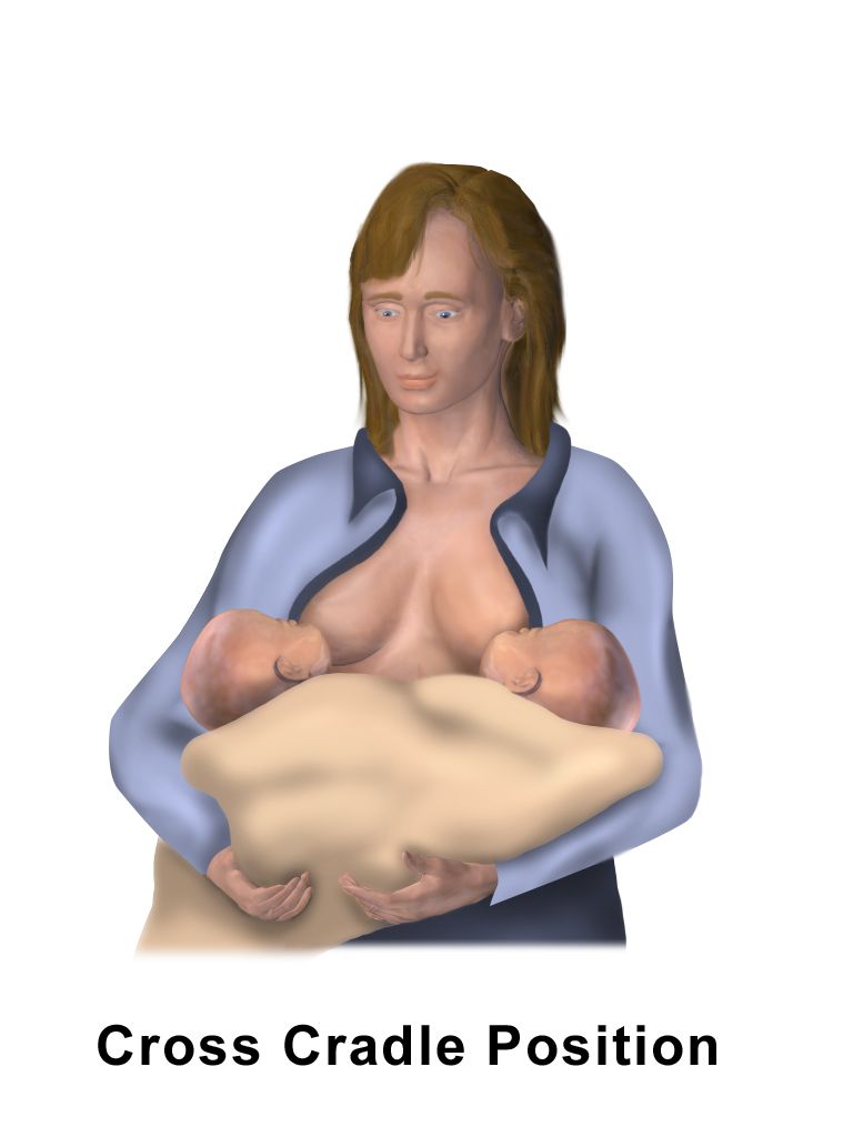
Expect 8 to 12 feeds in 24 hours, driven by the infant’s cues — rooting, hand-to-mouth, stirring, lip-smacking — with crying as a late and inefficient cue. Assess the latch rather than the clock: mouth wide open, lips flanged out, chin indenting the breast, a substantial portion of the areola taken in with more below than above, audible swallowing, and no pinching pain after the first few sucks. Persistent nipple pain means a shallow latch or a positioning problem — break the suction with a clean finger and start again rather than enduring it. Judge adequacy by output: by day 4 to 5 expect at least six wet nappies and three to four yellow, seedy stools a day. Newborns normally lose up to 7 to 10 per cent of birth weight, with the nadir at day 3 to 4 and return to birth weight by day 10 to 14.
Engorgement peaks at day 3 to 5 as vascular and lymphatic congestion accompanies secretory activation. Both breasts are firm, hot, tight and painful, and a low-grade temperature can occur. Manage it by feeding frequently, applying cold between feeds to reduce oedema, using brief warmth or reverse-pressure softening just before a feed so the infant can latch onto a softened areola, and expressing only enough to relieve pressure. Mastitis is different and unilateral: a wedge-shaped area of redness, heat and tenderness with fever of 38.5 °C or higher and flu-like aching. The key teaching, and the one most often got wrong, is that breastfeeding continues on the affected side — the milk is safe for the infant and drainage is treatment. Add rest, fluids, analgesia and a ten- to fourteen-day course of a penicillinase-resistant antibiotic such as dicloxacillin 500 mg four times daily or cephalexin 500 mg four times daily; an untreated mastitis can become a breast abscess. For a parent who is not lactating, suppression means a well-fitting supportive bra, cold packs, avoiding any nipple stimulation or expression, and analgesia; symptoms settle in 24 to 72 hours.
Medications at the couplet’s bedside
The postpartum nurse gives medications to two patients with a roughly twentyfold difference in body mass, and the newborn’s doses are never improvised from adult practice. Routine newborn prophylaxis illustrates the discipline: vitamin K1 (phytonadione) 0.5 to 1 mg intramuscularly into the vastus lateralis within 6 hours of birth to prevent vitamin K deficiency bleeding, and erythromycin 0.5 per cent ophthalmic ointment as a 1 cm ribbon to each lower conjunctival sac within an hour or two of birth. Both are fixed newborn doses, verified against the order and the infant’s weight before they are drawn up.
For the birthing parent, analgesia is chosen with lactation in mind: scheduled ibuprofen 600 mg every 6 hours with paracetamol 650–1000 mg every 6 hours (maximum 3000–4000 mg per 24 hours) controls most postpartum pain, while codeine and tramadol are contraindicated during breastfeeding because ultra-rapid CYP2D6 metabolisers can transfer dangerous morphine concentrations into milk and cause infant respiratory depression. Ibuprofen is safe with breastfeeding and treats afterpains and perineal pain well; time the dose about thirty minutes before a feed when afterpains are severe.
Psychological adaptation and mood
Rubin’s phases still describe the arc usefully. In taking-in (the first 24 to 48 hours) the birthing parent is dependent, focused on her own needs, hungry, sleepy and given to retelling the birth story — which is integration work, not chatter. In taking-hold (roughly day 2 to day 10) she becomes independent, task-focused, eager to master care and highly receptive to teaching. This is the teaching window; instruction delivered during taking-in is rarely retained. Letting-go follows over weeks as the family reorganises around the new member and grieves the relationships and identity that existed before. Watch also for bonding behaviours — en face gaze, fingertip-to-palm progression of touch, calling the infant by name, engrossment in a partner — and for sibling and cultural patterns, which vary widely and are not deficits.
Distinguish three conditions that are not on a single continuum. Postpartum blues affect 50 to 80 per cent of birthing parents, begin around day 3 to 5, peak at day 5, and resolve spontaneously by day 10 to 14. They present as tearfulness, mood lability, irritability, anxiety and fatigue, with intact function and no thoughts of harm; treatment is rest, nutrition, reassurance and practical support. Postpartum depression affects roughly one in seven, can begin any time within the first year, and lasts beyond two weeks with pervasive low mood, anhedonia, guilt, disturbed sleep even when the baby sleeps, poor concentration, appetite change and difficulty bonding. Screen with the Edinburgh Postnatal Depression Scale; a score of 10 or more warrants follow-up, 13 or more is a strong positive, and any positive response to item 10 — thoughts of self-harm — requires immediate assessment for safety before the visit ends. Treatment includes psychotherapy, sertraline or another lactation-compatible SSRI, and support. Postpartum psychosis occurs in about 1 to 2 per 1000, typically within the first two weeks and often within 72 hours, and is a psychiatric emergency: delusions frequently concerning the infant, hallucinations, confusion, disorganisation, and grossly impaired reality testing, with substantial risk of suicide and of harm to the infant. Do not leave the parent alone with the baby, ensure continuous supervision, and arrange urgent psychiatric admission. Bipolar disorder is the strongest risk factor, and every patient with that history should be flagged antenatally.
Going home: teaching, contraception and follow-up
Discharge teaching is the last safety net, and it works better as a short list of alarms than as a lecture. The POST-BIRTH warning signs give the patient and her family concrete thresholds. Call emergency services for pain in the chest, obstructed breathing or shortness of breath, seizures, or thoughts of hurting oneself or the baby. Call the provider for bleeding that soaks more than one pad in an hour or clots the size of an egg or larger, an incision that is not healing, a red or swollen leg that is painful or warm, a temperature of 38.0 °C (100.4 °F) or higher, or a headache that does not improve or that comes with visual changes. Teach the patient and a support person together, teach back rather than ask “any questions?”, and give the numbers to call in writing.
Fertility returns before periods do. Ovulation can precede the first menses, so contraception is discussed before discharge. Progestin-only methods (the progestin-only pill, the implant, depot medroxyprogesterone, the levonorgestrel intrauterine device) and copper intrauterine devices are compatible with breastfeeding and can be started immediately postpartum, including immediate post-placental IUD insertion. Combined hormonal contraceptives are avoided in the first 21 days postpartum for everyone because of thromboembolic risk, and until 42 days if additional VTE risk factors are present. The lactational amenorrhoea method is about 98 per cent effective only when all three conditions hold simultaneously: exclusive breastfeeding day and night, amenorrhoea, and an infant under six months old. Advise resuming intercourse when lochia has stopped and the perineum is comfortable, and warn about vaginal dryness during lactation. Finally, follow the fourth-trimester model: contact within the first three weeks — within 72 hours for hypertensive disorders — and a comprehensive visit no later than 12 weeks that reviews mood, bleeding, blood pressure, feeding, contraception, chronic disease and the plan for future pregnancies.
The fourth trimester is nursing work: protect the feeding relationship, give newborn prophylaxis and parental analgesia with lactation in mind, screen the mood rather than assume it, and send the family home with specific alarms rather than general reassurance.
The chapter in one breath
The puerperium reverses nine months of adaptation in six weeks. The uterus involutes through myometrial retraction, autolysis and regeneration — falling from about 1000 g to 60–80 g, descending roughly a centimetre a day, and shedding lochia rubra, serosa and alba. The plasma volume that protected the patient in labour now unloads through diuresis and diaphoresis, producing a benign bradycardia of 40–60 and a leucocytosis to 30,000 that make ordinary vital-sign rules useless; tachycardia over 100, oliguria under 35 mL per hour or a saturation under 95 per cent are the real warnings. Assessment is structured (BUBBLE-HE), quantified (weigh blood, 1 g equals 1 mL) and never delegated. In haemorrhage the sequence is the treatment — massage the fundus first, call for help, empty the bladder, then choose a uterotonic that the patient’s blood pressure and airway will tolerate. A firm fundus with bright red bleeding is a laceration; concealed pain is a haematoma; no palpable fundus with disproportionate shock is inversion, and uterotonics stop. Magnesium toxicity takes reflexes before respirations, so hourly reflexes buy the time that calcium gluconate then uses. Hypertension, sepsis and thromboembolism stalk the weeks after discharge, and mood must be screened, not assumed. Newborn prophylaxis and parental analgesia are given with lactation in mind, and every family goes home with named alarms.
Words worth knowing
| Term | What it means |
|---|---|
| Involution | Return of the uterus toward its pre-pregnancy size and position through retraction, autolysis and endometrial regeneration; complete by about six weeks. |
| Subinvolution | Delayed or arrested involution — a boggy, higher-than-expected uterus with persistent or recurrent rubra, usually from retained fragments or infection. |
| Living ligature | The interlacing myometrial fibres that mechanically clamp the spiral arterioles of the placental site when the uterus contracts. |
| Lochia | Postpartum uterine discharge of blood, decidua, leucocytes and exudate, progressing rubra (days 1–3) to serosa (days 4–10) to alba (day 10 to six weeks). |
| Afterpains | Cramping from intermittent uterine contractions, stronger in multiparas, over-distended uteri and during breastfeeding because suckling releases oxytocin. |
| Quantitative blood loss (QBL) | Measured rather than estimated blood loss, obtained by weighing soaked materials and subtracting dry weight; 1 gram equals 1 millilitre. |
| Postpartum haemorrhage | Cumulative blood loss of 1000 mL or more, or any loss with signs of hypovolaemia, within 24 hours of birth regardless of mode of birth. |
| Uterine atony | Failure of the myometrium to contract after birth; the cause of 70–80 per cent of primary postpartum haemorrhage. |
| Four Ts | Tone, Trauma, Tissue, Thrombin — the diagnostic framework for postpartum haemorrhage. |
| Shock index | Heart rate divided by systolic blood pressure; a value of 0.9 or higher in a postpartum patient suggests significant haemorrhage before hypotension appears. |
| Uterine inversion | The fundus turning inside out into the uterine cavity or vagina; managed by stopping uterotonics, not removing the placenta, and replacing the uterus before restarting uterotonics. |
| Puerperal bradycardia | Normal heart rate of 40–60 beats per minute for the first six to ten days, caused by the sudden rise in stroke volume after birth. |
| Puerperal morbidity | Temperature of 38.0 °C or higher on any two of the first ten postpartum days, excluding the first 24 hours — the classic definition of postpartum infection. |
| Endometritis | Infection of the uterine lining, usually on day 2–3, with fever, uterine tenderness, subinvolution and foul lochia; more common after caesarean birth. |
| Lactogenesis II | Secretory activation — the copious milk production that begins 30 to 72 hours after birth once progesterone falls with placental delivery. |
| REEDA | Perineal wound assessment: Redness, Ecchymosis, Oedema, Discharge, Approximation. |
| BUBBLE-HE | The structured postpartum assessment: Breasts, Uterus, Bladder, Bowel, Lochia, Episiotomy/perineum, Homans/lower extremities, Emotional status. |
| Taking-hold | Rubin’s second phase, roughly day 2 to day 10, when the birthing parent becomes independent and task-focused — the optimal window for teaching. |
| Lactational amenorrhoea method | Contraception that is about 98 per cent effective only when exclusive breastfeeding, amenorrhoea and an infant under six months are all present. |