Neurologic Disorders: Seizures, Headache and CNS Infection
Three complaints in this chapter arrive looking almost identical — a patient who is confused, a patient whose head hurts, a patient who has just shaken on the floor — and the whole of your nursing judgement lies in telling the benign version from the one that kills within hours. A seizure is a burst of synchronous cortical firing; whether it is a nuisance or an emergency depends entirely on whether it stops. A headache is the commonest human symptom and the presentation of subarachnoid haemorrhage, giant cell arteritis and bacterial meningitis. An infection of the meninges is treatable with a drug you already have on the unit, but the outcome is measured in minutes to first antibiotic dose. So learn the mechanism briefly, then learn the timed algorithms: five minutes for status epilepticus, one hour for meningitis, and “before biopsy” for temporal arteritis. Your assessment, not the scan, is what starts those clocks.
By the end of this chapter you can
- Classify seizures as focal or generalized onset, describe the ictal and postictal phases, and recognise Todd paralysis as a mimic of stroke.
- Perform correct seizure precautions and bedside management, and document the ten observations that determine the diagnosis.
- Define status epilepticus by the five-minute rule and execute the timed algorithm — benzodiazepine first, then fosphenytoin, levetiracetam or valproate.
- Match antiseizure medications to their mechanism, monitoring parameter and signature toxicity, including phenytoin levels, lamotrigine rash and valproate teratogenicity.
- Distinguish migraine, tension-type and cluster headache, and apply the SNOOP red flags that mandate imaging before treatment.
- Interpret cerebrospinal fluid results against real reference values to separate bacterial from viral, fungal and tuberculous meningitis.
- Prioritise the first hour of suspected bacterial meningitis: cultures, dexamethasone, empiric antibiotics, droplet precautions and contact chemoprophylaxis.
- Recognise herpes simplex encephalitis and brain abscess, and state why acyclovir is started before the polymerase chain reaction result returns.
- Describe older-adult variation in all three presentations and construct an SBAR escalation for each red flag.
Section 1Seizures: What the Cortex Is Doing and What You Must See
The Mechanism in One Paragraph
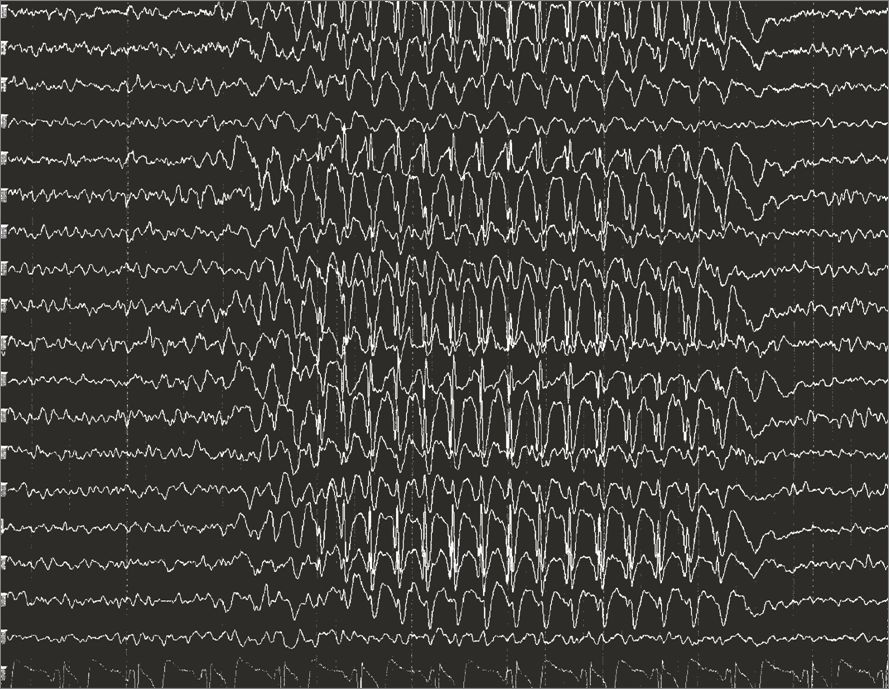
A normal cortex balances excitation carried by glutamate against inhibition carried by gamma-aminobutyric acid, and it keeps neighbouring neurons out of step with each other. A seizure is what happens when that balance tips and a population of neurons fires together, repetitively and without the usual braking. If the discharge stays in one region, the patient has a focal seizure and the symptoms are a caricature of whatever that region normally does — the motor strip produces jerking of a limb, the temporal lobe produces a rising epigastric sensation, an odd smell, lip-smacking or an intense sense of familiarity. If the discharge recruits networks in both hemispheres from the outset, the patient has a generalized seizure and consciousness is lost immediately.
That single mechanism explains almost every finding you will chart. Tonic stiffening is sustained motor cortex discharge; the clonic phase is that discharge being interrupted by returning inhibition. Cyanosis occurs because the diaphragm and intercostals are tonically contracted and the patient is not moving air — it is expected, it is brief, and it is not a reason to force an airway between clenched teeth. Incontinence follows loss of sphincter control. Tongue biting is lateral, not on the tip, because the jaw closes on a tongue caught between molars. The postictal state — sleepiness, confusion, headache, muscle soreness — is the cortex exhausted of neurotransmitter and running an oxygen debt, and it may last minutes to hours.
One postictal finding gets mistaken for catastrophe every week in every hospital. Todd paralysis is focal weakness of the limb or side that was seizing, present after the seizure ends and resolving within minutes to as long as forty-eight hours. It looks exactly like a stroke. It is not your job to decide which it is; it is your job to know that a new focal deficit after a witnessed seizure still gets a stroke evaluation, because a stroke can also cause the seizure.
Naming What You Saw
Modern classification asks three questions in order. Where did it start — focal, generalized or unknown onset? Was awareness preserved? Were the first prominent features motor or non-motor? A focal aware seizure is what students were taught to call an aura; the patient is awake and remembers it, and it is the most useful piece of localising information anyone will ever collect. A focal impaired-awareness seizure (previously complex partial) is the staring, unresponsive patient with automatisms — picking at bedding, chewing, fumbling — who afterwards cannot account for the last two minutes. Any focal seizure can spread; when it does, you chart a focal to bilateral tonic-clonic seizure, and the fact that it began focally is diagnostically precious.
Generalized types include tonic-clonic, myoclonic (brief shock-like jerks, often on waking), atonic (sudden loss of tone, the “drop attack” that produces facial and dental injury and often means a helmet at home), tonic, clonic and absence. Absence seizures are seconds of blank staring with abrupt onset and abrupt return, no postictal confusion, and often dozens per day — the school-age child labelled inattentive. Note the contrast that examinations love: the absence patient resumes mid-sentence, the focal impaired-awareness patient is groggy and disoriented for several minutes.
Epilepsy is a diagnosis, not an event. It means two unprovoked seizures more than twenty-four hours apart, or one unprovoked seizure with a recurrence risk of at least sixty per cent (for example a seizure arising from an old infarct visible on MRI), or a defined epilepsy syndrome. A single provoked seizure — from hypoglycaemia, alcohol withdrawal or hyponatraemia — is not epilepsy, and treating it with a lifelong drug instead of correcting the provocation is a classic error.
What You Do During and After — and What You Never Do
The first move is airway and safety, in that order and simultaneously if you can. Stay with the patient and call for help. Note the time on the clock — not “it felt like ages”, the actual time, because the entire treatment algorithm is timed. Ease the patient to the floor if standing, or lower the bed and raise the padded rails if in bed. Turn the patient onto the side so secretions drain, loosen anything tight at the neck, place something soft under the head and clear hard objects out of the arc of the limbs. Give oxygen and have suction at hand. Stay until the airway is patent and the patient is oriented.
Now the prohibitions, because they appear on every examination and, more importantly, because they injure people. Do not restrain the limbs — you will cause fractures and dislocations and you will not shorten the seizure. Do not put anything in the mouth, including an oral airway, a padded tongue blade or your fingers; the tongue cannot be swallowed, and forcing objects between clenched teeth breaks teeth and bites fingers. Do not attempt to insert an oral temperature probe on any patient with seizure precautions. Do not leave to fetch equipment; use the call bell.
Documentation is your clinical contribution, and it is more diagnostic than any scan. Chart what the patient was doing beforehand and any warning; the exact time of onset and of cessation; where the movement started and how it spread; head and eye deviation and to which side; whether awareness was preserved; automatisms; colour change; incontinence; tongue or other injury; the postictal state including orientation, speech and focal weakness; vital signs and capillary glucose. Ten observations, one minute of charting, and the neurologist can classify the seizure from your note.
| Seizure precautions | What it means at the bedside |
|---|---|
| Bed and rails | Bed in lowest position, side rails up and padded per facility policy, call bell within reach. |
| Airway equipment | Suction set up and tested, oxygen and a bag-valve-mask at the bedside — not down the hall. |
| Access | Patent IV for patients at risk, so a benzodiazepine can be given without a search for a vein. |
| Temperature route | Tympanic, temporal or axillary — never oral. |
| Ambulation and hygiene | Supervised showers, no tub baths, no locked bathroom door, assistance for transfers. |
| Medication timing | Antiseizure doses given on time, never held for “NPO” without contacting the prescriber for an IV equivalent. |
Provoked Seizures, the Workup, and the Older Adult
Every first seizure earns a search for a cause, and the reversible causes are ones you can identify from the chart before the neurologist arrives. Capillary glucose is the first test, always, because hypoglycaemia is instantly correctable and instantly fatal if missed. Serum sodium below roughly 120 mEq/L (normal 135–145) seizes; so does severe hypocalcaemia and severe hypomagnesaemia. Alcohol withdrawal seizures cluster six to forty-eight hours after the last drink. Drugs lower the threshold — bupropion, tramadol, meperidine through its metabolite normeperidine, high-dose beta-lactams in renal failure, and stimulant or cocaine intoxication. In pregnancy after twenty weeks and up to six weeks postpartum, a seizure is eclampsia until disproved, and the drug is magnesium sulfate, not lorazepam.
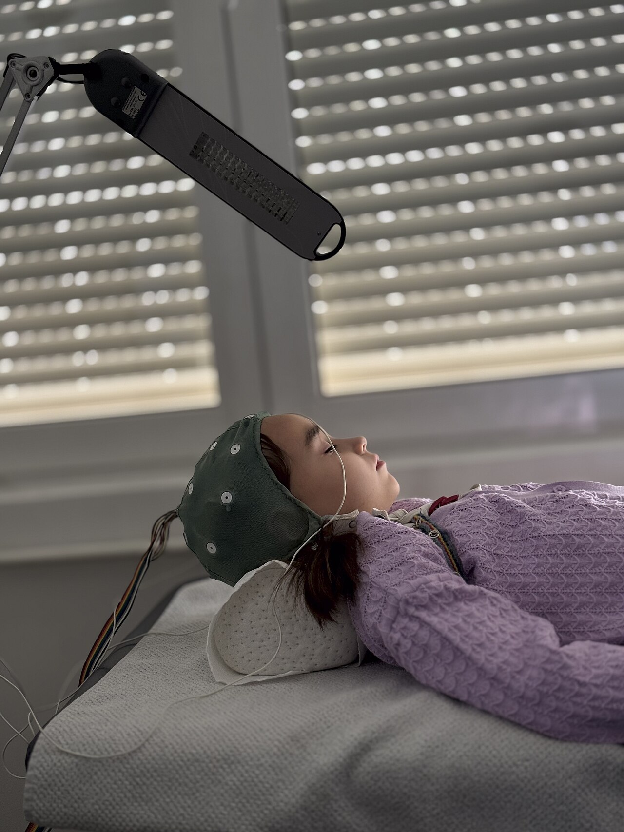
The structural workup is neuroimaging — MRI with epilepsy protocol where available, CT acutely to exclude haemorrhage or mass — plus electroencephalography. Remember what EEG can and cannot do: a normal interictal EEG never excludes epilepsy, because the study samples thirty minutes of a lifetime. Sleep deprivation, photic stimulation and hyperventilation are used to provoke discharges; prolonged video EEG is the gold standard when the diagnosis is uncertain. Add lumbar puncture when fever or immunosuppression suggests infection, and a toxicology screen when the history is thin.
Older adults are different and the difference is dangerous. New-onset seizures after age sixty are most often caused by cerebrovascular disease — a remote infarct is the single commonest substrate — followed by tumour, trauma, dementia-related change and drug effects. Presentation is frequently non-convulsive: hours to days of fluctuating confusion, staring, or unexplained aphasia rather than a dramatic convulsion. This is non-convulsive status epilepticus, it is missed because it is charted as “sundowning”, and the only way to find it is to think of it and ask for an EEG. Postictal states last far longer in older patients, and interactions with warfarin, antihypertensives and hepatically cleared drugs make antiseizure prescribing a careful business.
Delegation. Assistive personnel can pad rails, keep the bed low, place suction and oxygen at the bedside, take vital signs on a stable patient, assist with hygiene and stay with the patient while calling out for help. What may not be delegated: the neurologic assessment, interpreting what the seizure was, teaching about triggers and medications, evaluating whether care worked, and any care of an actively seizing or postictal unstable patient. Those are RN work, every time.
During a seizure your only jobs are time it, protect the airway and the head, and never restrain or place anything in the mouth — and afterwards your observations, not the scan, are what classify it.
Section 2Status Epilepticus and the Drugs That Stop Seizures
The Five-Minute Rule
Status epilepticus is five minutes or more of continuous seizure activity, or two or more seizures between which the patient does not return to baseline awareness. The old thirty-minute definition was abandoned for a practical reason: almost no untreated tonic-clonic seizure that has lasted five minutes will stop on its own, and the longer it runs the harder it becomes to stop. Receptors that benzodiazepines target are internalised within minutes, so a drug that works beautifully at four minutes may fail at forty. Mortality for convulsive status is roughly twenty per cent and climbs with duration, age and cause.
The damage is done three ways. Neurons die from excitotoxic calcium entry driven by relentless glutamate. Systemically, the patient develops hyperthermia from sustained muscle activity, lactic acidosis, hypoxaemia, hyperkalaemia, hypoglycaemia and, in prolonged cases, rhabdomyolysis with acute kidney injury. Autonomic surge produces tachycardia, hypertension then hypotension, and neurogenic pulmonary oedema. This is why the resuscitation is not just anticonvulsant — it is airway, oxygen, glucose, temperature and circulation at the same time.
The commonest precipitant on a medical floor is not new epilepsy. It is a known epileptic whose drug was held for a procedure, whose absorption failed with vomiting, or who ran out of a prescription. Second is acute brain injury — stroke, haemorrhage, trauma, CNS infection. Third is metabolic derangement and alcohol withdrawal. Ask, in this order: what medication was missed, what is the glucose, what is the sodium, and when did the drinking stop.
The Timed Algorithm
Zero to five minutes is stabilisation. Position, open the airway, high-flow oxygen by non-rebreather, suction, pulse oximetry, cardiac monitor, two large-bore IVs. Capillary glucose immediately; if below 60 mg/dL give dextrose, and in a patient with alcohol use disorder or malnutrition give thiamine 100 mg IV before or with the glucose to avoid precipitating Wernicke encephalopathy. Draw sodium, calcium, magnesium, renal and hepatic panel, complete blood count, toxicology and levels of any antiseizure drug the patient takes. Check temperature and start cooling if hyperthermic.
Five to twenty minutes is first-line therapy, and first line is always a benzodiazepine, which enhances GABA-mediated chloride influx and hyperpolarises the neuron. Give IV lorazepam 0.1 mg/kg, maximum 4 mg per dose, repeated once after three to five minutes if seizing continues. If there is no IV, give midazolam 10 mg intramuscularly for an adult over 40 kg — the intramuscular route is as effective as intravenous and faster to deliver in the field. Diazepam 0.15–0.2 mg/kg IV to a maximum of 10 mg is the alternative. The nursing watch is respiratory depression, hypotension and over-sedation; bag-valve-mask at the bedside, not in the supply room. Under-dosing is the commonest error — two milligrams of lorazepam given once is not a treated patient.
Twenty to forty minutes is second-line therapy, given even if the seizure has stopped, to prevent recurrence. Three agents are considered equivalent: fosphenytoin 20 mg phenytoin-equivalents/kg IV (maximum 1500 mg PE, infused no faster than 150 mg PE/min) with continuous cardiac monitoring and blood pressure checks for bradycardia, hypotension and arrhythmia; levetiracetam 60 mg/kg IV (maximum 4500 mg), the friendliest option because it has essentially no cardiac toxicity and no drug interactions; or valproate 40 mg/kg IV (maximum 3000 mg), which is avoided in hepatic disease, pancreatitis, mitochondrial disease and pregnancy.
Beyond forty minutes the patient is in refractory status: repeat a second-line agent or move to anaesthetic infusions of midazolam, propofol or pentobarbital, which means intubation, an intensive care bed and continuous EEG. That last point matters to floor nurses too. Once a patient is paralysed or deeply sedated, convulsions stop but electrical seizures may not, so “the shaking stopped” is no longer evidence of anything. Only the EEG tells you.
Maintenance Antiseizure Medications by Mechanism
Nearly every antiseizure drug does one of three things: block voltage-gated sodium channels so a neuron cannot fire repetitively, enhance GABA-mediated inhibition, or reduce glutamate release and calcium entry. Group them that way and the adverse effects become predictable rather than memorisable. Sodium-channel blockers (phenytoin, carbamazepine, oxcarbazepine, lamotrigine, lacosamide) produce dose-related dizziness, diplopia and ataxia because the cerebellum and brainstem use those same channels. GABA enhancers (benzodiazepines, phenobarbital) sedate. All of them can cause rash, and a few of those rashes are fatal.
| Drug | Mechanism | Therapeutic level | Signature toxicity and nursing monitoring |
|---|---|---|---|
| Phenytoin | Sodium-channel blocker | Total 10–20 mcg/mL; free 1–2 mcg/mL | Nystagmus near 20, ataxia above 30, lethargy and coma above 40 mcg/mL. Gingival hyperplasia — teach meticulous oral care and dental visits. Hirsutism, coarse facies, folate and vitamin D deficiency, osteoporosis. Rash may herald Stevens–Johnson syndrome. Highly protein-bound — check a free level in hypoalbuminaemia or renal failure. Potent enzyme inducer: reduces efficacy of oral contraceptives and warfarin control. Teratogenic. |
| Carbamazepine | Sodium-channel blocker | 4–12 mcg/mL | Hyponatraemia from SIADH — check sodium. Agranulocytosis and aplastic anaemia — baseline and serial CBC, report sore throat, fever, bruising. Stevens–Johnson syndrome and toxic epidermal necrolysis strongly linked to HLA-B*15:02; screen patients of Asian ancestry before starting. Autoinduces its own metabolism, so levels fall after a few weeks. Avoid grapefruit juice. |
| Valproate | Multiple: sodium channels, GABA | 50–100 mcg/mL | Hepatotoxicity — baseline and serial LFTs, report anorexia, vomiting, jaundice. Pancreatitis — report severe abdominal pain. Hyperammonaemia with encephalopathy even when LFTs are normal (ammonia 15–45 mcg/dL). Thrombocytopenia. Weight gain, tremor, hair loss. Neural tube defects — not for patients who can become pregnant without effective contraception and full counselling. |
| Levetiracetam | SV2A synaptic vesicle protein | Not routinely monitored | Behavioural adverse effects are the whole story: irritability, agitation, aggression, depression, occasionally suicidal ideation. Ask the family, not just the patient. Renally cleared — dose reduced in kidney disease. Minimal interactions, which is why it is so widely used. |
| Lamotrigine | Sodium-channel blocker | Not routinely monitored | Serious rash including Stevens–Johnson syndrome; risk rises with rapid titration. Titrate slowly over weeks; when combined with valproate the level roughly doubles, so the starting dose is halved and titration slowed further. Any rash stops the drug and calls the prescriber. Good mood profile, favoured in pregnancy. |
| Phenobarbital | GABA-A enhancer | 15–40 mcg/mL | Sedation, cognitive slowing, respiratory depression, dependence. Strong enzyme inducer. Still first-line for neonatal seizures. |
| Topiramate | Mixed: sodium channels, GABA, carbonic anhydrase | Not routinely monitored | Word-finding difficulty and cognitive slowing, weight loss, paraesthesia, metabolic acidosis, kidney stones (push fluids), reduced sweating with heat intolerance, and acute angle-closure glaucoma — sudden eye pain and blurred vision is an emergency. |
| Oxcarbazepine | Sodium-channel blocker | Monitor sodium, not level | Hyponatraemia more common than with carbamazepine, especially in older adults on diuretics. Fewer haematologic effects; cross-reactive rash with carbamazepine in about one quarter of patients. |
Two rules cut across the whole table. First, never stop an antiseizure medication abruptly — abrupt withdrawal is itself a leading cause of status epilepticus, and that includes holding doses for a procedure without arranging a parenteral equivalent. Second, the enzyme inducers (phenytoin, carbamazepine, phenobarbital) accelerate the metabolism of oral contraceptives, warfarin, direct oral anticoagulants, statins, some antiretrovirals and many chemotherapy agents; every new prescription for these patients needs an interaction check.
Living With Epilepsy: Teaching and Escalation
Adherence is the whole game, and the reasons people miss doses are boring and fixable: cost, forgetting, sedation, and stopping because they “feel fine”. Teach a pill organiser and phone alarms, a written seizure diary with dates, duration and suspected triggers, and never running the last week of a prescription without a refill. Triggers to control are sleep deprivation, missed doses, alcohol, febrile illness, physiologic stress and, in a minority, flashing lights. Teach the family how to time a seizure, when to give a rescue benzodiazepine if one is prescribed (rectal diazepam gel, intranasal midazolam), and precisely when to call emergency services: a seizure lasting more than five minutes, a second seizure without recovery, injury, difficulty breathing, or a first-ever seizure.
Discuss safety honestly. Shower rather than bathe, do not swim alone, use the back burners of the stove, avoid ladders and heights, and consider a helmet for atonic seizures. Driving is regulated by the state and typically requires a seizure-free interval of three to twelve months with prescriber notification. Discuss pregnancy planning early: folic acid supplementation, avoidance of valproate, and never stopping medication on discovering pregnancy without speaking to the neurologist, because convulsive seizures harm a fetus more than most drugs do. And discuss SUDEP — sudden unexpected death in epilepsy — because the biggest modifiable risk factor is uncontrolled generalized tonic-clonic seizures, and patients who understand that take their tablets.
For drug-resistant epilepsy, defined as failure of two appropriately chosen and tolerated drugs, options include epilepsy surgery (resection of a defined focus after video EEG localisation), vagus nerve stimulation, responsive neurostimulation and the ketogenic diet. Referral to a comprehensive epilepsy centre after two failed drugs is the standard, and it is often the nurse who notices that the patient is on their fourth agent and still seizing weekly.
A seizure lasting five minutes is status epilepticus and needs an adequately dosed benzodiazepine immediately, followed by a second-line agent even if the convulsing has stopped.
Section 3Headache: Sorting the Miserable From the Catastrophic

The Three Primary Headaches and How They Differ
Primary headache means the headache is the disease; there is nothing structurally wrong. Migraine is a disorder of a hyperexcitable brain in which a slow wave of neuronal depolarisation followed by suppression — cortical spreading depression — produces the aura, and activation of the trigeminovascular system with release of calcitonin gene-related peptide (CGRP) produces the pain, the meningeal vasodilation and the sensitisation that makes light, sound and even hair-brushing intolerable. Clinically: four to seventy-two hours, unilateral more often than not, pulsating, moderate to severe, worse with routine activity, with nausea or vomiting and photophobia and phonophobia. Roughly a quarter have aura — visual most commonly, sensory or speech occasionally — developing over five or more minutes and lasting under an hour.
Tension-type headache is the opposite phenotype and the commonest headache overall: bilateral, pressing or tightening like a band, mild to moderate, not worsened by walking upstairs, without vomiting and with at most one of photophobia or phonophobia. Patients function through it. It responds to simple analgesia, sleep, hydration, posture correction and stress management, and it is the headache most often over-treated into a bigger problem.
Cluster headache is the most severe pain in clinical medicine and the easiest to recognise if you have seen one. Strictly unilateral, orbital or temporal, boring or stabbing, lasting fifteen to a hundred and eighty minutes untreated, occurring one to eight times daily in bouts of weeks to months, often waking the patient at the same hour each night. The give-away is the pairing of ipsilateral autonomic features — tearing, conjunctival redness, nasal congestion or rhinorrhoea, eyelid oedema, ptosis and miosis — with restlessness. The migraine patient lies still in a dark room; the cluster patient paces, rocks and cannot keep still. It affects men more than women, and alcohol reliably triggers attacks during a bout.
Treating Migraine and Cluster
Abortive migraine therapy works best taken early, at the first sign of pain, and works poorly once central sensitisation has set in — teach that explicitly, because patients are trained to “wait and see”. Mild attacks: NSAIDs or aspirin or acetaminophen, with an antiemetic such as metoclopramide or prochlorperazine, which treats the nausea and also has independent antimigraine effect (watch for akathisia and acute dystonia; diphenhydramine is the antidote at hand). Moderate to severe attacks: a triptan, a serotonin 1B/1D agonist that constricts dilated cranial vessels and inhibits CGRP release. Sumatriptan comes oral, nasal and subcutaneous; the subcutaneous route works in ten minutes when vomiting defeats tablets.
The triptan contraindications are the ones you must know cold, because they are vascular: known coronary artery disease, prior myocardial infarction, uncontrolled hypertension, peripheral vascular disease, ischaemic stroke, hemiplegic or brainstem-aura migraine, and use of an ergot within twenty-four hours. Teach the patient that chest tightness, jaw or throat tightness and flushing can occur and must be reported. Where triptans are unsafe, the newer classes are a genuine advance: gepants (ubrogepant, rimegepant) are small-molecule CGRP receptor antagonists that do not vasoconstrict, and lasmiditan is a 5-HT1F agonist without vascular effect — but it causes dizziness and sedation and the patient must not drive for eight hours after a dose.
Prevention is offered when attacks occur four or more days a month, or are disabling. Classes: beta-blockers (propranolol, metoprolol — avoid in asthma and check heart rate), topiramate, valproate, amitriptyline or venlafaxine, candesartan, and the monoclonal antibodies against CGRP or its receptor (erenumab, fremanezumab, galcanezumab, eptinezumab) given monthly or quarterly. OnabotulinumtoxinA is licensed for chronic migraine, defined as fifteen or more headache days a month. Every preventive needs a two-to-three-month trial at an adequate dose before it is called a failure — tell the patient that at the first visit or they will abandon it in week three.
Cluster headache is treated differently and the difference is examinable. The acute treatment is high-flow oxygen, 12–15 L/min via a non-rebreather mask for fifteen to twenty minutes, which aborts most attacks, plus subcutaneous sumatriptan 6 mg or intranasal zolmitriptan. Oral drugs are useless — the attack is over before they absorb. Preventive treatment during a bout is verapamil at high dose, which requires a baseline and serial ECG because of heart block, with a steroid taper or occipital nerve block as a bridge. Galcanezumab is approved for episodic cluster. Lithium remains an option.
Secondary Headache: The Red Flags You Cannot Miss
Secondary headache means something else is causing it, and the screening mnemonic is SNOOP. Systemic features — fever, weight loss, immunosuppression, cancer, pregnancy. Neurologic signs — any focal deficit, papilloedema, seizure or altered mentation. Onset that is sudden — the thunderclap headache reaching maximum intensity within one minute. Older — a new headache after age fifty. Pattern change, Positional worsening, Precipitation by Valsalva, cough or exertion, Papilloedema, Pregnancy or postpartum state, and Painkiller overuse. Any of these means imaging and a full evaluation before symptomatic treatment.
A thunderclap headache — “the worst headache of my life”, maximal in under a minute, often with vomiting, neck stiffness or brief loss of consciousness — is subarachnoid haemorrhage until proven otherwise. Non-contrast CT within six hours of onset detects over ninety-five per cent; beyond that a negative CT is followed by lumbar puncture looking for xanthochromia, or by CT angiography. Other thunderclap causes include cervical artery dissection, cerebral venous sinus thrombosis (think postpartum, oral contraceptives, thrombophilia, dehydration), reversible cerebral vasoconstriction syndrome and pituitary apoplexy. Your action at the bedside is the same for all of them: stop, do not give a triptan, take vital signs and a neurologic check, and escalate immediately.
A new headache in a patient over fifty with scalp tenderness, jaw claudication when chewing, shoulder-girdle aching or any visual change is giant cell arteritis until proved otherwise. The erythrocyte sedimentation rate is usually well above 50 mm/hr and C-reactive protein is elevated; temporal artery biopsy or vascular ultrasound confirms it. The critical teaching point is sequencing: high-dose corticosteroids are started immediately, before the biopsy, because the untreated disease causes permanent, irreversible blindness and biopsy remains informative for one to two weeks after steroids begin. If vision is already affected, treatment is intravenous methylprednisolone.
Two more patterns worth holding. A headache that is worse lying flat and on waking, worse with cough or straining, with vomiting and papilloedema, suggests raised intracranial pressure from a mass or from idiopathic intracranial hypertension — classically a young woman with obesity, papilloedema, pulsatile tinnitus and a CSF opening pressure above 25 cm H2O, treated with weight loss and acetazolamide. And in an older adult, especially one on an anticoagulant, a headache that has crept up over days to weeks after a minor knock — sometimes a bump they have forgotten — is a chronic subdural haematoma; look for gait change, personality change or fluctuating confusion.
| Feature | Migraine | Tension-type | Cluster |
|---|---|---|---|
| Site | Unilateral, may alternate | Bilateral, band-like | Strictly unilateral, orbital or temporal |
| Quality and severity | Pulsating, moderate to severe | Pressing, mild to moderate | Boring or stabbing, excruciating |
| Duration | 4–72 hours | 30 minutes to 7 days | 15–180 minutes, 1–8 per day |
| Associated features | Nausea, vomiting, photophobia, phonophobia, aura in about 25 per cent | Few or none | Ipsilateral tearing, nasal congestion, ptosis, miosis |
| Behaviour | Lies still in a dark, quiet room | Continues activity | Restless, pacing, cannot sit still |
| Acute treatment | NSAID, triptan, gepant, antiemetic | Simple analgesia, non-drug measures | Oxygen 12–15 L/min plus subcutaneous sumatriptan |
Treat headache as primary only after the SNOOP red flags are clear — thunderclap onset, new headache over fifty, fever with neck stiffness, focal deficit or papilloedema all buy imaging and escalation, not analgesia.
Section 4Meningitis: The Hour That Decides the Outcome
Mechanism and Presentation
Bacteria reach the subarachnoid space by colonising the nasopharynx and invading the bloodstream, by contiguous spread from sinusitis, otitis media or mastoiditis, or directly through a skull fracture, neurosurgical wound or shunt. Once inside, they replicate in a space with almost no immune surveillance. The inflammatory response — not the organism — does most of the damage: cytokines increase blood–brain barrier permeability, producing vasogenic cerebral oedema; purulent exudate obstructs cerebrospinal fluid outflow, producing hydrocephalus and rising intracranial pressure; and inflamed vessels thrombose, producing infarcts and cranial nerve palsies. That is why steroids given before the antibiotic matter, and it is why the patient can deteriorate in the first hours of apparently correct treatment.
The organisms follow age and host. Streptococcus pneumoniae is the commonest cause in adults and the most likely to leave deafness or focal deficit behind. Neisseria meningitidis favours adolescents and young adults in congregate settings and produces the petechial or purpuric rash that must never be dismissed as a viral exanthem. Listeria monocytogenes matters in adults over fifty, pregnancy, alcohol use disorder, malignancy and immunosuppression — and it is the reason ampicillin is added to the empiric regimen in those patients. Gram-negative bacilli and staphylococci dominate after neurosurgery or head trauma. Viral (aseptic) meningitis, most often enteroviral, is far commoner overall and far milder.
The classic triad of fever, nuchal rigidity and altered mental status is present in fewer than half of adults, so do not wait for it. Almost all patients have at least two of headache, fever, neck stiffness and altered mentation. Add photophobia, vomiting, seizures in roughly one in five, and focal deficits. Kernig sign (resistance and pain on extending the knee with the hip flexed) and Brudzinski sign (involuntary hip and knee flexion on passive neck flexion) are famous but insensitive — their absence proves nothing. In older adults the presentation is often confusion alone, with a low-grade or absent fever and a supple neck, which is precisely why the diagnosis is missed in the population with the highest mortality.
Diagnostics: Reading the Cerebrospinal Fluid
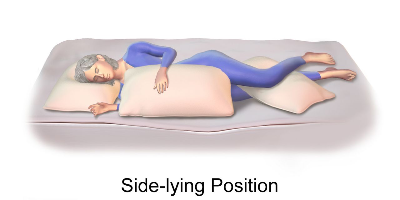
Lumbar puncture is the definitive test. Normal CSF is crystal clear with an opening pressure of 6–20 cm H2O, 0–5 white cells/mm3 (lymphocytes), protein 15–45 mg/dL and glucose 45–80 mg/dL, which should be about two-thirds of a simultaneous serum glucose. Nursing role: have the patient empty the bladder, position curled in lateral decubitus or sitting leaning forward to open the interspaces, provide a consent check and support during the procedure, label tubes in order, send them promptly, and afterwards position the patient for comfort — routine flat bed rest after the tap does not prevent post-dural-puncture headache and is no longer required — encourage fluids, and monitor the site for leakage and the patient for post-dural-puncture headache (positional, worse upright, treated with fluids, caffeine and if persistent an epidural blood patch).
| CSF parameter | Normal | Bacterial | Viral | Fungal or tuberculous |
|---|---|---|---|---|
| Appearance | Clear, colourless | Cloudy, turbid, purulent | Clear | Clear or slightly cloudy |
| Opening pressure (cm H2O) | 6–20 | Elevated, often above 30 | Normal or mildly raised | Elevated, sometimes markedly |
| White cells per mm3 | 0–5 | 1000–5000, neutrophils above 80 per cent | 50–1000, lymphocyte predominant | 100–500, lymphocyte predominant |
| Protein (mg/dL) | 15–45 | 100–500 | Normal to about 100 | Very high, often above 200 |
| Glucose (mg/dL) | 45–80; CSF-to-serum ratio at or above 0.6 | Below 40; ratio below 0.4 | Normal | Low |
| Other | Sterile | Gram stain positive in 60–90 per cent; culture | Enterovirus or HSV PCR | India ink, cryptococcal antigen, acid-fast stain, PCR |
Head CT is obtained before lumbar puncture only when herniation risk is real: immunocompromise, known CNS disease, new-onset seizure, papilloedema, reduced or fluctuating consciousness, focal neurologic deficit, or age sixty and above. Blood cultures — two sets — are drawn in every case, and they are positive in roughly half to three-quarters of bacterial cases when they are drawn before antibiotics. Prior antibiotic exposure sharply reduces the yield of both blood and CSF cultures, which is exactly why cultures precede the first dose in the first-hour sequence. Expect elevated white count with left shift, elevated CRP and procalcitonin, and check sodium at baseline because SIADH is common.
The First Hour
Here is the sequence that saves lives, and the order is the point. Recognise the patient. Draw blood cultures. Give dexamethasone 0.15 mg/kg IV ten to twenty minutes before or together with the first antibiotic dose, continued every six hours for four days — it reduces hearing loss and mortality in pneumococcal disease, and it works only if it precedes the bacterial lysis the antibiotic causes. Give empiric antibiotics. Then arrange CT and lumbar puncture if they are indicated. Antibiotics are never delayed for a scan or a tap. A student who answers “get the LP first” has just cost a patient their hearing.
Empiric therapy for community-acquired bacterial meningitis in an adult is vancomycin plus ceftriaxone 2 g IV every 12 hours (or cefotaxime), with ampicillin 2 g IV every 4 hours added for patients over fifty, pregnant patients and the immunocompromised to cover Listeria, which cephalosporins do not touch. Add acyclovir if encephalitis is in the differential. Vancomycin is dosed to a trough of 15–20 mcg/mL or by AUC, with renal function and infusion-rate monitoring — infuse over at least an hour to avoid the histamine flushing reaction. Therapy is narrowed once cultures return and typically runs seven days for meningococcus, ten to fourteen for pneumococcus and twenty-one for Listeria or gram-negatives.
Nursing management runs in parallel. Neurologic checks with Glasgow Coma Scale and pupils at least hourly while unstable. Head of bed at 30 degrees with the neck midline to aid venous drainage. A quiet, dimly lit room, with nursing activities spaced rather than clustered so intracranial pressure can settle back to baseline between them, because photophobia and hyperacusis are genuine and stacked stimulation drives the pressure up. Seizure precautions in place from admission. Antipyretics and cooling for fever, which increases cerebral metabolic demand. Strict intake and output with daily weights and serum sodium, because SIADH presents as falling sodium with concentrated urine and may require fluid restriction. Pain management, oral care, deep-vein thrombosis prophylaxis and pressure-injury prevention for the patient who cannot move.
Isolation, Prophylaxis and What Is Left Behind
Place any patient with suspected bacterial meningitis on droplet precautions immediately — private room, surgical mask donned on entry to the room rather than only when you come within a metre, mask on the patient during transport — and maintain them until twenty-four hours of effective antibiotic therapy have been completed. Do not wait for confirmation of the organism; you are protecting staff and other patients against meningococcus, which is transmitted by respiratory droplets.
Close contacts of a meningococcal case need chemoprophylaxis within twenty-four hours: household members, childcare contacts, anyone directly exposed to oral secretions, and healthcare workers who performed mouth-to-mouth resuscitation, intubation or unprotected airway suctioning. Simply being in the room, or handing a chart across the desk, does not qualify. Regimens are ciprofloxacin 500 mg orally as a single dose, rifampin 600 mg orally twice daily for two days (warn about orange discoloration of urine, sweat and tears, permanent staining of soft contact lenses, and reduced oral contraceptive efficacy), or ceftriaxone 250 mg intramuscularly once, which is preferred in pregnancy. Prevention long-term is vaccination: MenACWY and MenB for adolescents and at-risk adults, pneumococcal conjugate vaccine for older adults and the immunocompromised, and Hib in childhood.
Complications to anticipate and chart against: rising intracranial pressure and herniation, seizures, communicating hydrocephalus, cerebral infarction from vasculitis, cranial nerve palsies, septic shock, disseminated intravascular coagulation and — the commonest permanent sequela — sensorineural hearing loss. Every survivor of bacterial meningitis needs formal audiology testing before or shortly after discharge, and families need to know that a child who seems fine may have lost hearing.
In suspected bacterial meningitis the order is cultures, dexamethasone, antibiotics, then imaging and lumbar puncture — the tap never delays the drug, and droplet precautions start on suspicion.
Section 5Encephalitis, Brain Abscess and the Long Tail of CNS Infection
Encephalitis: When the Brain Itself Is Inflamed
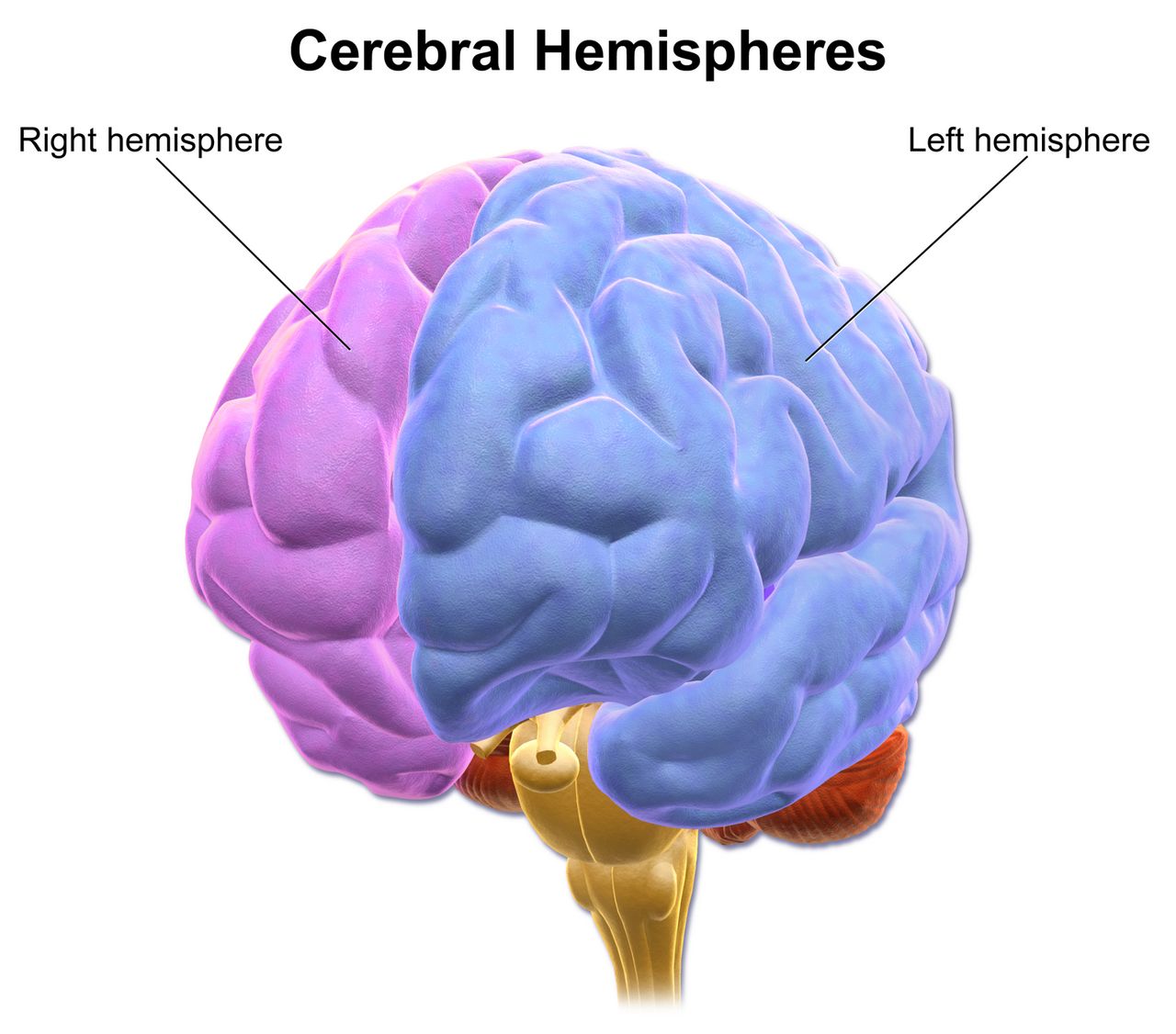
Meningitis inflames the coverings; encephalitis inflames the brain substance. That single distinction predicts the clinical difference. Meningitis gives you headache, fever, neck stiffness and a patient who is irritable but whose cortex works. Encephalitis gives you altered mental status as the defining feature — personality change, psychosis, disorientation, aphasia, focal deficits, movement disorders and seizures — often with fever and often with meningeal signs too, since the two overlap as meningoencephalitis.
Herpes simplex virus type 1 is the commonest sporadic cause in adults and the one that must never be missed, because it is treatable and untreated mortality approaches seventy per cent. It has a predilection for the temporal and inferior frontal lobes, which is why it presents with bizarre behaviour, olfactory or gustatory hallucinations, memory failure, aphasia and focal seizures. MRI shows temporal lobe hyperintensity; EEG may show periodic lateralised discharges; CSF shows a lymphocytic pleocytosis, elevated protein, normal glucose, often red cells, and a positive HSV polymerase chain reaction. Intravenous acyclovir 10 mg/kg every 8 hours for fourteen to twenty-one days is started empirically on suspicion and never held for the PCR result — every hour of delay costs neurologic function. The nursing watch is renal: acyclovir crystallises in the tubules, so infuse over at least one hour, hydrate generously, and monitor creatinine and urine output. Phlebitis at the site is common.
Other causes worth recognising. Arboviral encephalitis follows mosquito season: West Nile virus classically adds an asymmetric flaccid paralysis resembling polio; eastern equine and La Crosse viruses are regional. Rabies, once symptomatic, is essentially always fatal — the intervention is post-exposure prophylaxis at the time of the bite. And a category students miss entirely: autoimmune encephalitis, of which anti-NMDA-receptor encephalitis is the archetype — typically a young woman with a viral prodrome, then psychiatric symptoms severe enough to reach a psychiatric ward, then seizures, orofacial dyskinesias and autonomic instability, sometimes with an underlying ovarian teratoma. Treatment is immunotherapy and tumour removal, not antivirals, and the diagnosis is made only when someone thinks of it.
Brain Abscess and Other Space-Occupying Infection
A brain abscess begins as a focal cerebritis that walls itself into a pus-filled cavity with a capsule. Bacteria arrive by contiguous spread from otitis media, mastoiditis, sinusitis or dental infection, by haematogenous seeding from endocarditis, lung abscess or a right-to-left cardiac shunt, or directly from penetrating trauma or surgery. The classic triad of headache, fever and focal deficit is complete in a minority; headache is nearly universal, fever occurs in about half, and the tempo is days to weeks rather than hours. New focal seizures, progressive hemiparesis or a papilloedematous fundus in a patient with a chronic ear or sinus infection should prompt imaging.
Contrast-enhanced MRI or CT shows the ring-enhancing lesion with surrounding vasogenic oedema, and diffusion-weighted imaging distinguishes abscess from tumour. Lumbar puncture is contraindicated when there is mass effect, because withdrawing fluid below a pressure gradient invites herniation; the diagnosis is made by imaging and by culture of aspirated material. Management is stereotactic aspiration or excision plus prolonged intravenous antibiotics, commonly vancomycin with a third or fourth-generation cephalosporin and metronidazole for anaerobes, for six to eight weeks, with dexamethasone if oedema threatens and antiseizure prophylaxis in many cases. Nursing care is neurologic surveillance, long-line management, and the practical work of a six-week outpatient antibiotic course.
Two other patterns deserve a line. Cryptococcal meningitis presents subacutely in advanced HIV with a CD4 count usually below 100 cells/mm3, with headache and fever but often minimal neck stiffness; diagnosis is by cryptococcal antigen or India ink; treatment is amphotericin B with flucytosine followed by fluconazole, and the defining nursing issue is serial therapeutic lumbar punctures to control the very high opening pressure that causes blindness and death. Tuberculous meningitis is basilar, subacute over weeks, with cranial nerve palsies, very high CSF protein and low glucose, and requires four-drug therapy with adjunctive steroids. And any patient with a ventriculoperitoneal shunt who develops fever, headache, vomiting or altered mentation has a shunt infection or malfunction until proved otherwise.
Complications, the Older Adult, and Priority Setting
Whatever the organism, CNS infection kills through a small number of final common pathways, and these are what your assessments are hunting for. Raised intracranial pressure from oedema, hydrocephalus or abscess: declining level of consciousness first, then headache and vomiting, then the late and ominous Cushing triad of widening pulse pressure, bradycardia and irregular respirations, with a fixed dilated pupil signalling herniation. Seizures, including non-convulsive status in the persistently obtunded. SIADH with dilutional hyponatraemia. Septic shock and DIC. Cerebral infarction from inflamed, thrombosed vessels. And after recovery, cognitive impairment, hearing loss, epilepsy and fatigue that outlast the infection by months.
The priority framework does not change. Airway and breathing before circulation, circulation before neurologic status, and any change in level of consciousness outranks every other neurologic finding because it is the earliest and most sensitive sign of rising pressure. Unstable before stable; acute before chronic; actual problems before risks. Applied concretely: the postictal patient with noisy respirations is seen before the febrile confused patient, who is seen before the migraine patient waiting for her second dose of analgesia, who is seen before the stable epilepsy patient wanting discharge teaching — but the teaching still happens before the shift ends, because it is what prevents the next admission.
Older adults change every one of these presentations. Meningitis appears as confusion without fever or neck stiffness, and Listeria coverage becomes mandatory. New seizures are usually cerebrovascular and often non-convulsive. New headache demands consideration of giant cell arteritis, subdural haematoma and mass lesion before anyone says migraine, since primary headache rarely begins after fifty. Antiseizure drugs interact with polypharmacy, and drugs like phenytoin and phenobarbital add sedation, ataxia and fall risk to a patient who already has both. Renal and hepatic clearance are reduced, so start low and go slow, and reassess gait and orthostatic vitals after every dose change.
| Red flag you observe | What it may mean | Your first action |
|---|---|---|
| Seizure lasting past five minutes, or a second seizure without waking | Status epilepticus | Airway, oxygen, glucose, IV access; call for a benzodiazepine order now |
| Falling GCS, widening pulse pressure, bradycardia, irregular breathing | Raised ICP with impending herniation | Airway and oxygen first, then head of bed 30 degrees with neck midline; call rapid response, prepare hyperosmolar therapy |
| Fever with headache and any spreading petechial rash | Meningococcaemia | Droplet precautions, cultures, escalate for immediate antibiotics and fluid resuscitation |
| Thunderclap headache, maximal within one minute | Subarachnoid haemorrhage | Vital signs and neurologic check, escalate immediately for stat non-contrast CT; keep nothing by mouth and give no triptan |
| New headache over age fifty with jaw claudication or visual change | Giant cell arteritis | Stat ESR and CRP, escalate for high-dose steroids before biopsy |
| Any rash in a patient recently started on lamotrigine or carbamazepine | Stevens–Johnson syndrome | Hold the next dose and notify the prescriber before giving it |
| Serum sodium falling with concentrated urine after CNS infection | SIADH | Strict intake and output, daily weight, seizure precautions, notify for fluid restriction |
Every CNS infection converges on the same killers — rising intracranial pressure, seizures, SIADH and shock — so a falling level of consciousness is the single finding that outranks all others and triggers escalation.
The chapter in one breath
A seizure is synchronous cortical firing, and what you see depends on where it fires: focal seizures caricature the region involved, generalized seizures take consciousness at once. At the bedside you time it, protect the airway and head, never restrain and never put anything in the mouth, then document the ten observations that classify it. Five minutes of continuous seizing is status epilepticus, and the algorithm is timed — stabilise and check glucose, then an adequately dosed benzodiazepine, then fosphenytoin, levetiracetam or valproate whether or not the shaking has stopped. Maintenance drugs are grouped by mechanism, and their monitoring follows: phenytoin levels 10–20 mcg/mL and gum care, carbamazepine sodium and blood counts, valproate liver and ammonia and pregnancy risk, lamotrigine rash, levetiracetam behaviour. Headache is sorted by phenotype and screened by SNOOP: thunderclap means subarachnoid haemorrhage, new headache over fifty means giant cell arteritis and steroids before biopsy, fever with stiff neck means meningitis. In suspected bacterial meningitis the order is cultures, dexamethasone, antibiotics, then the scan and the tap — with droplet precautions from the moment you suspect it. And in encephalitis, acyclovir starts before the PCR returns.
Words worth knowing
| Term | What it means |
|---|---|
| Aura | A focal aware seizure that precedes spread; the patient is conscious and remembers it, and it localises the seizure onset. |
| Postictal state | The period after a seizure of confusion, drowsiness, headache and muscle soreness as the exhausted cortex recovers. |
| Todd paralysis | Transient focal weakness after a seizure, lasting minutes up to 48 hours, that mimics stroke and still warrants stroke evaluation. |
| Status epilepticus | Five minutes or more of continuous seizure activity, or repeated seizures without return to baseline awareness between them. |
| Non-convulsive status | Ongoing electrical seizure activity presenting as prolonged confusion or unresponsiveness without convulsions; diagnosed only by EEG. |
| Automatism | Repetitive purposeless movement — lip-smacking, chewing, picking at clothing — typical of focal impaired-awareness seizures. |
| SUDEP | Sudden unexpected death in epilepsy; risk rises with uncontrolled generalized tonic-clonic seizures and poor medication adherence. |
| Phenytoin equivalents (PE) | The dosing unit for fosphenytoin, expressing the amount of phenytoin the prodrug will yield; misreading it causes overdose. |
| Cortical spreading depression | A slow wave of neuronal depolarisation followed by suppression that produces migraine aura. |
| CGRP | Calcitonin gene-related peptide, the neuropeptide central to migraine pain and the target of gepants and the monoclonal antibodies. |
| Thunderclap headache | Headache reaching maximum intensity within one minute; subarachnoid haemorrhage until proved otherwise. |
| Medication-overuse headache | Daily or near-daily headache caused by frequent acute analgesic use; requires withdrawal plus a preventive agent. |
| Giant cell arteritis | Large-vessel vasculitis of older adults causing new headache, scalp tenderness, jaw claudication and irreversible blindness if steroids are delayed. |
| Nuchal rigidity | Resistance and pain on passive neck flexion from meningeal irritation; absent in many older adults with meningitis. |
| Kernig and Brudzinski signs | Classic meningeal irritation manoeuvres; highly specific when present but far too insensitive to rule meningitis out. |
| Opening pressure | Cerebrospinal fluid pressure measured at lumbar puncture; normal 6–20 cm H2O, markedly elevated in bacterial and cryptococcal disease. |
| Pleocytosis | An increased white cell count in cerebrospinal fluid; neutrophilic suggests bacterial, lymphocytic suggests viral, fungal or tuberculous. |
| Waterhouse–Friderichsen syndrome | Bilateral adrenal haemorrhage with vascular collapse and DIC complicating fulminant meningococcaemia. |
| Ring-enhancing lesion | A rounded lesion whose capsule takes up contrast on CT or MRI; in the febrile patient it suggests brain abscess. |
| Aseptic meningitis | Meningeal inflammation with negative routine bacterial cultures, most often enteroviral and usually self-limiting. |