Neurologic Disorders: Degenerative and Neuromuscular Disease
Stroke announces itself; these diseases do not. Parkinson disease, multiple sclerosis, myasthenia gravis, Guillain–Barré syndrome, amyotrophic lateral sclerosis and Alzheimer disease arrive over months or slide in over a weekend, and the nurse is usually the person who notices that something has changed. What unites them is a single motor pathway — cortex to corticospinal tract to anterior horn cell to peripheral nerve to neuromuscular junction to muscle — and a single shared endpoint that kills people: the diaphragm gets weak, secretions pool, the swallow fails and the patient aspirates or stops ventilating. This chapter teaches you to localise the lesion from the bedside exam, to read the diagnostics that confirm it, to give the drugs safely (levodopa is time-critical; magnesium can put a myasthenic on a ventilator), and to recognise the moment when a chronic disease becomes an airway emergency. Everything else — teaching, mobility, dignity — is built on getting that moment right.
By the end of this chapter you can
- Differentiate upper motor neuron, lower motor neuron, neuromuscular junction and muscle lesions using reflexes, tone, atrophy, fasciculations and sensory findings.
- Monitor neuromuscular respiratory failure with forced vital capacity, negative inspiratory force and bedside screens, and state the values that trigger escalation.
- Explain the dopaminergic basis of Parkinson disease and administer carbidopa–levodopa and adjuncts safely, including the dopamine blockers that are contraindicated.
- Apply the McDonald criteria concept of dissemination in space and time to multiple sclerosis, and distinguish a true relapse from a pseudo-relapse.
- Compare myasthenic crisis and cholinergic crisis and prioritise airway assessment over diagnostic labelling.
- Describe albuminocytologic dissociation, autonomic instability and the immunotherapy of Guillain–Barré syndrome, including why corticosteroids are not used.
- Plan care for amyotrophic lateral sclerosis and Alzheimer disease, including noninvasive ventilation timing, enteral access decisions and behavioural management without restraints.
- Identify red flags in each disorder and escalate using SBAR, and delegate correctly — assessment, teaching, evaluation and unstable patients stay with the RN.
- Describe how older adults present differently, and how polypharmacy and anticholinergic burden change the risk calculation.
Section 1Reading the failing motor unit
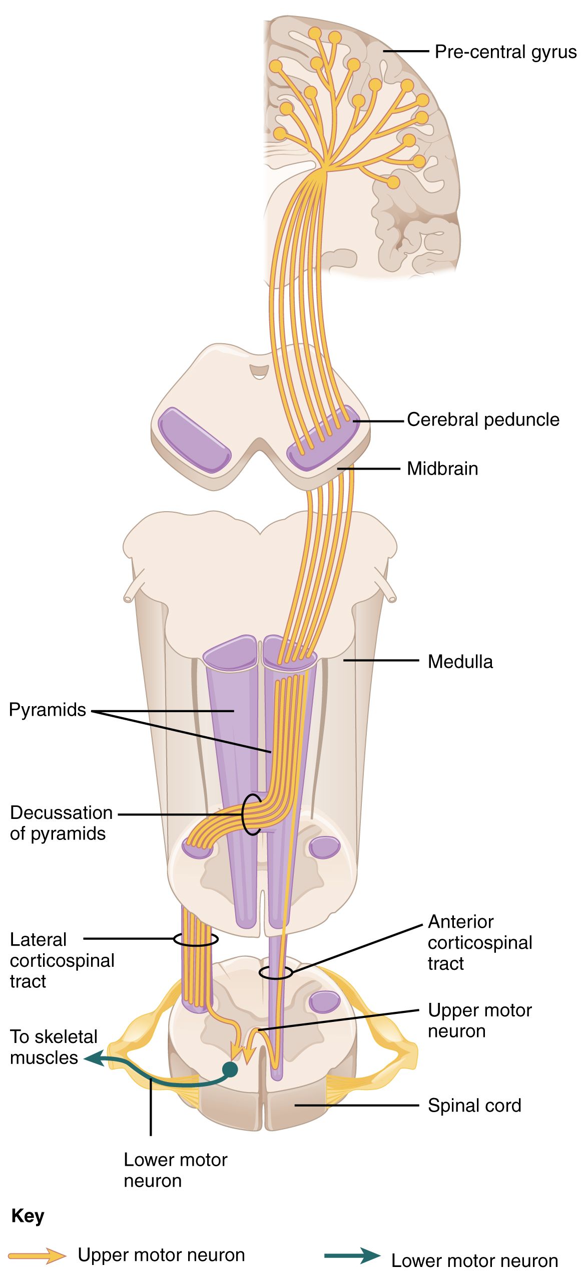
Upper motor neuron, lower motor neuron, and everything between
Voluntary movement travels a two-neuron relay. The upper motor neuron begins in the precentral gyrus, descends through the internal capsule, brainstem and corticospinal tract, and synapses in the anterior horn of the spinal cord. The lower motor neuron leaves the anterior horn, becomes the peripheral nerve, crosses the neuromuscular junction and contracts the muscle. Damage anywhere along that relay produces weakness — but weakness with a different accent depending on where the break is. Learning that accent is the single most useful skill in this chapter, because it lets you turn a vague complaint (“my legs feel heavy”) into a localisation, and a localisation into a plan.
Upper motor neuron lesions release the cord from cortical inhibition. The result is spasticity, hyperreflexia, clonus, a positive Babinski sign (upgoing great toe on plantar stimulation) and weakness in a pyramidal pattern — arm extensors and leg flexors give out first. Muscle bulk is preserved early because the muscle is still innervated; any atrophy is from disuse and appears late. Lower motor neuron lesions cut the muscle off from its nerve entirely: tone falls, reflexes diminish or vanish, the muscle wastes, and denervated fibres twitch spontaneously as fasciculations visible under the skin.
Two further levels have their own signatures. Neuromuscular junction disease produces fatigable weakness that worsens with repeated use and improves with rest, spares sensation entirely, leaves reflexes normal, and preferentially strikes the extraocular, eyelid and bulbar muscles — ptosis and diplopia at the end of the day. Primary muscle disease produces symmetric proximal weakness (trouble rising from a chair, trouble reaching a shelf) with normal sensation and a creatine kinase raised above the usual reference range, which in most laboratories runs about 30 to 200 U/L.
The presence or absence of sensory findings is a powerful discriminator that students consistently under-use. Myasthenia gravis and amyotrophic lateral sclerosis do not cause numbness. If a patient with suspected ALS reports glove-and-stocking sensory loss, something else is going on — vitamin B12 deficiency (level below roughly 200 pg/mL), diabetic polyneuropathy, cervical myelopathy or a paraneoplastic process. Guillain–Barré syndrome and multiple sclerosis, by contrast, involve sensory pathways and produce paraesthesia, numbness and pain alongside weakness.
| Level of lesion | Tone | Reflexes | Atrophy / fasciculations | Sensation | Classic example |
|---|---|---|---|---|---|
| Upper motor neuron | Increased (spastic, clasp-knife) | Brisk, clonus, Babinski present | Late disuse atrophy; no fasciculations | Variable, depends on tracts involved | Stroke, MS spinal cord plaque |
| Lower motor neuron | Decreased (flaccid) | Diminished or absent | Early marked atrophy; fasciculations | Intact if pure anterior horn | ALS anterior horn loss, polio |
| Peripheral nerve | Decreased | Absent, often distal first | Atrophy over weeks | Impaired — numbness, pain | Guillain–Barré, diabetic neuropathy |
| Neuromuscular junction | Normal | Normal | None | Normal | Myasthenia gravis |
| Muscle | Normal or decreased | Normal until severe | Proximal wasting; no fasciculations | Normal | Inflammatory myopathy, statin myopathy |
The vital sign these patients share: respiratory reserve
Whatever the diagnosis on the whiteboard, the neuromuscular patient dies of the same thing — ventilatory failure and aspiration. The diaphragm is driven by the phrenic nerve from C3, C4 and C5 (“C3, 4, 5 keeps the diaphragm alive”). When intercostal and diaphragmatic strength fall, tidal volume falls, the patient compensates with a fast shallow pattern, carbon dioxide climbs, and only at the very end does oxygen saturation drop. This is the trap: pulse oximetry is reassuring right up until it is catastrophic, particularly if the patient is on supplemental oxygen. Never chart “respiratory status stable, sat 96 percent” in a patient whose neck flexors will not lift their head off the pillow.
Bedside measures that actually detect the problem are forced vital capacity (FVC) and negative inspiratory force (NIF, also called maximal inspiratory pressure). Normal FVC is roughly 60 to 70 mL/kg of ideal body weight, so about 4 to 5 L in an average adult. Normal NIF is more negative than −60 cm H2O. In Guillain–Barré syndrome and myasthenic crisis, the widely taught “20/30/40 rule” flags impending failure: vital capacity below 20 mL/kg, NIF less negative than −30 cm H2O, or maximal expiratory pressure below 40 cm H2O. Elective intubation is usually undertaken by the time FVC falls below about 15 mL/kg or NIF is less negative than roughly −20 to −25 cm H2O — and elective is the word that matters, because emergent intubation in a patient with a full stomach, a weak cough and autonomic instability is a far more dangerous event.
You will not always have a spirometer in your hand at 0300. Two bedside screens buy you information immediately. Ask the patient to take a full breath and count aloud at a normal pace — counting comfortably into the twenties suggests reasonable reserve, while stopping in the low teens or below, or needing to breathe mid-count, is a warning. Then watch them speak in a full sentence, watch for accessory muscle use, paradoxical abdominal movement when supine, and a weak or wet cough. A patient who cannot lift their head off the bed for five seconds has neck flexor weakness that correlates with diaphragmatic weakness, and that patient does not go to a monitored floor bed — they go where an airway can be secured.
When a single nerve fails: Bell palsy and trigeminal neuralgia
Not every cranial nerve problem is a stroke, but the differentiation must be deliberate. Bell palsy is an acute idiopathic lower motor neuron palsy of cranial nerve VII, often preceded by a viral prodrome and thought to involve herpes simplex reactivation with oedema of the nerve in the facial canal. Because the lower facial nucleus receives only contralateral cortical input while the forehead receives bilateral input, a peripheral (Bell) lesion paralyses the entire half of the face — the patient cannot wrinkle the forehead or close the eye — whereas a central lesion such as a stroke spares the forehead. Patients may also report hyperacusis, loss of taste on the anterior two-thirds of the tongue and decreased tearing. Onset is over hours to about 72 hours; anything slower, bilateral, or accompanied by limb weakness or a rash deserves urgent reconsideration.
Management is prednisone, typically 60 to 80 mg daily for about a week with a short taper, started within 72 hours of onset because that window drives recovery; an antiviral such as valaciclovir may be added in severe cases. The nursing priority is the cornea. An eye that cannot close will dry, ulcerate and scar. Teach artificial tears every hour while awake, lubricating ointment and taping or patching the eye at night, and protective eyewear outdoors. Reassure the patient that roughly seven in ten recover completely, most within three to six months, and that facial exercises and warm compresses help comfort even if they do not change the timeline. Screen for the emotional impact — a paralysed face is disfiguring, and patients frequently withdraw socially.
Trigeminal neuralgia is the opposite complaint: not weakness but pain. Brief, electric, lancinating attacks in the V2 or V3 distribution, triggered by chewing, talking, a cold breeze, brushing teeth or even light touch to a specific point. Most cases stem from vascular compression of the nerve root; in a patient under 40 or with bilateral symptoms, suspect multiple sclerosis and image the brain. First-line therapy is carbamazepine, which requires real monitoring: baseline and periodic CBC (aplastic anaemia and agranulocytosis), sodium (hyponatraemia from a syndrome of inappropriate antidiuretic hormone effect — normal sodium is 135 to 145 mEq/L), liver enzymes, and HLA-B*15:02 screening in patients of Han Chinese, Thai and other Southeast Asian ancestry before starting because of severe cutaneous reactions. Oxcarbazepine, gabapentin and baclofen are alternatives; microvascular decompression and gamma-knife radiosurgery are options when medication fails. Nursing care means clustering care to avoid triggering an attack, offering room-temperature fluids and soft foods, avoiding drafts, and never casually touching the patient’s face.
Localise the lesion from tone, reflexes, atrophy, fasciculations and sensation — then monitor the one thing every neuromuscular patient can lose overnight, which is the strength to breathe and swallow.
Section 2Parkinson disease and disorders of movement
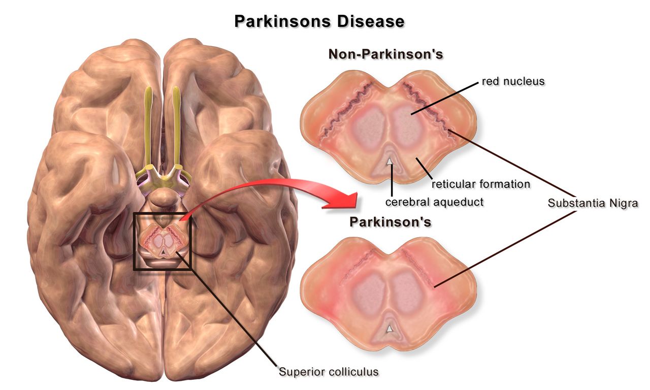
Dopamine, the basal ganglia and TRAP
Parkinson disease is the progressive loss of dopamine-producing neurons in the substantia nigra pars compacta, with intracellular aggregates of misfolded alpha-synuclein called Lewy bodies. The nigra normally feeds dopamine to the striatum, where it biases the basal ganglia circuits toward permitting movement. Strip that input away and the inhibitory output wins: movements become smaller, slower and harder to initiate. Because the system has enormous reserve, symptoms do not appear until roughly 60 to 80 percent of nigral neurons are gone — which is why the disease is already well advanced on the day of diagnosis, and why patients often say in hindsight that their handwriting shrank or their arm stopped swinging years earlier.
The cardinal motor features are remembered as TRAP. Tremor at rest, classically a 4 to 6 Hz pill-rolling tremor of the hand that damps down with purposeful movement and worsens with stress — the opposite of essential tremor, which appears with action and posture and often improves transiently with alcohol. Rigidity, felt as ratcheting cogwheel resistance when you passively flex the wrist or elbow, present in flexors and extensors equally. Akinesia and bradykinesia — slow, small movements, masked facies, decreased blink, hypophonic monotone speech, micrographia, difficulty turning in bed. Postural instability, which appears later and produces the retropulsion, festinating shuffle and freezing at doorways that generate falls.
The non-motor features are what actually degrade life and frequently precede the tremor by a decade: hyposmia, constipation, REM sleep behaviour disorder (acting out dreams), depression and anxiety, orthostatic hypotension, urinary urgency, drooling from reduced automatic swallowing, and eventually cognitive impairment or dementia in a substantial proportion of long-standing cases. Diagnosis remains clinical — bradykinesia plus rest tremor or rigidity, asymmetric onset, and a clear response to levodopa. A dopamine transporter SPECT scan (DaTscan) supports the diagnosis when the picture is muddy, and MRI is used to exclude vascular parkinsonism, normal-pressure hydrocephalus and structural mimics. There is no blood test.
Drug therapy: mechanism, monitoring and the doses you must not miss
Levodopa remains the most effective drug in neurology. Dopamine itself cannot cross the blood–brain barrier, but its precursor can; carbidopa is added to block peripheral decarboxylation so more levodopa reaches the brain and far less becomes circulating dopamine causing nausea and hypotension. Over years, as the storage capacity of remaining neurons fails, the smooth response fragments: patients develop wearing off before the next dose, unpredictable on–off fluctuations, and peak-dose dyskinesias — writhing involuntary movements that families often mistake for the disease worsening when they actually signal too much drug at the peak.
Two administration facts belong in your muscle memory. First, dietary protein competes with levodopa for the same large neutral amino acid transporter in the gut and at the blood–brain barrier, so doses work best about 30 minutes before meals; if nausea is limiting, a few crackers — not a protein shake — are the workaround. Second, carbidopa–levodopa is a time-critical medication, meaning it must be given within about 30 minutes either side of the scheduled time rather than within the usual one-hour administration window. A dose given “with the 0900 pass” when it was due at 0730 can leave a patient rigid, immobile, unable to swallow and at risk of aspiration for hours; abrupt discontinuation can precipitate a parkinsonism-hyperpyrexia syndrome resembling neuroleptic malignant syndrome, with fever, severe rigidity, autonomic instability and rhabdomyolysis. Home schedules are individualised and must be reconciled and honoured, not folded into standard hospital times.
| Class / example | Mechanism | What to monitor and teach |
|---|---|---|
| Carbidopa–levodopa | Dopamine precursor plus peripheral decarboxylase inhibitor | Give on time and before meals; nausea, orthostatic hypotension, dyskinesia, vivid dreams, darkened sweat and urine. Do not crush extended-release tablets. Never stop abruptly. |
| Dopamine agonists — pramipexole, ropinirole, rotigotine patch | Direct postsynaptic D2/D3 stimulation | Impulse-control disorders (gambling, hypersexuality, shopping — ask directly and ask the family), sudden sleep attacks, hallucinations, ankle oedema, orthostasis. Higher confusion risk in older adults. |
| MAO-B inhibitors — selegiline, rasagiline, safinamide | Block central dopamine breakdown | Insomnia; avoid meperidine and use caution with SSRIs, SNRIs and tramadol because of serotonin syndrome; teach tyramine awareness at higher doses. |
| COMT inhibitors — entacapone, opicapone | Extend levodopa half-life peripherally | Only useful with levodopa; brownish-orange urine discolouration is harmless; diarrhoea; may amplify dyskinesia. Tolcapone requires liver enzyme monitoring. |
| Amantadine | NMDA antagonist with dopaminergic effects | Best drug for levodopa-induced dyskinesia; livedo reticularis, ankle oedema, confusion and hallucinations especially in older adults; renal dose adjustment. |
| Anticholinergics — trihexyphenidyl, benztropine | Rebalance striatal acetylcholine | Tremor-predominant younger patients only; dry mouth, blurred vision, urinary retention, constipation, confusion. Listed on Beers criteria — avoid in older adults. |
| Apomorphine subcutaneous / sublingual | Rescue dopamine agonist for sudden off episodes | Profound hypotension and vomiting; contraindicated with 5-HT3 antagonists — ondansetron with apomorphine can cause severe hypotension and loss of consciousness. |
Deep brain stimulation of the subthalamic nucleus or globus pallidus internus is offered to patients with good levodopa response but disabling fluctuations or dyskinesia. It does not cure the disease and does not reliably help axial symptoms such as freezing or falls. Post-implant nursing points: the patient carries a controller and identification card, MRI is restricted per device labelling, and the device may need to be turned off for certain procedures — check before diathermy or cardioversion.
Nursing care: falls, swallowing, and the two conditions that share the ward
Aspiration pneumonia is the leading cause of death in Parkinson disease, and it grows out of quiet dysphagia that no one screened for. Any Parkinson patient who coughs during meals, holds food in the cheek, takes a long time to eat, has a wet voice after drinking, or has lost weight needs a bedside swallow screen and a speech-language pathology evaluation before you feed them. Practical measures: sit fully upright, remain upright 30 to 60 minutes after eating, small bites with a chin-tuck if recommended, no straws if that is the plan of care, no distractions or conversation during swallowing, and timing meals to the medication “on” period. Oral care twice daily meaningfully reduces aspiration pneumonia risk and is one of the highest-value tasks on the unit.
Falls occur at predictable moments — turning, standing up, starting to walk, passing through a doorway, and reaching for something while walking. Teach turning in a wide arc rather than pivoting, one task at a time, no carrying objects while ambulating, and cueing strategies for freezing: count a marching rhythm, step over an imaginary line or a laser projected from a cane, rock side to side to initiate. Check orthostatic vital signs — a fall of 20 mm Hg systolic or 10 mm Hg diastolic within three minutes of standing defines orthostatic hypotension and is extremely common here from both the disease and its drugs. Constipation is managed with fluid, fibre, activity and a scheduled regimen; it also delays levodopa absorption, so it is a motor issue, not just a comfort one.
Two other movement disorders appear on medical floors. Huntington disease is an autosomal dominant trinucleotide repeat disorder (40 or more CAG repeats in the HTT gene on chromosome 4) producing chorea, behavioural change and dementia in midlife. Nursing focus is safety from injurious involuntary movement, enormous caloric need (choreic patients may require several thousand extra calories daily), aspiration precautions, suicide risk screening, and genetic counselling for children who have a 50 percent risk. VMAT2 inhibitors such as tetrabenazine and deutetrabenazine reduce chorea but carry a boxed warning for depression and suicidality — screen mood at every encounter. Essential tremor, by contrast, is benign, symmetric, action-related, often familial, and treated with propranolol or primidone; the most important nursing act is telling the patient this is not Parkinson disease.
Older adults deserve a specific caution. Bradykinesia, stooped posture and a soft voice are frequently written off as “just getting old,” delaying diagnosis by years. At the same time older patients tolerate dopamine agonists and anticholinergics poorly, developing confusion, hallucinations and urinary retention where a 55-year-old would not. When an older Parkinson patient becomes acutely confused in hospital, work the delirium algorithm — infection, retention, constipation, pain, hypoxia, new drug — before assuming disease progression.
In Parkinson disease the two nursing acts that change outcomes are giving levodopa exactly on the home schedule and protecting the airway from a swallow that is failing silently.
Section 3Multiple sclerosis and demyelinating disease
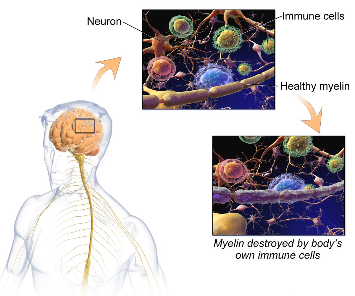
Demyelination, and why the symptoms wander
Multiple sclerosis is an immune-mediated attack on central nervous system myelin. Autoreactive T and B lymphocytes cross the blood–brain barrier and destroy oligodendrocytes and the myelin sheaths they maintain, leaving plaques of demyelination in periventricular white matter, the optic nerves, brainstem, cerebellum and spinal cord. Because myelin exists to make saltatory conduction fast, demyelinated axons conduct slowly, unreliably, or not at all — which is why symptoms appear, partially remit as inflammation subsides and remyelination occurs, then recur elsewhere. Over years, repeated inflammation causes irreversible axonal transection, and that is what converts relapsing disease into fixed disability.
Onset is typically between ages 20 and 40, with women affected two to three times as often as men. Risk rises with distance from the equator, low vitamin D, smoking, obesity in adolescence, and prior Epstein–Barr virus infection. Roughly 85 percent begin with a relapsing–remitting course; many eventually transition to secondary progressive disease with steady accumulation of disability, while about 10 to 15 percent have primary progressive MS from the outset with no distinct relapses.
The clinical picture is genuinely scattered, because a plaque can land anywhere. Optic neuritis — unilateral blurring or loss of central vision with pain on eye movement and a relative afferent pupillary defect — is a common first event. Internuclear ophthalmoplegia produces double vision with impaired adduction on lateral gaze. Sensory complaints, spasticity, ataxia, intention tremor, scanning dysarthria, bladder urgency or retention, bowel dysfunction, cognitive slowing and depression all occur. Two signs are worth memorising: Lhermitte sign, an electric shock down the spine on neck flexion, and Uhthoff phenomenon, the temporary worsening of symptoms with any increase in body temperature — a hot shower, a fever, exercise, a summer afternoon. Fatigue, which patients describe as a wall rather than tiredness, is consistently rated the most disabling symptom of all.
Diagnostics and telling a relapse from an imposter
Diagnosis rests on demonstrating lesions disseminated in space (multiple central nervous system sites) and in time (new lesions appearing, or the coexistence of enhancing and non-enhancing lesions), with no better explanation — the substance of the McDonald criteria. MRI of brain and spinal cord with gadolinium is the anchor: gadolinium enhancement indicates a lesion less than roughly four to six weeks old with an open blood–brain barrier, while T2/FLAIR hyperintensities map old and new burden. Lumbar puncture is no longer mandatory but is used when the picture is atypical; cerebrospinal fluid shows oligoclonal bands — two or more immunoglobulin bands present in CSF but not in serum — and an elevated IgG index. CSF protein is normal or only mildly raised (normal roughly 15 to 45 mg/dL) and cell counts are normal or show mild lymphocytosis. Evoked potentials can reveal clinically silent slowed conduction. Blood work exists to exclude mimics: vitamin B12, TSH, ANA, HIV, Lyme serology, and aquaporin-4 and MOG antibodies to rule out neuromyelitis optica spectrum disorder, which is treated very differently.
A true relapse is a new or clearly worsening neurologic symptom lasting more than 24 hours in the absence of fever or infection, developing over hours to days. A pseudo-relapse is the temporary re-emergence of old deficits caused by anything that raises body temperature or physiologic stress — most commonly a urinary tract infection, but also pneumonia, dehydration, heat, or poor sleep. The nursing implication is concrete and heavily tested: when an MS patient arrives with worsening weakness, check the temperature and obtain a urinalysis and culture before anyone reaches for high-dose steroids. Treating an infection with immunosuppression is the wrong direction.
Acute relapses that impair function are treated with methylprednisolone 1 g intravenously daily for three to five days, sometimes followed by an oral taper. Steroids shorten the relapse; they do not change long-term disability. Monitor capillary glucose (steroids will unmask or worsen hyperglycaemia, particularly in diabetes), blood pressure, mood and sleep — insomnia, irritability and frank steroid psychosis are common — provide gastric protection, and warn about metallic taste and facial flushing. Plasma exchange is used for severe steroid-refractory relapses.
Disease-modifying therapy and daily life
Disease-modifying therapies reduce relapse rate and new lesion formation in relapsing forms; they do not repair existing damage, which is why early initiation is emphasised. Adherence teaching is a nursing job, and so is anticipatory guidance about side effects, because patients who are surprised by side effects stop the drug.
| Therapy | Mechanism in one line | Key monitoring / teaching |
|---|---|---|
| Interferon beta-1a / beta-1b (injection) | Immunomodulation, reduced lymphocyte trafficking | Flu-like symptoms — dose at bedtime, pre-treat with acetaminophen or ibuprofen; rotate injection sites; monitor CBC and liver enzymes; screen for depression. |
| Glatiramer acetate (injection) | Decoy antigen shifting T-cell response | Injection-site lipoatrophy; a self-limited post-injection reaction of chest tightness, flushing, palpitations and anxiety lasting 15 to 30 minutes — frightening but not cardiac. Teach it in advance. |
| Dimethyl fumarate / diroximel (oral) | Nrf2 antioxidant pathway activation | Flushing (take with food, aspirin may help) and GI upset; monitor CBC for lymphopenia and liver enzymes. |
| Teriflunomide (oral) | Pyrimidine synthesis inhibition | Boxed warning — hepatotoxicity and teratogenicity; effective contraception essential for both sexes; hair thinning; accelerated elimination protocol with cholestyramine if pregnancy occurs. |
| Sphingosine-1-phosphate modulators — fingolimod, siponimod, ozanimod (oral) | Trap lymphocytes in lymph nodes | First-dose bradycardia monitoring is drug-specific: fingolimod requires about 6 hours of observation with an ECG before and after the first dose in every patient; siponimod uses a titration pack and needs first-dose observation only with pre-existing sinus bradycardia, first- or second-degree AV block, or prior myocardial infarction or heart failure; ozanimod uses dose escalation with no routine first-dose observation. Across the class — macular oedema screening at 3 to 4 months; varicella titres and vaccination before starting; infection risk. |
| Natalizumab (IV monthly) | Blocks alpha-4 integrin lymphocyte entry to CNS | Progressive multifocal leukoencephalopathy risk — JC virus antibody testing and index monitoring; report any new cognitive, visual or behavioural change immediately; restricted distribution programme. |
| Anti-CD20 antibodies — ocrelizumab, ofatumumab, rituximab | B-cell depletion | Infusion reactions — premedicate and monitor; hepatitis B screening before therapy; hypogammaglobulinaemia and infection over time; hold live vaccines. |
Symptom management often matters more to the patient than the DMT. Spasticity responds to baclofen or tizanidine — tizanidine causes hypotension, sedation and requires liver enzyme monitoring, and oral baclofen must be tapered, while abrupt withdrawal of intrathecal baclofen is a medical emergency producing rebound spasticity, hyperthermia, altered mental status and rhabdomyolysis. Fatigue is approached with energy conservation, scheduled rest, cooling, exercise, and sometimes amantadine or modafinil. Neuropathic pain responds to gabapentin, pregabalin or duloxetine. Dalfampridine, a potassium-channel blocker, may improve walking speed but is contraindicated with any seizure history or creatinine clearance of 50 mL/min or less. Bladder dysfunction must be characterised before it is treated: a spastic bladder causes urgency and frequency and responds to antimuscarinics or mirabegron, while a flaccid bladder retains urine and needs post-void residual measurement and intermittent catheterisation — giving an anticholinergic to a retaining bladder causes exactly the infection you are trying to prevent.
Teaching priorities: avoid heat (hot tubs, saunas, hot yoga), use cooling vests and cool showers, treat fevers promptly, keep vaccinations current but coordinate live vaccines with the DMT, stop smoking, maintain vitamin D, exercise in a cool pool, plan pregnancy with the neurologist because most DMTs are stopped beforehand, and report any new symptom lasting more than a day. Older adults with long-standing MS carry an added burden of osteoporosis from steroids and immobility, and their DMTs are often de-escalated because infection risk rises while relapse rate falls with age.
Multiple sclerosis is inflammation scattered in space and time; nurses change outcomes by separating true relapse from infection-driven pseudo-relapse, protecting patients from heat, and making disease-modifying therapy tolerable enough to continue.
Section 4Neuromuscular emergencies: myasthenia gravis and Guillain–Barré syndrome
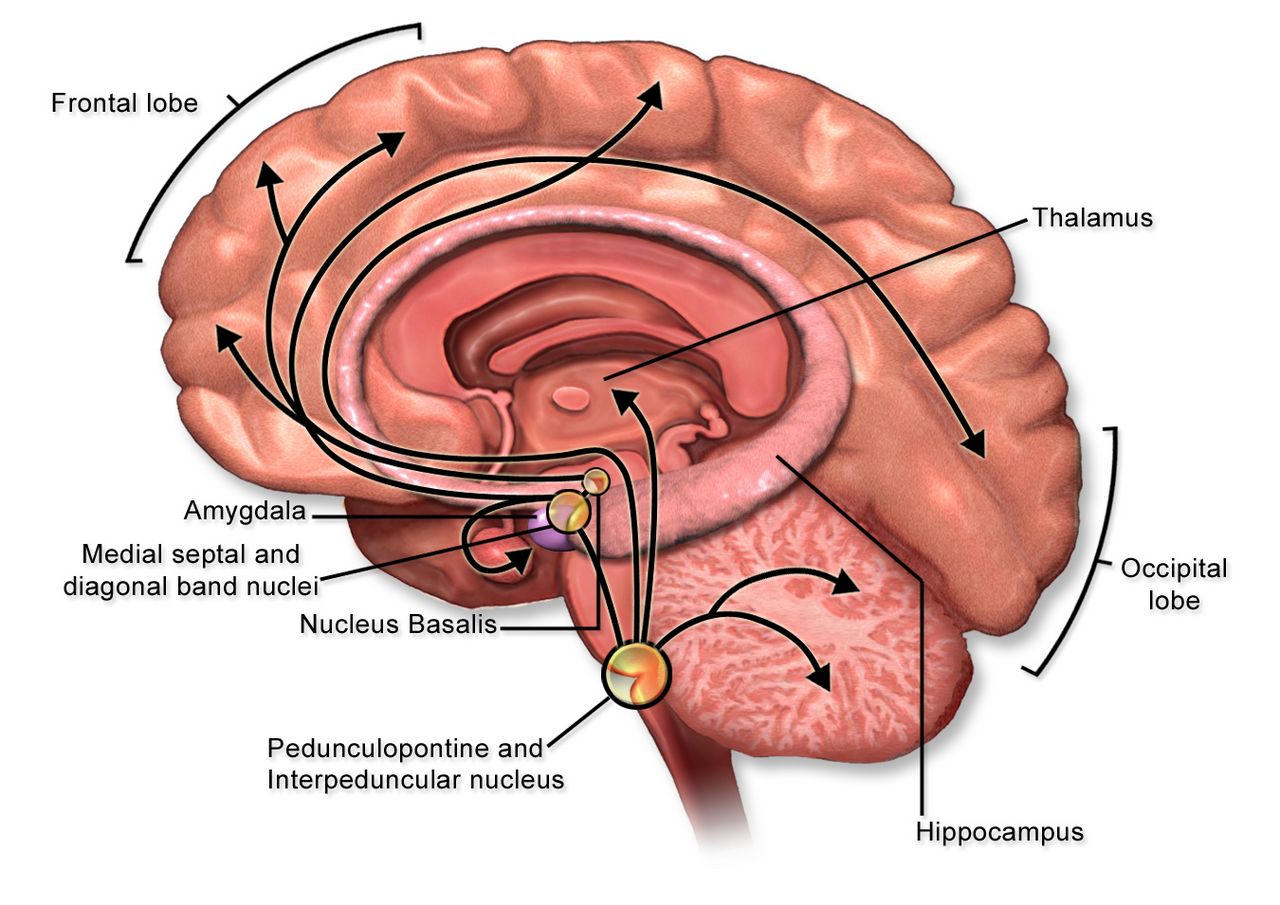
Myasthenia gravis: the junction that runs out
Myasthenia gravis is an autoimmune attack on the postsynaptic membrane of the neuromuscular junction. Antibodies against the nicotinic acetylcholine receptor — present in roughly 85 percent of generalised cases and about half of purely ocular cases — cross-link and internalise receptors, activate complement and flatten the postsynaptic folds. Fewer receptors means a smaller endplate potential; add the physiologic run-down of acetylcholine release with repeated stimulation and you get the defining clinical feature: weakness that worsens with sustained use and recovers with rest. A subset of patients instead carry MuSK antibodies (about 5 to 8 percent), which cause more bulbar and respiratory involvement and respond less well to cholinesterase inhibitors. Thymic abnormality is central — hyperplasia in about two-thirds and thymoma in 10 to 15 percent.
Assessment findings follow the fatigability rule. Ptosis and diplopia are the presenting complaint in roughly half and are worse in the evening, after reading or after driving. Bulbar involvement gives a nasal or slurred voice that deteriorates during a conversation, jaw fatigue partway through a meal, difficulty chewing and swallowing, and nasal regurgitation of liquids. Proximal limb weakness makes stairs and hair-brushing hard; neck extensor weakness produces a dropped head. Crucially, sensation is normal, reflexes are normal and pupils are normal — a distinction that separates myasthenia from botulism, which produces a descending paralysis with fixed dilated pupils and prominent dry mouth. On exam, sustained upgaze for 60 seconds reproduces ptosis; an ice pack applied to the closed lid for two minutes improves it, because cooling slows acetylcholinesterase.
Diagnostics: serum acetylcholine receptor binding antibodies first, then MuSK if negative. Repetitive nerve stimulation shows a decremental response greater than about 10 percent; single-fibre electromyography is the most sensitive test. CT of the chest is mandatory to look for thymoma. Check TSH, because autoimmune thyroid disease frequently travels with myasthenia, and screen for other autoimmune conditions. The edrophonium (Tensilon) test is largely historical and no longer generally available.
Treatment has two arms. Symptomatic therapy is pyridostigmine, a cholinesterase inhibitor typically dosed around 60 mg orally every 4 to 6 hours, with onset in 30 to 60 minutes and peak effect at about one to two hours — time the dose 30 to 45 minutes before meals in a patient with chewing or swallowing weakness, and time physical therapy to the peak. Its side effects are muscarinic: abdominal cramping, diarrhoea, salivation, lacrimation, bradycardia, increased bronchial secretions. Disease-modifying therapy is immunosuppression: corticosteroids (which can transiently worsen weakness during initiation, so severe bulbar patients are started in hospital), azathioprine or mycophenolate for maintenance with a slow onset over many months, rituximab particularly for MuSK disease, complement inhibitors such as eculizumab and ravulizumab (meningococcal vaccination is required before the first dose), and neonatal Fc receptor blockers such as efgartigimod that accelerate IgG clearance. Thymectomy is indicated for any thymoma and improves outcomes in many acetylcholine-receptor-positive generalised patients.
| Drug class | Examples | Why it matters |
|---|---|---|
| Aminoglycosides | Gentamicin, tobramycin, amikacin | Impair presynaptic acetylcholine release — can precipitate crisis. Flag the order. |
| Fluoroquinolones and macrolides | Ciprofloxacin, levofloxacin, azithromycin; telithromycin is contraindicated | Fluoroquinolones carry a boxed warning for exacerbation of myasthenia; macrolides can also worsen it. Choose an alternative when possible. |
| Magnesium (especially IV) | Magnesium sulfate for eclampsia or repletion | Blocks calcium-dependent acetylcholine release — a classic cause of iatrogenic crisis. |
| Cardiac agents | Beta blockers, procainamide, quinidine, some calcium channel blockers | Blunt neuromuscular transmission; monitor closely if unavoidable. |
| Non-depolarizing blockers | Rocuronium, vecuronium | Marked sensitivity — a standard dose can produce profound, prolonged paralysis, so the dose is reduced substantially, quantitative train-of-four monitoring is essential and sugammadex is the preferred reversal. Anaesthesia must know the diagnosis. |
| Depolarizing blocker | Succinylcholine | The opposite problem — with fewer acetylcholine receptors these patients are relatively resistant, so a standard dose may not relax them and a higher dose is needed. Duration may still be prolonged in a patient taking pyridostigmine, because cholinesterase inhibition slows the drug’s breakdown. Anaesthesia must know the diagnosis. |
| Other | Penicillamine, checkpoint inhibitors, botulinum toxin, high-dose corticosteroid initiation | Can induce or unmask myasthenia, or transiently worsen it. |
Crisis: what you do first, and only then what you call it
Myasthenic crisis is respiratory failure from disease severity, usually triggered by infection, surgery, aspiration, pregnancy or the postpartum period, tapering of immunosuppression, or one of the drugs above. Cholinergic crisis is respiratory failure from excess cholinesterase inhibitor, and it is now uncommon. Both present as a weak patient who cannot breathe. Students burn precious minutes trying to decide which one it is; the correct first action is neither — it is to assess and support ventilation. Raise the head of the bed, suction, stop oral intake, apply continuous pulse oximetry and cardiac monitoring, obtain a vital capacity and NIF, call the provider and rapid response, and prepare for intubation. Diagnosis follows stabilisation.
| Myasthenic crisis | Cholinergic crisis | |
|---|---|---|
| Cause | Too little drug, or a trigger (infection, surgery, drug) | Too much cholinesterase inhibitor |
| Timing clue | Weakness improves after a dose; often a missed or late dose | Weakness begins within about an hour of a dose |
| Pupils | Normal | Constricted (miosis) |
| Autonomic signs | Dry, often tachycardic, anxious | SLUDGE — salivation, lacrimation, urination, diarrhoea, GI cramping, emesis; bradycardia, sweating, fasciculations |
| Secretions | Usually manageable | Copious oral and bronchial secretions |
| Treatment | Airway support plus IVIG or plasma exchange; corticosteroids; treat the trigger | Hold the cholinesterase inhibitor; atropine for muscarinic symptoms; airway support |
In crisis, intravenous immune globulin (commonly 2 g/kg divided over 2 to 5 days) or plasma exchange (typically five exchanges) produces improvement within days. IVIG carries risks of aseptic meningitis, acute kidney injury, thrombosis, volume overload and anaphylaxis in IgA-deficient patients — hydrate, infuse slowly at the start, monitor renal function and watch for headache. Plasma exchange requires large-bore or central access and causes citrate-induced hypocalcaemia (watch for perioral tingling, Chvostek and Trousseau signs), hypotension and coagulopathy. Pyridostigmine is usually held while the patient is intubated to reduce secretions and restarted as weaning begins.
Guillain–Barré syndrome: paralysis that climbs
Guillain–Barré syndrome is an acute immune-mediated polyradiculoneuropathy in which antibodies raised against a preceding infection cross-react with peripheral nerve myelin or axolemma — molecular mimicry. About two-thirds of patients report a respiratory or gastrointestinal illness one to three weeks earlier; Campylobacter jejuni is the most frequently identified trigger, followed by cytomegalovirus, Epstein–Barr virus, Mycoplasma, influenza, hepatitis E and Zika virus. The acute inflammatory demyelinating polyradiculoneuropathy (AIDP) form predominates in North America and Europe; axonal variants and the Miller Fisher variant (ophthalmoplegia, ataxia and areflexia, associated with anti-GQ1b antibodies) also occur.
The classic presentation is symmetric ascending flaccid weakness beginning in the legs, with early loss of deep tendon reflexes that is often more striking than the weakness itself. Paraesthesias precede or accompany the weakness, and severe aching back and limb pain occurs in a majority and is frequently under-treated. Progression is rapid — hours to two weeks, reaching nadir within four weeks by definition — and ascends toward the trunk, arms, and then bulbar and respiratory muscles. Roughly one in four patients requires mechanical ventilation. Facial diplegia and difficulty handling secretions are ominous.
Autonomic instability is the feature that kills the ones who do not die of respiratory failure: labile blood pressure swinging from hypertension to hypotension, tachyarrhythmias and sudden bradyarrhythmias, ileus, urinary retention, diaphoresis and abnormal thermoregulation. Practical consequences — keep the patient on cardiac monitoring, change position slowly, be cautious with suctioning (vagal stimulation can precipitate profound bradycardia and asystole in these patients), avoid abrupt dosing of antihypertensives because the pressure may reverse in minutes, listen for bowel sounds, and monitor for retention.
Diagnosis is clinical and supported by lumbar puncture and nerve conduction studies. After about the first week, CSF shows the hallmark albuminocytologic dissociation: elevated protein (above the usual upper limit of about 45 mg/dL, often 100 to several hundred) with a normal white cell count, typically fewer than 10 cells/mm3. A high CSF cell count should redirect you toward HIV seroconversion, Lyme disease, sarcoidosis or malignant infiltration. Nerve conduction studies show slowed conduction, prolonged distal latencies and conduction block.
Treatment is intravenous immune globulin 0.4 g/kg daily for five days or plasma exchange — they are equally effective, they are not combined, and neither reverses damage already done; both shorten the course. Corticosteroids are ineffective in Guillain–Barré syndrome and are not recommended, which is a favourite examination point precisely because steroids are used in so many other neuroimmune conditions. Everything else is supportive and it is almost entirely nursing: serial vital capacity every 2 to 4 hours during the progressive phase, elevation of the head of bed, aspiration precautions and swallow assessment, venous thromboembolism prophylaxis (these patients are immobile and at high clot risk), pressure injury prevention with a turning schedule, passive range of motion and splinting to prevent contractures, eye care if the lids will not close, bowel and bladder programmes, and aggressive neuropathic pain management with gabapentin or pregabalin, using opioids cautiously because of ileus.
Do not overlook the psychological dimension. A patient with severe Guillain–Barré syndrome may be fully alert, oriented and terrified inside a body that will not respond, unable to speak around an endotracheal tube. Establish a communication method early — an eye-blink code, a letter board, a call system adapted to whatever movement remains — explain every procedure before touching them, orient them to time and progress, and keep family present. Most patients recover: roughly 80 percent walk independently by six months, though fatigue and residual weakness can persist for a year or more, and mortality remains around 3 to 7 percent, higher in older adults, who also recover more slowly and less completely.
Myasthenic crisis and Guillain–Barré syndrome are the same emergency wearing different masks — serial vital capacity and negative inspiratory force, not pulse oximetry, tell you when to secure the airway before the patient decompensates.
Section 5ALS, dementia and the long arc of progressive disease
Amyotrophic lateral sclerosis: both motor neurons, intact senses
ALS is the progressive degeneration of upper and lower motor neurons simultaneously, which is why it produces a combination of findings that occurs in no other common disease: spasticity and hyperreflexia in a limb that is also wasting and fasciculating. Roughly 90 percent of cases are sporadic; about 10 percent are familial, with C9orf72 repeat expansions and SOD1 mutations among the most common. Sensation, extraocular movement, bowel and bladder control, and sexual function are characteristically spared, and most cognition is preserved — although about 15 percent develop frontotemporal dementia and up to half show some executive or behavioural change.
Presentation is limb-onset in about two-thirds — asymmetric foot drop with tripping, or a hand that drops objects and loses fine dexterity — and bulbar-onset in about a third, with slurred speech, difficulty swallowing, and a wasted fasciculating tongue. Bulbar onset carries a worse prognosis. Pseudobulbar affect, uncontrollable laughing or crying disproportionate to feeling, distresses families and is treatable. Median survival is around three to five years from symptom onset, with respiratory failure the usual cause of death.
Diagnosis is clinical, supported by electromyography showing active and chronic denervation across multiple body regions, and requires excluding treatable mimics: cervical spondylotic myelopathy, myasthenia gravis, vitamin B12 deficiency, thyroid disease, lead toxicity, Lyme disease and multifocal motor neuropathy. Creatine kinase may be modestly elevated. Disease-modifying options are limited — riluzole 50 mg orally every 12 hours, taken one hour before or two hours after meals, with liver enzymes checked at baseline, monthly for the first three months and periodically thereafter, extends survival by a few months; edaravone is an antioxidant option; tofersen targets SOD1-associated disease. The real work is multidisciplinary symptom management.
| Problem | Approach | Nursing focus |
|---|---|---|
| Respiratory decline | Noninvasive ventilation, usually initiated as FVC falls below about 50 percent predicted or with orthopnoea and nocturnal hypoventilation | Serial FVC at each visit; NIV improves survival and quality of life; teach mask fit and troubleshoot early intolerance so the patient stays with it. Cough assist devices clear secretions. |
| Dysphagia and weight loss | Diet texture modification, then gastrostomy tube — placed before FVC drops below roughly 50 percent, when the procedure is safer | Weigh at every visit; a falling weight predicts poorer survival. Raise the enteral access conversation early, not at the crisis. |
| Sialorrhoea and thick secretions | Glycopyrrolate, amitriptyline, atropine drops, botulinum toxin; nebulised saline and mucolytics for thick secretions | Portable suction at home; teach caregivers to distinguish thin drooling from thick plugging, which need opposite treatments. |
| Spasticity, cramps, pain | Baclofen, tizanidine, mexiletine for cramps; range of motion and positioning | ALS pain is largely musculoskeletal from immobility — repositioning and stretching are as valuable as drugs. |
| Pseudobulbar affect | Dextromethorphan–quinidine, or an SSRI | Explain to the family that the emotion displayed is not the emotion felt. |
| Communication loss | Voice banking early, speech-generating and eye-gaze devices | Start before speech fails. A patient who can still speak clearly is the one who should be recording their voice. |
Alzheimer disease and other dementias
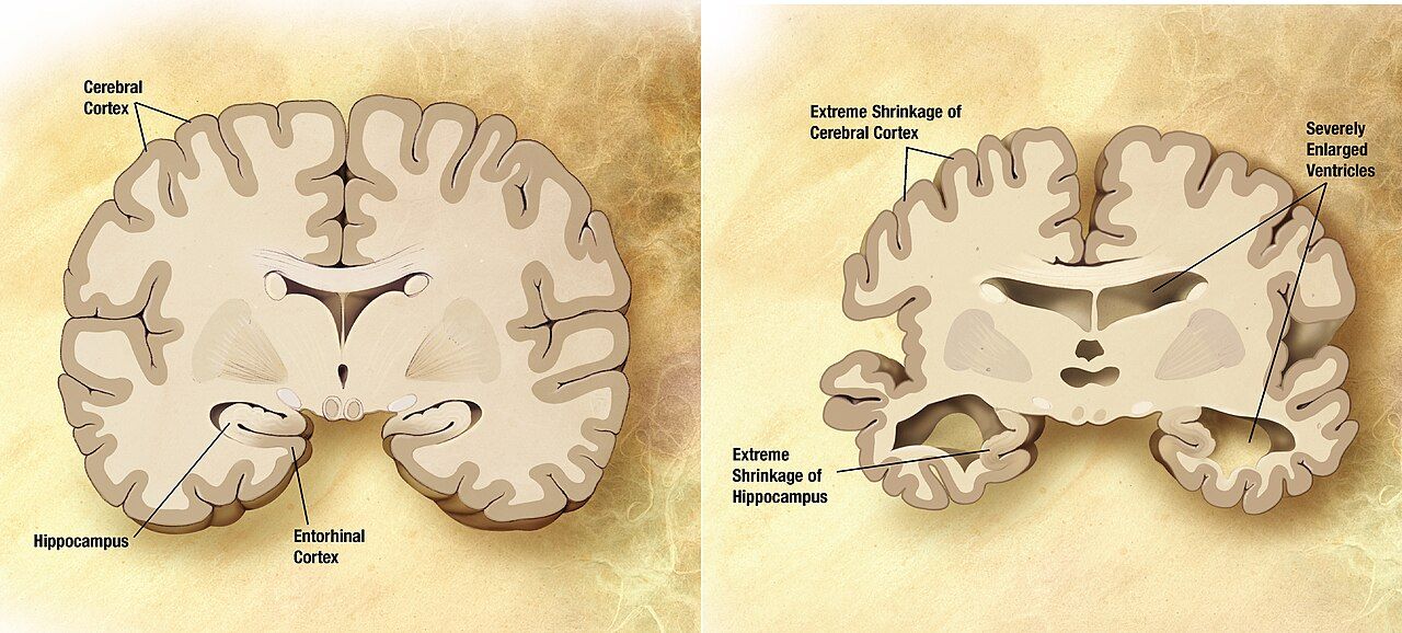
Alzheimer disease is a progressive neurodegeneration characterised by extracellular beta-amyloid plaques and intraneuronal neurofibrillary tangles of hyperphosphorylated tau, with early hippocampal and entorhinal degeneration spreading to association cortex, and loss of cholinergic projections from the basal forebrain. Clinically it begins with episodic memory failure — the same question asked repeatedly, misplaced objects, missed appointments — and advances through the four A’s: amnesia, aphasia (word-finding failure, then empty speech), apraxia (inability to perform learned motor tasks such as dressing) and agnosia (failure to recognise objects and eventually people). Other dementias have their own signatures worth recognising: vascular dementia progresses in steps with focal findings, dementia with Lewy bodies brings visual hallucinations, fluctuating cognition, parkinsonism and severe neuroleptic sensitivity, and frontotemporal dementia presents in a younger patient with behavioural disinhibition or progressive aphasia rather than memory loss.
Workup exists to find what is reversible. Obtain a structured cognitive assessment — the Montreal Cognitive Assessment is commonly used, with a score of 26 or above out of 30 considered normal — along with CBC, comprehensive metabolic panel, TSH, vitamin B12, and neuroimaging; depression, hypothyroidism, B12 deficiency, normal-pressure hydrocephalus, subdural haematoma and medication effects all masquerade as dementia. Amyloid PET imaging and CSF or plasma biomarkers such as phosphorylated tau are increasingly used to confirm Alzheimer pathology, particularly when an anti-amyloid therapy is being considered.
Pharmacology is modest. Cholinesterase inhibitors — donepezil, rivastigmine (including a patch), galantamine — raise synaptic acetylcholine and may slow functional decline; they cause nausea, diarrhoea, anorexia, vivid dreams, bradycardia and syncope, so check the apical pulse before administration and be cautious in patients on beta blockers or with conduction disease. Memantine, an NMDA receptor antagonist, is added in moderate to severe disease and requires renal dose adjustment. Anti-amyloid monoclonal antibodies such as lecanemab and donanemab are available for early symptomatic Alzheimer disease with confirmed amyloid; their defining safety issue is amyloid-related imaging abnormalities (ARIA) — brain oedema or microhaemorrhage — requiring scheduled surveillance MRI, extra caution in APOE ε4 homozygotes and in patients on anticoagulation, and immediate reporting of new headache, confusion, visual change, dizziness or seizure.
Most of what helps is not a drug. For agitation, wandering and sundowning, work non-pharmacologically first: consistent routine and caregivers, orientation cues, adequate daytime light and activity, limited napping and caffeine, reduced noise and clutter, familiar objects, simple one-step instructions, validation and redirection rather than argument, and a systematic search for an unmet need — pain, hunger, thirst, a full bladder, constipation, or being too hot or cold. Antipsychotics carry a boxed warning for increased mortality in older adults with dementia-related psychosis and are a last resort requiring an explicit risk conversation; physical restraints increase injury, delirium and death and are not a solution. Home safety planning covers wandering and identification, stove and firearm access, driving cessation, medication supervision and fall-proofing, and financial and legal planning — including advance directives and health care proxy designation — should be completed while the patient still has capacity.
The long arc: prioritising, delegating and planning
These diseases overlap on a busy floor, and prioritisation follows the same hierarchy every time: airway and ventilation, then circulation and acute neurologic deterioration, then safety, then chronic disease management and teaching. If you hold assignments for a Parkinson patient whose 0730 levodopa is due, a myasthenic patient with a new nasal voice, a Guillain–Barré patient asking for pain medication, and a dementia patient calling out, you see the myasthenic first (progressing bulbar and respiratory weakness), then give the levodopa before it is late (a time-critical medication whose omission causes rigidity and dysphagia), then assess the dementia patient for a treatable cause of the calling out, then treat pain. Acute beats chronic, unstable beats stable, and physiologic need beats comfort — but note that the chronic drug given late becomes an acute problem, which is why levodopa outranks routine comfort measures.
Delegation in this population is unforgiving. Unlicensed assistive personnel may take vital signs, assist with hygiene and repositioning, perform passive range of motion under direction, apply splints already fitted, ambulate a stable patient with a documented plan, and feed a patient whose swallow has already been assessed and whose plan is established. The RN retains all assessment including neurologic and swallow screening, vital capacity and NIF measurement, interpretation of any change, all teaching, evaluation of response, the care of any unstable patient, IVIG and plasma exchange management, and the first dose of a new medication. Reporting expectations must be explicit: tell the assistive personnel exactly what to report immediately — coughing during meals, a new change in voice, difficulty being understood, a heart rate below a stated threshold, any fall or near-fall.
Older adults change every equation in this chapter. They present atypically (myasthenia mistaken for a stroke or for “ageing eyelids”, ALS mistaken for arthritis or deconditioning, Parkinson mistaken for depression); they carry an anticholinergic burden from bladder drugs, antihistamines and antidepressants that compounds every centrally acting agent you add; they have less physiologic reserve for aspiration, immobility and infection; and they are more likely to live alone with a caregiver who is themselves elderly. Assess the caregiver as deliberately as you assess the patient — burden, sleep, depression, finances and respite — because caregiver collapse is the most common reason a plan of care fails.
Finally, plan for the trajectory rather than the visit. Every one of these diagnoses warrants advance care planning early, while the patient can speak for themselves: health care proxy, advance directive, and specific decisions about noninvasive ventilation, tracheostomy and long-term ventilation in ALS, and about enteral feeding in advanced dementia — where the evidence is clear that feeding tubes do not prolong survival, do not prevent aspiration and do not improve pressure injuries, so careful hand feeding is preferred. Palliative care runs alongside disease-modifying therapy from the beginning, not after it fails; Chapter 14 develops that conversation in full.
Progressive neurologic disease is managed by anticipating the next loss — ventilation, swallowing, communication, safety — and having the equipment, the teaching and the goals-of-care conversation in place before the loss arrives.
The chapter in one breath
Every disorder here breaks the same motor relay at a different point, and the exam tells you where: spasticity with brisk reflexes points upstream to the upper motor neuron, flaccid areflexic wasting with fasciculations points to the lower motor neuron and peripheral nerve, fatigable painless weakness with normal reflexes and sensation points to the neuromuscular junction. Parkinson disease is nigral dopamine loss producing tremor, rigidity, bradykinesia and postural instability — give levodopa exactly on the home schedule, keep dopamine blockers such as metoclopramide and haloperidol away from the patient, and guard the swallow, because aspiration is what kills. Multiple sclerosis scatters demyelinating plaques in space and time, confirmed by MRI and oligoclonal bands, treated acutely with methylprednisolone and long term with disease-modifying therapy — but new weakness is a urinary tract infection until proven otherwise. Myasthenia gravis and Guillain–Barré syndrome are airway emergencies waiting to happen: trend vital capacity and negative inspiratory force rather than oxygen saturation, remember IVIG or plasma exchange (and no steroids in Guillain–Barré), and screen every order for magnesium, aminoglycosides and fluoroquinolones. ALS and Alzheimer disease demand anticipation — ventilation, nutrition, communication, safety, caregiver support and advance directives arranged before the crisis, never during it.
Words worth knowing
| Term | What it means |
|---|---|
| Albuminocytologic dissociation | Elevated cerebrospinal fluid protein (above about 45 mg/dL) with a normal cell count (under 10 cells/mm3) — the hallmark CSF finding of Guillain–Barré syndrome after the first week. |
| ARIA | Amyloid-related imaging abnormalities — brain oedema or microhaemorrhage caused by anti-amyloid monoclonal antibodies, monitored with scheduled MRI. |
| Babinski sign | Upgoing great toe with fanning on plantar stimulation; abnormal after infancy and indicates an upper motor neuron lesion. |
| Bradykinesia | Slowness and progressive reduction in the amplitude of voluntary movement; the required feature for a Parkinson disease diagnosis. |
| Bulbar weakness | Weakness of muscles supplied by the lower cranial nerves — producing dysarthria, dysphagia, nasal speech and impaired secretion handling. |
| Cogwheel rigidity | Ratcheting resistance felt during passive limb movement, caused by tremor superimposed on increased tone in Parkinson disease. |
| Fasciculation | Visible spontaneous twitching of a denervated motor unit; a lower motor neuron sign, prominent in ALS. |
| Festinating gait | Short, accelerating shuffling steps with a forward-leaning trunk, seen in Parkinson disease and associated with falls. |
| Fatigable weakness | Weakness that worsens with sustained or repeated use and improves after rest — the defining feature of neuromuscular junction disease. |
| Lhermitte sign | An electric-shock sensation running down the spine and into the limbs on neck flexion, indicating cervical cord demyelination. |
| Lower motor neuron | The neuron from anterior horn cell to muscle; damage causes flaccidity, hyporeflexia, atrophy and fasciculations. |
| Myasthenic crisis | Respiratory failure from myasthenia gravis itself, usually triggered by infection, surgery or a contraindicated drug, requiring ventilatory support plus IVIG or plasma exchange. |
| Negative inspiratory force (NIF) | Maximal negative pressure generated on inspiration against an occluded airway; normally more negative than −60 cm H2O, with values less negative than −30 cm H2O signalling impending failure. |
| Oligoclonal bands | Two or more immunoglobulin bands found in cerebrospinal fluid but not in serum, indicating intrathecal antibody production; supports multiple sclerosis. |
| On–off phenomenon | Abrupt, sometimes unpredictable swings between mobility and immobility in advanced Parkinson disease on long-term levodopa. |
| Plasma exchange (plasmapheresis) | Extracorporeal removal of circulating antibodies; used in myasthenic crisis, Guillain–Barré syndrome and steroid-refractory MS relapse. Watch for hypocalcaemia, hypotension and coagulopathy. |
| Pseudo-relapse | Temporary re-emergence of old MS deficits caused by fever, infection, heat or metabolic stress rather than new demyelination; treat the cause, not with steroids. |
| Uhthoff phenomenon | Transient worsening of multiple sclerosis symptoms with any rise in body temperature, including exercise, fever or a hot bath. |
| Upper motor neuron | The neuron from motor cortex to anterior horn cell; damage causes spasticity, hyperreflexia and a positive Babinski sign without early atrophy. |
| Vital capacity | The maximum volume exhaled after a maximal inspiration; normally about 60 to 70 mL/kg, and the most useful serial measure of neuromuscular respiratory reserve. |