Body Structure, Directional Terms and Planes
Everything you have learned so far — roots, suffixes, prefixes — has to land somewhere on an actual body. This chapter gives you that map. You will learn the vocabulary of organization (cell, tissue, organ, system), the shared compass every clinician uses to say where something is (superior, distal, medial, ipsilateral), the flat planes that slice the body for imaging and surgery, and the cavities, quadrants and regions that turn “stomach pain” into “RLQ tenderness with rebound.” These are not decorative words. A misplaced medial or a confused ilium and ileum changes which structure a surgeon expects to find. Learn the word parts here and you will decode positional language for the rest of the book — because every body-system chapter that follows assumes you already know where retroperitoneal, epigastric and anterolateral point.
By the end of this chapter you can
- List the levels of organization from cell to organism and decode terms built on cyt/o, hist/o, kary/o, somat/o and viscer/o.
- Name the four primary tissue types and distinguish my/o (muscle) from myel/o (bone marrow or spinal cord).
- Describe the anatomical position and explain why every directional term assumes it.
- Use paired directional terms — superior/inferior, anterior/posterior, medial/lateral, proximal/distal, superficial/deep — correctly in a sentence about a patient.
- Identify the midsagittal, frontal (coronal), transverse and oblique planes and match each to how an image or a section is produced.
- Define common clinical positions including supine, prone, Fowler, Trendelenburg, lithotomy and Sims.
- Locate the dorsal and ventral body cavities and the organs of each, and distinguish parietal from visceral membranes.
- Map the four abdominopelvic quadrants and the nine regions, and name the major organ found in each.
- Apply singular-to-plural rules (-a/-ae, -us/-i, -um/-a, -is/-es, -ix/-ices, -nx/-nges) to structural terms.
Section 1From Cell to Organism: the Levels of Organization
Anatomy, physiology and the words that name them
Start with the two words on the front of every course catalog. Anatomy (ah-NAT-oh-mee) comes from ana- (up, apart) and -tomy (process of cutting): literally cutting apart, because that is how the field began. Physiology (fiz-ee-OL-oh-jee) is physi/o (nature, function) plus -logy (study of): the study of how the parts function. Anatomy is the parts list; physiology is the operating manual. Pathology (pah-THOL-oh-jee) — path/o (disease) plus -logy — is what happens when the operating manual fails.
Those three words already demonstrate the habit this book is training. You did not memorize anatomy as a whole; you read ana/tomy and understood it. Do that to every new term and your vocabulary compounds instead of piling up.
The body is organized in nested levels, and each level has its own root. The chemical level (atoms and molecules) builds the cell, the smallest living unit. Similar cells form a tissue. Two or more tissues working together form an organ. Organs cooperating toward a shared job form an organ system. All eleven systems together form the organism. When you read cytology, histology and systemic in a chart, the writer is telling you which level of that ladder is in question.
Word parts for cells and tissues
Two roots carry most of the load. Cyt/o means cell, and it works as a root or as the suffix -cyte. Hist/o (and histi/o) means tissue. So cyt/o/logy = cell + study of = the study of cells, which is exactly what a Pap test is: cytology. And hist/o/logy (hiss-TOL-oh-jee) = tissue + study of, the microscopic study of tissue, which is what a biopsy specimen goes for. A leuk/o/cyte (LOO-koh-site) is a white cell; an erythr/o/cyte (eh-RITH-roh-site) is a red cell.
| Word part | Meaning | Example term | What the example means |
|---|---|---|---|
| cyt/o, -cyte | cell | cytoplasm (SIGH-toh-plazm) | The substance formed within a cell, outside the nucleus |
| hist/o, histi/o | tissue | histology (hiss-TOL-oh-jee) | Study of tissue under the microscope |
| kary/o, nucle/o | nucleus | karyotype (KAR-ee-oh-type) | A picture of a cell’s chromosomes, sorted by type |
| somat/o, -some | body | somatic (soh-MAT-ik) | Pertaining to the body (as opposed to the mind) |
| viscer/o | internal organs | visceral (VISS-er-al) | Pertaining to the internal organs |
| organ/o | organ | organomegaly (or-gan-oh-MEG-ah-lee) | Enlargement of an organ |
| -plasm | formation, growth, substance | neoplasm (NEE-oh-plazm) | A new growth — a tumor |
| blast/o, -blast | immature cell, germ cell | osteoblast (OSS-tee-oh-blast) | An immature bone-forming cell |
| morph/o | shape, form | morphology (mor-FOL-oh-jee) | Study of shape and structure |
| gen/o, -genesis | producing, origin | pathogenesis (path-oh-JEN-eh-sis) | The origin and development of a disease |
Notice how -plasm behaves. Cyt/o/plasm is the substance inside a cell. Neo/plasm is a new formation. Hyper/plasia (high-per-PLAY-zhah) is excessive formation — too many cells. A/plasia (ah-PLAY-zhah) is absent formation, and dys/plasia (dis-PLAY-zhah) is bad or disordered formation, the word a pathologist uses on a cervical or colon biopsy that is not yet cancer but is not normal either. One suffix family, four clinically distinct meanings, all readable from the prefix.
The four tissue types
Every organ in the body is assembled from four primary tissues: epithelial tissue (covering and lining, including glands), connective tissue (support and binding — bone, cartilage, fat, blood), muscle tissue (contraction) and nervous tissue (conduction of impulses). Each type owns a set of combining forms that will reappear in every system chapter, so learning them once pays for itself ten times.
| Word part | Meaning | Example term | What the example means |
|---|---|---|---|
| epitheli/o | epithelium, surface tissue | epithelial (ep-ih-THEE-lee-al) | Pertaining to the tissue that covers and lines surfaces |
| my/o, muscul/o | muscle | myalgia (my-AL-jee-ah) | Muscle pain |
| myel/o | bone marrow; spinal cord | myelitis (my-eh-LYE-tis) | Inflammation of the spinal cord |
| neur/o | nerve | neuropathy (noo-ROP-ah-thee) | Disease of the nerves |
| oste/o | bone | osteocyte (OSS-tee-oh-site) | A mature bone cell |
| chondr/o | cartilage | chondromalacia (kon-droh-mah-LAY-shah) | Softening of cartilage |
| adip/o, lip/o, steat/o | fat | adipose (AD-ih-pohs) | Fatty; pertaining to fat tissue |
| fibr/o | fiber, fibrous tissue | fibrosis (fye-BROH-sis) | Abnormal condition of fibrous (scar) tissue |
| hemat/o, hem/o | blood | hematology (hee-mah-TOL-oh-jee) | Study of blood |
| angi/o, vas/o | vessel | angiogram (AN-jee-oh-gram) | A record (image) of a vessel |
The my/o — myel/o trap. One letter group separates two completely different tissues. My/o is muscle: my/algia is muscle pain, my/o/pathy is muscle disease, my/o/cardium is heart muscle. Myel/o is bone marrow or spinal cord: myel/oma is a marrow tumor, myel/o/gram is an image of the spinal canal, and oste/o/myel/itis (oss-tee-oh-my-eh-LYE-tis) is inflammation of bone and marrow — a bone infection, not a muscle problem. Which meaning of myel/o applies is decided by context: pair it with blood words and it is marrow; pair it with spine or nerve words and it is spinal cord. Read the whole term before you commit.
Above tissue sits the organ, and above the organ, the system. Two adjectives describe how far a problem has spread. Local means confined to one area; systemic (siss-TEM-ik) means affecting the body as a whole. A local infection is a red, warm finger; a systemic infection has fever, tachycardia and a rising white count. And two more roots sort a symptom by the tissue it comes from — somat/o (body wall) and viscer/o (internal organs): somatic pain is well-localized pain from skin, muscle or bone, while visceral pain is the dull, hard-to-point-to ache of an internal organ. That distinction is not academic — it is how a triage nurse begins to separate a pulled muscle from an inflamed gallbladder.
The body is organized cell to tissue to organ to system to organism, and each level owns combining forms — cyt/o, hist/o, viscer/o, somat/o — that tell you which level a term is describing.
Section 2Anatomical Position and Directional Terms
Why one starting posture matters
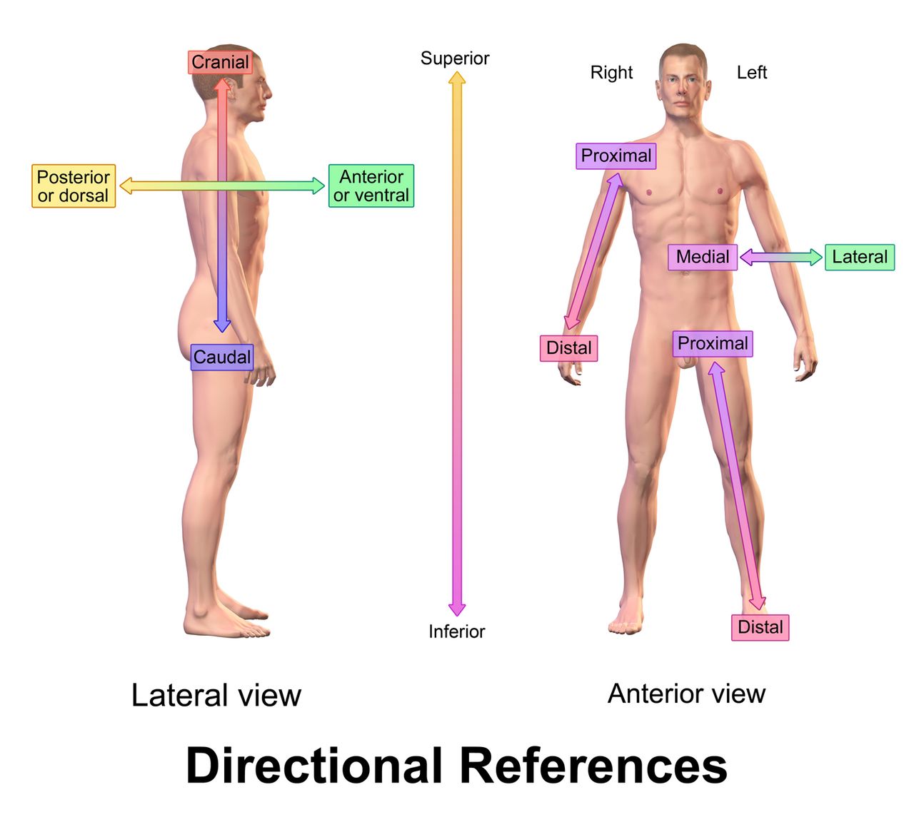
Say a patient has a wound “above the elbow.” If the arm is raised, above the elbow is the forearm. If the arm hangs down, above the elbow is the upper arm. Ordinary language collapses under a body that moves, so anatomy fixes the body in one agreed posture and describes everything from there. That posture is the anatomical position: standing erect, facing forward, arms at the sides, palms turned forward, feet slightly apart with toes pointing forward.
Every directional term in every chart, textbook and operative note assumes that position, whether or not the patient is actually in it. A patient lying face-down on a stretcher still has an anterior chest and a posterior back. The terms describe the body, not the moment.
Two other conventions ride along with it. First, right and left always mean the patient’s right and left, never the observer’s. Second, the palms-forward detail is not trivia: it is what makes the radius the lateral forearm bone and the thumb the lateral digit. Turn the palm inward and the bones cross; anatomical position uncrosses them so descriptions stay stable.
The paired terms
Directional terms come in opposing pairs, which is the easiest way to learn them. Each pair answers one question about location, and each has a combining form you can use to build adjectives.
| Word part | Meaning | Example term | What the example means |
|---|---|---|---|
| super/o | above, upward | superior (soo-PEER-ee-or) | Above, or toward the head — the lungs are superior to the diaphragm |
| infer/o | below, downward | inferior (in-FEER-ee-or) | Below, or toward the feet — the stomach is inferior to the heart |
| anter/o | front | anterior (an-TEER-ee-or) | Toward the front of the body |
| poster/o | back, behind | posterior (pos-TEER-ee-or) | Toward the back of the body |
| ventr/o | belly side | ventral (VEN-tral) | Pertaining to the belly side — in humans, the same as anterior |
| dors/o | back side | dorsal (DOR-sal) | Pertaining to the back side — in humans, the same as posterior |
| medi/o | middle | medial (MEE-dee-al) | Toward the midline — the great toe is medial |
| later/o | side | lateral (LAT-er-al) | Away from the midline, toward the side |
| proxim/o | near (the point of attachment) | proximal (PROK-sih-mal) | Nearer the trunk — the elbow is proximal to the wrist |
| dist/o | far, distant | distal (DISS-tal) | Farther from the trunk — the fingers are distal to the wrist |
| cephal/o | head | cephalad (SEF-ah-lad) | Toward the head |
| caud/o | tail, lower part of the body | caudal (KAW-dal) | Toward the tailbone; inferior |
Two more pairs describe depth rather than compass direction. Superficial (soo-per-FISH-al), also called external, means near the surface; deep, also called internal, means away from the surface. A superficial laceration involves skin; a deep laceration reaches muscle or tendon. And parietal (pah-RYE-eh-tal) versus visceral (VISS-er-al) describes the two layers of a serous membrane: parietal lines the wall of a cavity (think parietal, from the Latin for wall), visceral hugs the organ. That pair returns in Section 4 and again in the cardiovascular and respiratory chapters.
The suffix -ad means toward and turns a direction into motion. Cephal/ad is toward the head, caud/ad toward the tail, medi/ad toward the midline, later/ad toward the side. A surgeon who writes “the incision was extended cephalad” means it was extended upward toward the head. Contrast that with -al, -ic and -ior, which simply mean pertaining to: cephalic is of the head; cephalad is toward the head.
Combining directions and taking sides
Directional roots stack, and when they do, the combining vowel does the joining. Anter/o + poster/ior gives anteroposterior (an-ter-oh-pos-TEER-ee-or), abbreviated AP: from front to back. Poster/o + anter/ior gives posteroanterior (PA): from back to front. In radiography the order names the path of the beam, and it matters. A standard chest film is a PA view — the patient faces the plate and the beam enters the back — because that keeps the heart close to the film and true to size. A portable AP film taken on a patient too sick to stand puts the heart farther from the plate and magnifies it, which is why an AP film alone should never be used to declare a heart enlarged.
The same stacking gives you anterolateral (front and to the side), posterolateral, superolateral, inferomedial and craniocaudal. Read them left to right and the meaning assembles itself.
| Word part | Meaning | Example term | What the example means |
|---|---|---|---|
| uni- | one | unilateral (yoo-nih-LAT-er-al) | Affecting one side only |
| bi- | two | bilateral (bye-LAT-er-al) | Affecting both sides |
| ipsi- | same | ipsilateral (ip-sih-LAT-er-al) | On the same side as the finding in question |
| contra- | opposite, against | contralateral (kon-trah-LAT-er-al) | On the opposite side |
| ambi- | both | ambilateral (am-bih-LAT-er-al) | Pertaining to both sides |
| peri- | around | periumbilical (per-ee-um-BILL-ih-kal) | Around the navel |
| epi- | above, upon | epigastric (ep-ih-GAS-trik) | Above (upon) the stomach |
| sub-, infra- | under, below | subcutaneous (sub-kyoo-TAY-nee-us) | Under the skin |
| supra- | above | suprapubic (soo-prah-PYOO-bik) | Above the pubic bone |
| retro- | behind, backward | retrosternal (reh-troh-STER-nal) | Behind the sternum |
| endo-, intra- | within, inside | intramuscular (in-trah-MUSS-kyoo-lar) | Within the muscle |
| trans- | across, through | transdermal (trans-DER-mal) | Through the skin |
Watch ipsilateral and contralateral closely, because neurology lives on them. A stroke in the right cerebral hemisphere produces weakness on the contralateral — left — side of the body, because the motor tracts cross. Charting the wrong one sends the next clinician looking for a lesion in the wrong hemisphere.
Right is the patient’s right — and on a film, that is your left. Two habits prevent most laterality errors. First, in every written and spoken description, right and left refer to the patient as if they were standing in anatomical position facing you; you never describe a limb from your own point of view. Second, a conventional AP or PA radiograph is displayed as though you were facing the patient, so the patient’s right side appears on the left of the image. This is exactly why surgical time-outs require the site to be marked on the patient and read aloud from the consent — laterality errors are among the most preventable and most serious mistakes in health care, and precise language is the first defense.
One more caution about ventral/dorsal. In four-legged animals, dorsal means the back that faces the sky and ventral means the belly that faces the ground, so dorsal is genuinely up. In an upright human, dorsal became the back surface and ventral the front. That is why human sources treat ventral as a synonym for anterior and dorsal for posterior, but the terms still hold their animal meaning in the hand and foot: the dorsum of the hand is the back of the hand, and the dorsum of the foot is its top surface, with the sole called plantar and the palm palmar or volar.
Directional terms are defined from the anatomical position and come in fixed pairs — superior/inferior, anterior/posterior, medial/lateral, proximal/distal, superficial/deep — and right and left always belong to the patient.
Section 3Planes, Sections and Clinical Positions
The three standard planes
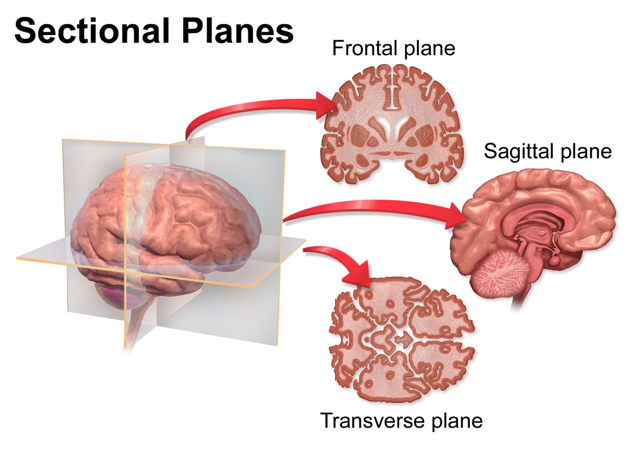
A plane is an imaginary flat surface passing through the body, and a section is the actual cut made along it. Imaging machines, anatomy atlases and operative notes all speak in planes, so knowing three of them unlocks a great deal of language.
The sagittal plane (SAJ-ih-tal) runs vertically front to back and divides the body into right and left portions. When the cut is exactly in the middle, producing equal right and left halves, it is called midsagittal or median. Any sagittal cut off the midline — producing unequal portions — is parasagittal (para- = beside). The name comes from the Latin sagitta, arrow, because the plane follows the sagittal suture that runs front to back across the top of the skull like an arrow shaft.
The frontal plane, also called the coronal plane (KOR-oh-nal), runs vertically side to side and divides the body into anterior and posterior portions. Coronal comes from corona, crown — the plane follows the coronal suture, where a crown would sit. The transverse plane, also called horizontal or axial, runs parallel to the ground and divides the body into superior and inferior portions. This is the plane of a standard CT slice, which is why CT images are described as axial. An oblique plane is any slanted cut that is none of the three.
Cutting words: tom/o and its family
The root tom/o means to cut, and it produces one of the most confused suffix families in medicine. Learn the four together and you will never mix them again.
| Word part | Meaning | Example term | What the example means |
|---|---|---|---|
| tom/o | to cut, section, slice | tomography (toh-MOG-rah-fee) | Imaging that records the body in slices |
| -tome | an instrument that cuts; a segment | dermatome (DER-mah-tohm) | An instrument for cutting skin; also the skin area supplied by one spinal nerve |
| -tomy | process of cutting, incision | osteotomy (oss-tee-OT-oh-mee) | Cutting into a bone |
| -ectomy | surgical removal, excision | appendectomy (ap-en-DEK-toh-mee) | Surgical removal of the appendix |
| -ostomy (-stomy) | surgical creation of an opening | colostomy (koh-LOSS-toh-mee) | A surgically created opening from the colon to the body surface |
| -centesis | surgical puncture to withdraw fluid | thoracentesis (thor-ah-sen-TEE-sis) | Puncture of the chest wall to drain fluid |
| -scopy | process of visually examining | laparoscopy (lap-ah-ROSS-koh-pee) | Visual examination inside the abdomen with a scope |
| -graphy / -gram | process of recording / the record | myelography (my-eh-LOG-rah-fee) | Recording an image of the spinal canal |
| sect/o | to cut | dissection (dih-SEK-shun) | Cutting apart to separate structures |
| lapar/o | abdomen, abdominal wall | laparotomy (lap-ah-ROT-oh-mee) | A surgical incision into the abdominal wall |
-tome, -tomy, -ectomy, -ostomy. Four look-alikes, four different events in an operating room. A -tome is a thing that cuts (an osteotome, a microtome). A -tomy is cutting into and the structure stays: trache/otomy is an incision into the trachea. An -ectomy takes it out: tonsill/ectomy, hyster/ectomy, append/ectomy. An -ostomy makes a mouth — a permanent or semi-permanent opening, usually to the outside or between two structures: col/ostomy, ile/ostomy, gastr/ostomy. Test yourself with one root: gastrotomy = cut into the stomach; gastrectomy = remove the stomach; gastrostomy = create an opening into the stomach for a feeding tube. Same root, three very different consent forms.
Now decode a real imaging term. Computed tomography is computer-assisted imaging that records the body in slices; add the plane and you can read any report line. Ultrason/o/graphy is recording with sound beyond the range of hearing. Magnetic resonance imaging uses no cutting root at all, but its images are still labeled sagittal, coronal and axial, because the planes belong to the body, not the machine.
Positioning the patient
Planes describe how the body is divided; positions describe how the body is placed. In nursing and allied health these words are orders, not descriptions — “place the patient in high Fowler” is an instruction with a measurable result — so precision here has immediate consequences for breathing, perfusion, aspiration risk and skin integrity.
| Position | What it looks like | Where it is used |
|---|---|---|
| Supine (soo-PINE), dorsal recumbent | Lying on the back, face up | Most physical exams, abdominal assessment, CPR |
| Prone (prone) | Lying on the abdomen, face down or turned to one side | Back and spine exams; proning for severe hypoxemia |
| Lateral recumbent | Lying on one side | Comfort, pressure relief, lumbar puncture |
| Fowler (FOW-ler) | Head of bed raised roughly 45 to 60 degrees | Breathing, eating, reducing aspiration risk |
| Semi-Fowler | Head of bed raised roughly 30 to 45 degrees | Routine bed rest, tube feedings |
| High Fowler | Head of bed raised roughly 60 to 90 degrees | Severe dyspnea, meals, respiratory treatments |
| Trendelenburg (tren-DEL-en-berg) | Supine with the whole bed tilted head-down | Some pelvic surgeries, central line placement |
| Reverse Trendelenburg | Supine with the whole bed tilted head-up | Upper abdominal surgery, reflux precautions |
| Lithotomy (lih-THOT-oh-mee) | Supine, hips and knees flexed, thighs apart, feet in stirrups | Pelvic exams, childbirth, urologic procedures |
| Sims (simz) | Left side-lying, right knee and thigh drawn up, left arm behind | Rectal exam, enema administration |
| Orthopneic (or-thop-NEE-ik) | Sitting upright, leaning forward on a table | Severe shortness of breath (tripod position) |
| Knee-chest (genupectoral) | Kneeling, chest and head resting on the surface | Selected rectal and gynecologic procedures |
Notice how many of those names are eponyms — terms built from a person’s name rather than from word parts. Fowler, Trendelenburg and Sims were surgeons; their names carry no decodable meaning, so eponyms have to be memorized outright. That is the whole argument against them, and modern terminology quietly replaces eponyms with descriptive terms where it can. Where they survive, style has shifted: most current style guides and the major medical dictionaries drop the possessive apostrophe, so you will increasingly see Fowler position, Sims position and Down syndrome rather than Fowler’s, Sims’ and Down’s. Both forms remain in circulation; write what your program and your facility use, and recognize both.
Two positional words are worth a second look because students reverse them constantly. Supine is face up — a useful memory hook is that you lie on your spine. Prone is face down, the position used for hours at a time in severe respiratory failure to improve oxygenation. And recumbent simply means lying down, so “dorsal recumbent” is lying on the dorsal surface: on the back.
Sagittal divides right from left, frontal (coronal) divides front from back, transverse divides upper from lower — and the -tome, -tomy, -ectomy, -ostomy family names what is cut, opened or removed along them.
Section 4Body Cavities, Quadrants and Regions
The two great cavities
Organs do not float loose. They sit in closed spaces called cavities, lined by membranes and separated by muscular or bony walls. The dorsal (posterior) cavity lies along the back and has two continuous parts: the cranial cavity, which holds the brain, and the spinal (vertebral) cavity, which holds the spinal cord. The ventral (anterior) cavity is far larger and is split by the diaphragm (DYE-ah-fram) into the thoracic cavity above and the abdominopelvic cavity below.
The thoracic cavity subdivides again. Two pleural cavities hold the lungs, and between them sits the mediastinum (mee-dee-ass-TYE-num), the central compartment containing the heart in its pericardial cavity, plus the esophagus, trachea, thymus and great vessels. The abdominopelvic cavity is one continuous space, but it is described in two halves: the abdominal cavity (stomach, small intestine, most of the large intestine, liver, gallbladder, spleen, pancreas, kidneys) and the pelvic cavity (urinary bladder, rectum, and the internal reproductive organs).
Some abdominal organs sit behind the peritoneal lining rather than inside it. These are retroperitoneal (reh-troh-per-ih-toe-NEE-al) — retro- (behind) + peritone/o (peritoneum) + -al — and the group includes the kidneys, adrenal glands, ureters, most of the pancreas and duodenum, the abdominal aorta and the inferior vena cava. That one word explains why kidney pain is felt in the back and why a surgeon reaching a kidney may approach from the flank rather than the front.
| Word part | Meaning | Example term | What the example means |
|---|---|---|---|
| crani/o | skull | craniotomy (kray-nee-OT-oh-mee) | Surgical incision into the skull |
| spin/o, vertebr/o | spine, backbone | vertebral (VER-teh-bral) | Pertaining to the backbone |
| thorac/o, steth/o | chest | thoracotomy (thor-ah-KOT-oh-mee) | Surgical incision into the chest wall |
| pleur/o | pleura, membrane around the lung | pleuritis (ploo-RYE-tis), also called pleurisy (PLOOR-ih-see) | Inflammation of the pleura |
| mediastin/o | mediastinum | mediastinitis (mee-dee-ass-tih-NYE-tis) | Inflammation of the mediastinum |
| pericardi/o | sac around the heart | pericarditis (per-ih-kar-DYE-tis) | Inflammation of the sac around the heart |
| abdomin/o, celi/o | abdomen | abdominocentesis (ab-dom-ih-noh-sen-TEE-sis) | Surgical puncture of the abdomen to withdraw fluid |
| periton/o, peritone/o | peritoneum | peritonitis (per-ih-toe-NYE-tis) | Inflammation of the peritoneum |
| pelv/i, pelv/o | pelvis | pelvimetry (pel-VIM-eh-tree) | Measurement of the pelvic dimensions |
| diaphragmat/o, phren/o | diaphragm | diaphragmatic (dye-ah-frag-MAT-ik) | Pertaining to the diaphragm |
| -al, -ic, -ac, -eal | pertaining to | peritoneal (per-ih-toe-NEE-al) | Pertaining to the peritoneum |
Decode peri/card/itis once more, slowly: peri- (around) + card (heart) + -itis (inflammation) = inflammation of the sac around the heart. Now the same logic elsewhere: periton/itis is inflammation of the membrane stretched around the abdominal organs, peri/oste/um (per-ee-OSS-tee-um) is the membrane around a bone, and peri/nat/al is the time around birth. One prefix, many locations, no memorization.
Peritoneum is not perineum. The peritoneum (per-ih-toe-NEE-um) is the serous membrane lining the abdominal cavity and draping the organs; peritonitis is a surgical emergency. The perineum (per-ih-NEE-um) is the external region between the thighs, from the pubis to the coccyx, containing the external genitalia and anus; it is the area assessed for tears after childbirth and cleaned in perineal care. The words differ by two letters and by an entire body region. Read syllable by syllable: perito-NEE-um has a t-o before the -neum; peri-NEE-um does not.
Four quadrants, nine regions
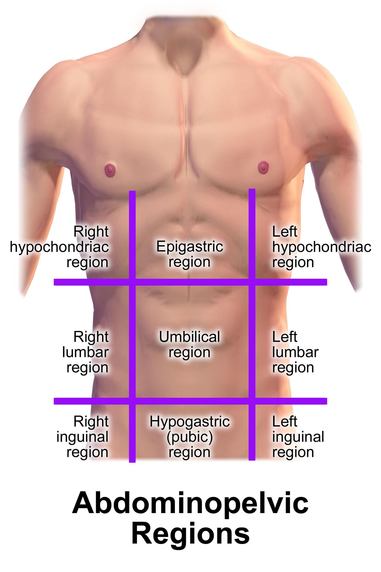
Clinically, the abdomen is divided two ways. The quick way draws one vertical line and one horizontal line through the umbilicus, producing four quadrants. This is the language of triage and daily charting.
| Quadrant | Abbreviation | Key structures | Classic pain there suggests |
|---|---|---|---|
| Right upper quadrant | RUQ | Liver, gallbladder, duodenum, head of pancreas, right kidney, hepatic flexure of colon | Cholecystitis, hepatitis |
| Left upper quadrant | LUQ | Stomach, spleen, body and tail of pancreas, left kidney, splenic flexure of colon | Splenic injury, gastritis, pancreatitis |
| Right lower quadrant | RLQ | Appendix, cecum, right ureter, right ovary and uterine tube | Appendicitis, ovarian torsion, ectopic pregnancy |
| Left lower quadrant | LLQ | Sigmoid colon, descending colon, left ureter, left ovary and uterine tube | Diverticulitis, ovarian cyst |
The precise way draws two vertical and two horizontal lines, producing nine regions in a three-by-three grid. Anatomists, radiologists and surgeons use these, and every name in the grid is decodable.
| Region | Word parts | Location | What lies there |
|---|---|---|---|
| Right and left hypochondriac (high-poh-KON-dree-ak) | hypo- (under) + chondr/o (cartilage) + -ac | Upper row, both sides — under the rib cartilage | Right: liver, gallbladder. Left: stomach, spleen |
| Epigastric (ep-ih-GAS-trik) | epi- (above) + gastr/o (stomach) + -ic | Upper row, center | Stomach, duodenum, pancreas, aorta |
| Right and left lumbar (LUM-bar) | lumb/o (lower back) + -ar | Middle row, both sides | Ascending and descending colon, kidneys |
| Umbilical (um-BILL-ih-kal) | umbilic/o (navel) + -al | Middle row, center | Small intestine, transverse colon |
| Right and left iliac or inguinal (ILL-ee-ak / ING-gwih-nal) | ili/o (hip bone) or inguin/o (groin) + -ac / -al | Lower row, both sides | Right: appendix, cecum. Left: sigmoid colon |
| Hypogastric (high-poh-GAS-trik), or pubic | hypo- (below) + gastr/o (stomach) + -ic | Lower row, center | Urinary bladder, uterus, rectum |
Hypochondriac earns a note. Literally it means under the cartilage, an anatomical location. The everyday meaning — a person convinced they are ill — came from an old belief that melancholy arose from organs beneath the ribs. Both meanings survive, so context decides: “right hypochondriac region” is a place, not a personality.
ile/o versus ili/o. Same pronunciation, adjacent territory, completely different structure. Ile/o is the ileum, the final segment of the small intestine — hence ileostomy, ileitis, and ileocecal valve. Ili/o is the ilium, the large flaring hip bone — hence iliac crest, iliac artery, and the iliac region. The spelling hook: intestine has an e, and so does ileum; the hip bone ilium is all i’s. Confusing them in a note can turn a bowel resection into a pelvic fracture. And do not let ileus (ILL-ee-us), a functional standstill of the bowel, pull you toward the bone either.
Cavities have their own membranes, and the parietal/visceral pair from Section 2 returns here in full force. Most ventral cavity organs sit inside a double-layered serous sac: the parietal layer lines the cavity wall and the visceral layer clings to the organ, with a thin film of fluid between them so surfaces glide instead of grate. In the chest that is parietal and visceral pleura; around the heart, parietal and visceral pericardium; in the abdomen, parietal and visceral peritoneum. Not every ventral structure is wrapped this way: the retroperitoneal organs named above — kidneys, adrenals, ureters, most of the pancreas and duodenum, the abdominal aorta and the inferior vena cava — lie behind the peritoneum rather than within a serous sac, and the mediastinal structures such as the esophagus, trachea and thymus have no serous covering of their own either. When the fluid becomes infected or blood or air enters the space, you get the conditions those chapters are built on: pleural effusion, pneumothorax, pericardial tamponade, peritonitis.
The dorsal cavity holds the brain and cord; the ventral cavity holds the thoracic and abdominopelvic organs — and the abdomen is charted in four quadrants for speed and nine regions for precision.
Section 5Putting It Together: Regional Terms, Plurals and the Chart
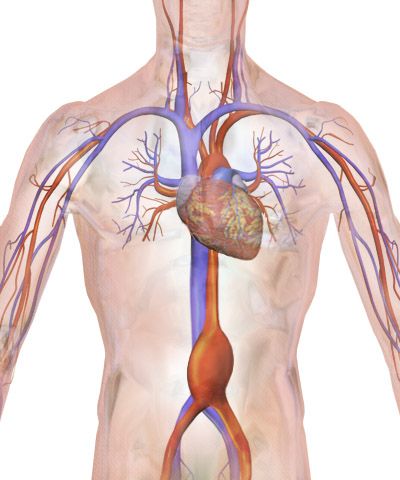
Combining forms for body regions
Between the whole-body compass of Section 2 and the cavities of Section 4 sits a middle layer: named surface regions. These roots turn up as adjectives everywhere — cervical collar, brachial pulse, femoral line, inguinal hernia, pedal edema — and each one is doing the same job of saying where.
| Word part | Meaning | Example term | What the example means |
|---|---|---|---|
| cephal/o | head | cephalalgia (sef-al-AL-jee-ah) | Headache |
| cervic/o | neck; neck of the uterus (cervix) | cervical (SER-vih-kal) | Pertaining to the neck — or to the cervix |
| acr/o | extremities, tip | acrocyanosis (ak-roh-sigh-ah-NOH-sis) | Bluish discoloration of the hands and feet |
| thorac/o | chest | thoracic (thoh-RASS-ik) | Pertaining to the chest |
| brachi/o | arm (upper arm) | brachial (BRAY-kee-al) | Pertaining to the arm — the pulse at the inner elbow |
| axill/o | armpit | axillary (AK-sih-lair-ee) | Pertaining to the armpit — where lymph nodes are checked |
| abdomin/o | abdomen | abdominal (ab-DOM-ih-nal) | Pertaining to the abdomen |
| lumb/o | lower back, loin | lumbar (LUM-bar) | Pertaining to the lower back |
| glute/o | buttock | gluteal (GLOO-tee-al) | Pertaining to the buttock — an injection site |
| inguin/o | groin | inguinal (ING-gwih-nal) | Pertaining to the groin |
| umbilic/o, omphal/o | navel | periumbilical (per-ee-um-BILL-ih-kal) | Around the navel |
| femor/o | thigh bone, thigh | femoral (FEM-or-al) | Pertaining to the femur or thigh |
| ped/o, pod/o | foot | pedal (PEE-dal) | Pertaining to the foot — as in pedal pulses |
| dactyl/o | finger or toe | polydactyly (pol-ee-DAK-tih-lee) | Condition of having extra fingers or toes |
| dors/o | back; upper surface | dorsiflexion (dor-sih-FLEK-shun) | Bending the foot upward toward its dorsal surface |
Two of those need a warning label. Cervic/o means neck, and the uterus has a neck, so cervical can describe either the cervical spine or the cervix — a cervical collar and a cervical biopsy are not in the same neighborhood. Context, and the rest of the sentence, resolve it every time. And ped/o is a genuine double agent: from Latin it means foot (pedal pulse, pedicure), while from Greek it means child (pediatrics, pedodontics). When you want an unambiguous root for foot, reach for pod/o — a podiatrist treats feet, and no one confuses that.
Singular and plural: the rules that actually appear
Medical terms carry Latin and Greek plural endings that English never adopted, and structural vocabulary is where they bite hardest. You will write vertebrae, bronchi and diagnoses in your first clinical week. Nine patterns cover nearly everything.
| Singular ending | Plural ending | Example (singular) | Example (plural) |
|---|---|---|---|
| -a | -ae | vertebra (VER-teh-brah), bursa, pleura | vertebrae (VER-teh-bree), bursae, pleurae |
| -us | -i | bronchus, thrombus, calculus, nucleus | bronchi (BRONG-kye), thrombi, calculi, nuclei |
| -um | -a | ovum, atrium, diverticulum, bacterium | ova, atria, diverticula, bacteria |
| -is | -es | diagnosis, prognosis, metastasis, testis | diagnoses (dye-ag-NOH-seez), prognoses, metastases, testes |
| -ix, -ex | -ices | appendix, cervix, apex, cortex, varix | appendices (ah-PEN-dih-seez), cervices, apices, cortices, varices |
| -nx | -nges | phalanx (FAY-lanks), larynx, pharynx, meninx | phalanges (fah-LAN-jeez), larynges, pharynges, meninges (meh-NIN-jeez) |
| -on | -a | ganglion, mitochondrion, spermatozoon | ganglia, mitochondria, spermatozoa |
| -oma | -omata (or -omas) | carcinoma, fibroma, sarcoma | carcinomata (kar-sih-NOH-mah-tah) or carcinomas |
| -y | -ies | biopsy, artery, ovary | biopsies, arteries, ovaries |
Never pluralize a plural. The most common written error in this whole area is adding an English -s to a word that is already plural, or treating a plural as a singular. There is no such word as diagnosises, phalanxes in anatomy, or metastasises. Equally, bacteria, data, criteria, viscera and meninges are plurals — the singulars are bacterium, datum, criterion, viscus and meninx. Two more traps: one vertebra, several vertebrae; one bronchus, two bronchi. And note that viscus (VISS-kus), the singular of viscera, is a real word used in surgery — “perforated viscus” means one hollow organ has ruptured, not several.
Decoding what a chart actually says
Everything in this chapter converges on a single skill: reading a line of documentation and knowing exactly where the writer means. Work through these the way you will in practice — find the suffix, find the prefix, then the root or roots, and reassemble from the end backward.
Retroperitoneal = retro- (behind) + peritone/o (peritoneum) + -al (pertaining to) = pertaining to the space behind the peritoneum, which is where the kidneys live. Anterolateral thoracotomy = anter/o (front) + later/o (side) + thorac/o (chest) + -tomy (incision) = a surgical incision into the chest made at the front and to the side. Subcostal = sub- (under) + cost/o (rib) + -al = under the rib. Intra-abdominal = intra- (within) + abdomin/o + -al = within the abdomen. Epigastric tenderness with periumbilical guarding tells you the examiner found pain above the stomach and protective muscle tightening around the navel — a description that, in a real patient, moves appendicitis up the list.
Now build in the other direction, which is the harder and more useful exercise. You want a word for surgical puncture of the chest to withdraw fluid: chest is thorac/o, surgical puncture to withdraw fluid is -centesis, so thoracentesis. You want visual examination of the abdomen: lapar/o plus -scopy gives laparoscopy. You want inflammation of the membrane around the lung: pleur/o plus -itis gives pleuritis. Each of these is a word you did not memorize and can now produce on demand — which is the entire point of learning terminology by parts rather than by lists.
Regional combining forms, correct plurals and part-by-part decoding turn a chart entry from a wall of unfamiliar words into a precise map of where the problem is.
The chapter in one breath
The body is organized in levels — cell, tissue, organ, system, organism — and each level has roots that name it: cyt/o and hist/o at the microscopic end, viscer/o and somat/o at the whole-body end. Four primary tissues (epithelial, connective, muscle, nervous) supply combining forms you will meet in every later chapter, including the pair students most often confuse, my/o for muscle and myel/o for marrow or spinal cord. Location is described from a single fixed posture, the anatomical position, using paired terms: superior and inferior, anterior and posterior, medial and lateral, proximal and distal, superficial and deep, with right and left always belonging to the patient. Three planes slice the body — sagittal for right and left, frontal or coronal for front and back, transverse for upper and lower — and the tom/o family names what is done along them: -tome cuts, -tomy incises, -ectomy removes, -ostomy creates an opening. Organs sit in the dorsal and ventral cavities, most of them wrapped in paired parietal and visceral membranes (the retroperitoneal and mediastinal structures are not), and the abdomen is charted in four quadrants or nine regions. Add the Latin plural rules and you can read position in any chart.
Words worth knowing
| Term | What it means |
|---|---|
| Anatomical position | Standing erect, facing forward, arms at the sides, palms forward, feet slightly apart — the reference posture for all directional terms |
| Superior / inferior | Above (toward the head) / below (toward the feet) |
| Anterior (ventral) / posterior (dorsal) | Toward the front of the body / toward the back of the body |
| Medial / lateral | Toward the midline / away from the midline |
| Proximal / distal | Nearer to the point of attachment or trunk / farther from it |
| Ipsilateral / contralateral | On the same side / on the opposite side |
| Midsagittal (median) plane | Vertical plane dividing the body into equal right and left halves |
| Frontal (coronal) plane | Vertical plane dividing the body into anterior and posterior portions |
| Transverse (axial) plane | Horizontal plane dividing the body into superior and inferior portions |
| Supine / prone | Lying face up / lying face down |
| Fowler position | Head of bed elevated roughly 45 to 60 degrees; semi-Fowler is lower, high Fowler is higher |
| Trendelenburg position | Supine with the entire bed tilted so the head is lower than the feet |
| Mediastinum (mee-dee-ass-TYE-num) | Central compartment of the thoracic cavity holding the heart, esophagus, trachea, thymus and great vessels |
| Peritoneum (per-ih-toe-NEE-um) | Serous membrane lining the abdominal cavity and covering the abdominal organs |
| Perineum (per-ih-NEE-um) | External region between the thighs, from pubis to coccyx — not the peritoneum |
| Retroperitoneal (reh-troh-per-ih-toe-NEE-al) | Located behind the peritoneum — kidneys, adrenals, ureters, most of the pancreas, aorta |
| Parietal / visceral | The membrane layer lining a cavity wall / the layer covering the organ itself |
| Epigastric region | Upper central abdominal region, above the stomach |
| Hypochondriac region | Upper right or left abdominal region, under the rib cartilage |
| Ileum vs ilium | Ileum is the last part of the small intestine (ile/o); ilium is the flaring hip bone (ili/o) |