The Endocrine System
The endocrine system is the body’s chemical messaging service — slower than nerves, but with reach that lasts hours to days. When one gland makes too much or too little of a hormone, the ripple effects show up everywhere: blood sugar, heart rate, blood pressure, mood, growth, and water balance. This chapter gives you the one idea that unlocks all of it — feedback — and then walks each major gland from healthy to what goes wrong, mechanism by mechanism.
By the end of this chapter you can
- Trace how the hypothalamus and pituitary command the other glands, and tell a tropic hormone from a direct one.
- Use negative feedback to predict a lab — and use a stimulating-hormone level to place a problem as primary (the gland) vs secondary (the pituitary).
- Contrast the too-much vs too-little state for the adrenal (Cushing’s vs Addison’s), thyroid, and ADH (SIADH vs DI).
- Read the electrolyte shifts that give a disorder away (Addison’s potassium; the sodium in SIADH and DI).
- Explain why Type 1 and Type 2 diabetes are different diseases, name the diagnostic numbers, and tell DKA from HHS.
- Recognize growth-hormone excess by timing (gigantism before the growth plates close, acromegaly after).
- Flag the endocrine emergencies: Addisonian crisis, thyroid storm, myxedema coma, DKA/HHS, and severe hyponatremia.
Section 1The chain of command
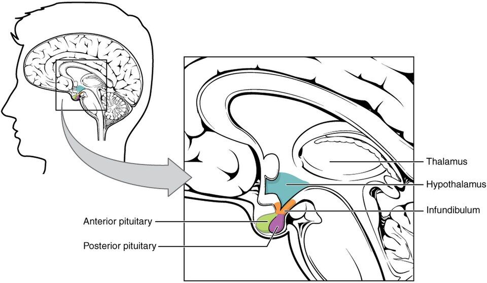
The normal wiring: how orders travel down
Picture the endocrine system as a company. The hypothalamus is the executive on the top floor. It constantly reads the body's status - temperature, blood pressure, stress, hunger, the levels of hormones already circulating - and decides what needs to change. Just beneath it sits its manager, the pituitary gland, tucked into a bony pocket at the base of the brain called the sella turcica. The two are connected by a short stalk (the infundibulum), and the way they talk to each other depends on which half of the pituitary we mean.
For the front half, the hypothalamus does not send nerve signals - it sends chemical memos. It makes tiny releasing hormones and inhibiting hormones (like TRH, CRH, GnRH, GHRH, and the inhibitor somatostatin) and drips them into a private set of blood vessels running down the stalk called the hypophyseal portal system. Think of it as a pneumatic tube between two offices: a dedicated capillary bed that carries the memo straight from the hypothalamus to the anterior pituitary without diluting it in the whole bloodstream first.
The anterior pituitary: a six-product factory
The anterior pituitary is a true factory - it manufactures six hormones. Several are tropic (from a Greek root meaning "to turn toward"): they don't act on body tissue directly, they turn on another gland. Others are direct hormones that act on the body's own tissues. A handy mnemonic is FLAT PiG: FSH, LH, ACTH, TSH (the tropic four) and Prolactin, GH (the two direct ones).
| Hormone | Target | Type |
|---|---|---|
| TSH | Thyroid gland | Tropic |
| ACTH | Adrenal cortex | Tropic |
| FSH & LH | Gonads (ovaries, testes) | Tropic |
| GH | Whole body & liver (via IGF-1) | Direct |
| Prolactin | Breast | Direct |
Notice that GH works partly by proxy: it tells the liver to release IGF-1 (insulin-like growth factor-1), which does much of the actual growth-promoting work in bone and muscle.
The posterior pituitary: a storage locker, not a factory
The back half is different, and students mix this up constantly. The posterior pituitary makes nothing. It is a storage locker. The two hormones it releases - ADH (antidiuretic hormone, also called vasopressin, which tells the kidneys to hold onto water) and oxytocin (uterine contraction in labor and milk let-down) - are actually built in the hypothalamus. Their parent neurons send long axons down the stalk, and the hormone simply travels down the nerve and waits in the terminals until a signal says "release." So the anterior lobe gets chemical memos through blood vessels, while the posterior lobe gets the finished product delivered down a nerve.
Negative feedback: the body's thermostat
The whole system governs itself with negative feedback, exactly like a home thermostat. Set the thermostat to 70 degrees: when the room warms past the setpoint, the furnace shuts off; when it cools, the furnace fires back up. Hormones do the same. Walk the thyroid axis: the hypothalamus releases TRH, which tells the pituitary to release TSH, which tells the thyroid to release T4 (and T3). When circulating T4 climbs high enough, it loops back and switches off both TRH and TSH - the furnace turns off. If T4 falls, the pituitary senses the shortage and cranks TSH up to push the thyroid harder. This is why the single most useful sentence in endocrine nursing is: low thyroid hormone usually means high TSH.
Positive feedback: the rare exception
Occasionally the body wants the opposite - a signal that amplifies itself until an event completes. This is positive feedback, and it is rare precisely because it is explosive. Two classic examples: the oxytocin surge in labor (stretch of the cervix drives more oxytocin, which drives stronger contractions, which stretches the cervix more - until the baby is delivered), and the LH surge at mid-cycle that triggers ovulation. Both shut off once the goal is reached.
The master skill: primary vs secondary vs tertiary
Here is the reasoning trick that separates strong students. When a hormone level is off, ask where the fault lives, and let the tropic hormone point you to it. A primary problem is in the target gland itself (the thyroid, adrenal, or gonad). A secondary problem is in the pituitary. A tertiary problem is up in the hypothalamus.
| Level | Broken part | Thyroid example (low T4) |
|---|---|---|
| Primary | Target gland | Low T4, high TSH (pituitary is shouting) |
| Secondary | Pituitary | Low T4, low TSH (no one is shouting) |
| Tertiary | Hypothalamus | Low T4, low TSH, low TRH |
Once you internalize the chain of command and the direction feedback pushes, endocrine disease stops being memorization and becomes detective work. Explore the wiring below.
- The hypothalamus sends releasing/inhibiting hormones through the hypophyseal portal blood vessels to the anterior pituitary, which manufactures six hormones (TSH, ACTH, FSH, LH, GH, prolactin).
- The posterior pituitary makes nothing - it only stores and releases ADH and oxytocin, which the hypothalamus builds and sends down the nerve.
- Tropic hormones (TSH, ACTH, FSH, LH) act on other glands; direct hormones (GH, prolactin) act on body tissue.
- Negative feedback works like a thermostat: when the final hormone rises it shuts off its own signals, so low thyroid hormone usually drives TSH up. Positive feedback (oxytocin in labor, the LH surge) is the rare, self-amplifying exception.
- Use the tropic hormone level to localize the lesion: primary = target gland, secondary = pituitary, tertiary = hypothalamus.
Section 2Adrenal glands — the cortisol problem
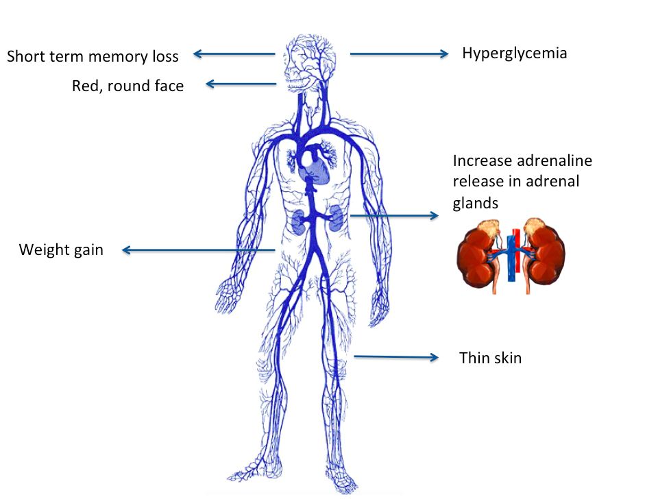
The normal adrenal gland: two organs stacked in one
Perched like little caps on top of each kidney, the adrenal glands are really two organs fused together. The outer cortex makes steroid hormones from cholesterol, and the inner medulla makes the fight-or-flight catecholamines (epinephrine and norepinephrine). The cortex is layered into three zones, and a handy way to remember what each makes is salt, sugar, sex as you travel inward. The outermost zona glomerulosa makes aldosterone (the mineralocorticoid that holds onto salt and water). The middle zona fasciculata makes cortisol (the glucocorticoid that manages sugar and stress). The innermost zona reticularis makes weak androgens (the sex steroids).
What cortisol actually does
Cortisol is your body's built-in stress hormone, released on a daily rhythm and surging whenever you are sick, injured, or frightened. It raises blood glucose by driving gluconeogenesis (the liver building new glucose) so your brain and muscles have fuel in a crisis. It is powerfully anti-inflammatory and immunosuppressive, which is exactly why we bottle it up as drugs like prednisone. And it helps maintain blood pressure by keeping blood vessels responsive to catecholamines. Control runs through the HPA axis: the pituitary releases ACTH, which tells the cortex to make cortisol, and rising cortisol switches ACTH back off (negative feedback). Hold onto that feedback loop, because it is the key to telling these diseases apart.
Cushing: too much cortisol
First, the vocabulary. Cushing syndrome is the umbrella term for any cause of excess cortisol. Cushing disease specifically means a pituitary ACTH-secreting adenoma driving the adrenals. But the single most common cause overall is neither of those: it is exogenous steroid medication taken long-term for asthma, autoimmune disease, or transplants. Adrenal tumors making cortisol directly are a third route.
Every classic sign traces back to too much cortisol. Because cortisol redistributes fat centrally, patients develop a rounded moon face, a buffalo hump between the shoulders, and truncal obesity with thin arms and legs (fat goes to the middle while cortisol breaks down limb muscle). Cortisol thins the skin and weakens collagen, so you see thin, fragile, easily-bruised skin and stretched purple striae across a belly that outgrew its weakened skin. Constant gluconeogenesis produces hyperglycemia (steroid-induced diabetes). Cortisol's mineralocorticoid spillover retains sodium and water, causing hypertension. Bone breakdown leads to osteoporosis, and the immunosuppression brings poor wound healing and infection risk. Mood swings, irritability, and even psychosis round out the picture.
Addison: too little cortisol (primary insufficiency)
Addison disease is primary adrenal insufficiency - the gland itself is destroyed, usually by autoimmune attack. Because the whole cortex fails, the patient loses all three outputs: cortisol, aldosterone, and adrenal androgens. Low aldosterone means sodium and water are lost while potassium is retained, so expect hyponatremia, HYPERkalemia, hypotension, and intense salt craving. Low cortisol brings hypoglycemia, profound weakness and fatigue, and weight loss. The tell-tale finding is bronze hyperpigmentation: with cortisol gone, feedback is lost and the pituitary floods the body with ACTH, and ACTH also stimulates skin melanocytes.
Secondary insufficiency: a pituitary problem
If the pituitary fails instead, ACTH is low. Two clues follow. There is no hyperpigmentation (no excess ACTH to darken skin), and aldosterone is largely spared because aldosterone is driven mainly by the renin-angiotensin system, not ACTH. So these patients get low cortisol but usually keep their sodium and potassium closer to normal.
Pheochromocytoma: the medulla's turn
A pheochromocytoma is a tumor of the adrenal medulla that dumps catecholamines in unpredictable surges. Remember the 5 P's: Pressure (episodic, often severe hypertension), Pain (pounding headache), Palpitations, Perspiration, and Pallor. Between surges the patient may feel completely normal, which is what makes it sneaky.
Putting it together
The fastest way to keep Cushing and Addison straight is a side-by-side look. Explore the interactive figure below, too.
| Feature | Cushing (excess) | Addison (deficiency) |
|---|---|---|
| Cortisol | High | Low |
| Blood glucose | High | Low |
| Blood pressure | High | Low |
| Sodium / Potassium | Na high / K low | Na low / K high |
| Weight & fat | Central gain, moon face | Loss, wasting |
| Skin | Thin, bruises, striae | Bronze hyperpigmentation |
One last feedback check that ties it all together: an adrenal tumor making its own cortisol pushes ACTH down (feedback shuts the pituitary off), while a failed adrenal gland lets ACTH climb up. The direction ACTH moves tells you where the problem lives.
- Cortex = salt (aldosterone), sugar (cortisol), sex (androgens); medulla = catecholamines. Cortisol raises glucose, calms inflammation, and supports blood pressure.
- Cushing = too much cortisol (most often from steroid medication): central obesity, moon face, striae, hyperglycemia, hypertension, thin skin, infection risk.
- Addison (primary) destroys the whole cortex: low cortisol AND aldosterone -> hypotension, hyponatremia, HYPERkalemia, hypoglycemia, salt craving, and bronze skin from high ACTH.
- Secondary insufficiency (pituitary) means low ACTH: no hyperpigmentation and aldosterone is spared. Never stop chronic steroids abruptly - taper to avoid an adrenal crisis (glucocorticoid-only, so potassium usually stays normal).
- Pheochromocytoma = medulla tumor with catecholamine surges (5 P's). Low ACTH points to an adrenal tumor; high ACTH points to a failed adrenal gland.
Section 3Thyroid — the metabolic thermostat
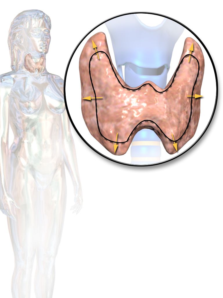
Think of your thyroid as the body's metabolic thermostat - a small butterfly-shaped gland sitting at the front of your neck that decides how fast every cell runs its engine. When it dials the setting up, your heart beats faster, you burn more fuel, and you feel warm. When it dials down, everything slows: heart, gut, brain, and body temperature all ease off. Almost every confusing thyroid symptom makes sense once you remember that one idea - this gland sets the pace of your metabolism.
The normal axis: a three-station relay
Control starts up in the brain. The hypothalamus releases TRH (thyrotropin-releasing hormone), which travels a short distance to the anterior pituitary and tells it to release TSH (thyroid-stimulating hormone). TSH then rides the bloodstream to the thyroid and says, "make hormone." The thyroid responds by producing mostly T4 (thyroxine) - the bulk of its output - and a smaller amount of T3 (triiodothyronine), which is the more active form. Much of the T4 gets converted to T3 out in the tissues, so think of T4 as the storage/delivery version and T3 as the one that actually revs the cell.
Two more facts anchor the whole system. First, the thyroid cannot build these hormones without iodine - it is a required raw ingredient, which is exactly why iodized salt exists. Second, the thyroid has a side job unrelated to metabolism: its C-cells secrete calcitonin, a hormone that gently lowers blood calcium by tucking it back into bone. It is a minor player in humans, but worth knowing it comes from the thyroid.
Hypothyroidism: the thermostat turned down
When the thyroid makes too little hormone, metabolism idles - and every symptom is that slowdown showing up in a different organ. In iodine-sufficient countries the number-one cause is Hashimoto's thyroiditis, an autoimmune attack that gradually destroys thyroid tissue. Worldwide, iodine deficiency is still the leading cause. Other causes include after treatment - post-thyroidectomy or radioactive iodine ablation - and certain medications (such as lithium and amiodarone).
Trace the mechanism and the signs write themselves. A slow metabolism means the body burns less fuel, so patients feel fatigued, gain weight despite a normal appetite, and cannot generate heat well - hence cold intolerance. Slowed cardiac pacing gives bradycardia. Sluggish skin turnover and reduced sweating leave skin dry and hair coarse and thinning. A slow gut means constipation. Reproductive hormones drift, causing menstrual changes (often heavier or irregular periods). Even the nervous system lags, producing the classic slowed, delayed deep-tendon reflexes. Severe, long-standing disease is called myxedema - a puffy, non-pitting swelling from mucin building up in the skin - and its life-threatening extreme is myxedema coma: hypothermia, low blood pressure, and a depressed level of consciousness. That is a true emergency.
Hyperthyroidism: the thermostat cranked up
Too much hormone and metabolism races. The leading cause is Graves' disease, an autoimmune condition in which antibodies against the TSH receptor (TSH-receptor antibodies, TRAb) - specifically the stimulating type, TSI (thyroid-stimulating immunoglobulins) - bind and switch that receptor on, flooring the gas pedal regardless of what the pituitary wants. Other causes include a toxic multinodular goiter (autonomous nodules that make hormone on their own) and thyroiditis (an inflamed gland leaking stored hormone).
The signs mirror hypothyroidism in reverse. Burning fuel too fast causes weight loss despite a big appetite and heat intolerance. The heart speeds up into tachycardia, palpitations, and sometimes atrial fibrillation. The overdriven nervous system brings anxiety, a fine tremor, and brisk hyperreflexia, while a fast gut causes diarrhea. Graves' adds two things unique to its autoantibody attack: bulging eyes, or exophthalmos (from tissue swelling behind the eye), and pretibial myxedema, a thickened patch of skin over the shins. The emergency here is thyroid storm - dangerous fever, extreme tachycardia, and agitation or delirium.
Reading the labs: let TSH tell the story
Because of negative feedback, the pattern of TSH versus T4 tells you both whether there is a problem and where it lives. In a primary problem the thyroid itself is the culprit; in a secondary problem the pituitary is failing to give proper orders, which flips the expected relationship.
| Pattern | TSH | T4 |
|---|---|---|
| Primary hypothyroid | High | Low |
| Primary hyperthyroid | Low | High |
| Secondary (pituitary) hypothyroid | Low or inappropriately normal | Low |
The tell for a secondary problem: a low T4 should drive TSH up, so a low T4 paired with a TSH that is low or "normal" is inappropriate - the pituitary is not doing its job.
Hypo vs hyper at a glance
| Feature | Hypothyroid | Hyperthyroid |
|---|---|---|
| Heart rate | Slow (bradycardia) | Fast (tachycardia, AFib) |
| Weight | Gain | Loss |
| Temperature | Cold intolerance | Heat intolerance |
| GI | Constipation | Diarrhea |
| Mood | Sluggish, low | Anxious, restless |
| Skin | Dry, coarse | Warm, moist |
Keep that one thermostat image in mind and the whole gland makes sense - explore the interactive figure below to see the axis and its feedback in motion.
- The thyroid sets metabolic speed: hypothyroid slows everything (fatigue, weight gain, cold, bradycardia, constipation); hyperthyroid speeds everything (weight loss, heat, tachycardia, anxiety, diarrhea).
- The axis runs TRH (hypothalamus) → TSH (pituitary) → T4 and active T3 (thyroid), and it needs iodine; negative feedback makes TSH the key lab.
- Hashimoto's is the top cause of hypothyroidism (iodine-sufficient areas); Graves' - via TSI/TRAb antibodies - is the top cause of hyperthyroidism and can add exophthalmos.
- Labs: primary hypothyroid = high TSH/low T4; primary hyperthyroid = low TSH/high T4; a low or inappropriately normal TSH with a low T4 points to a secondary (pituitary) cause.
- Emergencies are myxedema coma (hypo) and thyroid storm (hyper); after thyroid surgery, watch for hypocalcemia via Trousseau and Chvostek signs.
Section 4Diabetes mellitus
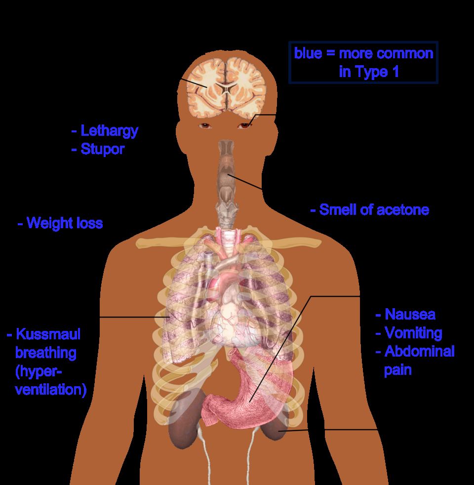
Before we talk about what breaks in diabetes, it helps to know how your body normally manages fuel. Think of your bloodstream as a delivery highway and glucose (blood sugar) as the cargo every cell wants for energy. The traffic controller is your pancreas, and specifically tiny clusters of cells inside it called the islets of Langerhans.
Normal fuel control: the fed state and the fasting state
Inside those islets, beta cells make insulin and alpha cells make glucagon. These two hormones pull in opposite directions, and that tug-of-war keeps blood sugar in a safe range. After a meal (the fed state), glucose rises, beta cells release insulin, and insulin acts like a key: it unlocks cells so glucose can move in to be burned or stored, and it tells the liver to bank extra glucose as glycogen. Between meals or overnight (the fasting state), glucose falls, so alpha cells release glucagon, which tells the liver to break glycogen back down and release glucose so your brain never runs dry. In short, insulin stores fuel and lets glucose into cells; glucagon raises blood sugar. Diabetes is what happens when the insulin side of that system fails.
Type 1: an absolute insulin deficiency
In type 1 diabetes, the immune system mistakenly attacks and destroys the beta cells. This is an autoimmune process, and we can often detect islet autoantibodies in the blood. Because the beta cells are gone, there is essentially no insulin at all, which we call an absolute insulin deficiency. It usually appears in younger, leaner people, and because the body cannot make insulin, these patients always require insulin injections to live. Without insulin, glucose cannot get into cells, so it piles up in the blood while the cells effectively starve.
Type 2: insulin resistance plus a relative shortfall
In type 2 diabetes, insulin is present, but the cells stop responding to it well. This is insulin resistance. The pancreas compensates by making extra insulin, but over time it cannot keep up, leaving a relative insulin deficiency. Type 2 is gradual, often silent for years, and strongly tied to obesity, metabolic syndrome, and older age. One physical clue is acanthosis nigricans, velvety dark patches in skin folds like the neck and armpits that signal chronic high insulin.
Why the three P's happen: osmotic diuresis
Whichever type, once blood glucose climbs high enough, the kidneys cannot reabsorb it all, so glucose spills into the urine. Sugar in the urine acts like a sponge, dragging water out with it. This is osmotic diuresis, and it explains the classic three P's: polyuria (peeing large volumes), which causes dehydration and triggers polydipsia (intense thirst), while starving cells drive polyphagia (hunger). Weight loss and blurry vision often ride along.
How we diagnose it
Diagnosis rests on standard numbers. Any one of these (usually confirmed on a repeat) makes the diagnosis:
- Fasting plasma glucose ≥ 126 mg/dL
- Random glucose ≥ 200 mg/dL with classic symptoms
- A1c ≥ 6.5 percent (a 3-month average of blood sugar)
- 2-hour OGTT ≥ 200 mg/dL (oral glucose tolerance test)
The long game: chronic complications
Years of high glucose damage blood vessels in two patterns. Microvascular (small-vessel) damage causes retinopathy (vision loss), nephropathy (kidney failure), and neuropathy (nerve damage, often numb, tingling feet). Macrovascular (large-vessel) damage speeds up coronary artery disease, stroke, and peripheral arterial disease. Combine numb feet with poor circulation and you get the feared diabetic foot ulcers and slow, poor wound healing.
Gestational diabetes
Pregnancy hormones naturally cause some insulin resistance. When the pancreas cannot keep up, gestational diabetes results. It is often silent, so we routinely screen pregnant patients (usually around 24 to 28 weeks). Extra maternal glucose crosses to the baby, who overproduces insulin and grows too large, called macrosomia. After delivery that sugar supply vanishes, but the baby's high insulin persists, causing neonatal hypoglycemia.
The two emergencies: DKA vs HHS
In type 1, with no insulin at all, the body burns fat for fuel and makes acidic ketones, tipping into diabetic ketoacidosis (DKA). In type 2, there is usually just enough insulin to block ketones, but sugar and dehydration climb to extremes in hyperosmolar hyperglycemic state (HHS).
| Feature | DKA | HHS |
|---|---|---|
| Usual type | Type 1 | Type 2 |
| Glucose | Usually > 250 mg/dL | Often > 600 mg/dL |
| Ketones | Present | Minimal or absent |
| Blood pH | Acidotic (low) | Near normal |
| Breathing | Kussmaul (deep, rapid) | Not Kussmaul |
| Onset | Rapid (hours) | Slow (days) |
| Core treatment | IV fluids, insulin, careful potassium | IV fluids, insulin, careful potassium |
Both are treated with the same core idea: rehydrate with IV fluids, give insulin to lower glucose and shut off ketone production, and replace potassium carefully (insulin drives potassium into cells, so levels can crash dangerously). Explore the comparison figure below.
- Insulin (from beta cells) lets glucose into cells and stores fuel; glucagon (from alpha cells) raises blood sugar between meals.
- Type 1 is autoimmune, an absolute insulin deficiency that always needs insulin and is DKA-prone; type 2 is insulin resistance plus a relative deficiency, tied to obesity and HHS-prone.
- High glucose spills into urine and drags water with it (osmotic diuresis), producing the three P's: polyuria, polydipsia, and polyphagia.
- Diagnose with fasting glucose ≥ 126, random ≥ 200 with symptoms, A1c ≥ 6.5 percent, or 2-hour OGTT ≥ 200 mg/dL; chronic highs cause microvascular and macrovascular damage.
- DKA (type 1) has ketones, acidosis, and Kussmaul breathing; HHS (type 2) has very high glucose with little ketosis; both need IV fluids, insulin, and careful potassium. Cold and clammy means give sugar fast.
Section 5Growth hormone — timing is everything
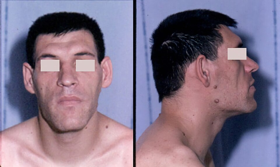
Somewhere near the base of your brain, a pea-sized gland called the anterior pituitary releases growth hormone (GH, somatotropin) in pulses, mostly overnight. GH is your body's build order: it tells bone, muscle, and soft tissue to grow. Like most hormones, it works on a push-pull system. The hypothalamus sends down GHRH (growth hormone-releasing hormone) to press the gas, and somatostatin to press the brake. The balance of those two, plus signals like sleep, stress, exercise, and low blood sugar, decides how much GH gets out.
How GH actually builds you
GH works two ways at once. It acts directly on tissues, and it acts indirectly by traveling to the liver and telling it to make IGF-1 (insulin-like growth factor 1). IGF-1 is the messenger that does much of the real growth work at the bone and muscle, which is why we lean on it for testing later. Together they lengthen and thicken bone at the growth zones, add muscle, and support organ and connective tissue. GH also nudges metabolism: it pushes the body to burn fat and it tends to raise blood glucose by opposing insulin. Hold onto that last fact, because it explains a lot of what goes wrong when there is too much GH.
When there is too little GH
In a child, not enough GH means the skeleton grows slowly but proportionally - a smaller child who is still nicely in proportion, head to limbs to trunk. This is proportional short stature, and it is different from conditions where the limbs and trunk are mismatched. In an adult, growth is already done, so GH deficiency shows up more quietly: fatigue, reduced muscle and strength, more central body fat, and a general sense of low vitality - changes in body composition rather than height.
Too much GH: why timing decides everything
Here is the single most important idea, and it is all about the epiphyseal (growth) plates - the cartilage zones at the ends of long bones where new bone is laid down to make you taller. While those plates are open (childhood and adolescence), bone can still lengthen. Once they fuse after puberty, that lengthening door is closed for good.
So excess GH lands very differently depending on when it strikes. Before the plates close, extra GH lets the whole skeleton keep lengthening - the child grows abnormally tall all over. This is gigantism. After the plates fuse, bone can no longer get longer, so the growth signal has nowhere to go but outward - bone and soft tissue thicken instead. This is acromegaly.
Acromegaly, sign by sign - and why
Because acromegaly builds outward, the clues are about things getting bigger and coarser over years, often so slowly the patient does not notice. As bone and soft tissue in the hands and feet expand, patients report a rising ring and shoe size. The face coarsens: the brow and jaw thicken, producing a protruding jaw called prognathism, and the teeth may spread apart as the jaw enlarges. The tongue enlarges (macroglossia), the voice may deepen, and internal organs enlarge too (organomegaly). Soft-tissue swelling in the wrist compresses the median nerve, giving carpal tunnel syndrome - numbness and tingling in the hand. Because GH opposes insulin, many patients develop glucose intolerance or frank diabetes. And overactive sweat glands cause noticeable sweating (hyperhidrosis).
The usual culprit - and its neighbors
Both conditions are almost always caused by a GH-secreting pituitary adenoma, a benign tumor of the anterior pituitary. Because that tumor sits in a tight bony pocket near important structures, it can cause mass effects as it grows: headache from local pressure, and classically bitemporal hemianopsia - loss of the outer (temporal) half of each visual field - when the tumor presses upward on the optic chiasm, where the nasal retinal fibers cross.
| Feature | Gigantism | Acromegaly |
|---|---|---|
| Timing of GH excess | Before growth plates close (childhood) | After growth plates fuse (adulthood) |
| Growth plates | Still open | Fused |
| What changes | Whole skeleton lengthens | Bone and tissue thicken |
| Main result | Abnormal tall height | Enlarging hands, feet, face (thickness) |
| Usual cause | GH-secreting pituitary adenoma | GH-secreting pituitary adenoma |
Picture a growth plate opening and closing and it all clicks into place - explore it below.
- GH from the anterior pituitary is balanced by GHRH (stimulates) and somatostatin (inhibits); it acts partly through liver-made IGF-1 and tends to raise blood glucose.
- The deciding factor for GH excess is whether the epiphyseal growth plates are open: open plates give gigantism (tall all over), fused plates give acromegaly (bones thicken).
- Acromegaly shows enlarging hands and feet, prognathism with spreading teeth, macroglossia, carpal tunnel syndrome, glucose intolerance, and sweating - all from outward growth.
- The usual cause is a GH-secreting pituitary adenoma, which can also cause headache and bitemporal hemianopsia from pressure on the optic chiasm.
- Diagnose with an elevated IGF-1 plus failure of GH to suppress after an oral glucose load.
Section 6ADH — the body’s water valve
The normal setup: one hormone, one job
Antidiuretic hormone (ADH), also called vasopressin, really does have a single mission: tell the kidneys to hang on to water. To understand its disorders, start with how the body decides when to hold water. Tucked in the hypothalamus are tiny sensors called osmoreceptors. Their whole job is to taste the blood - not for sugar or salt specifically, but for how concentrated it is (its osmolality). When you sweat on a hot day or forget to drink, the blood gets saltier and more concentrated. The osmoreceptors feel that and sound the alarm.
Here is a detail students love once it clicks: the hypothalamus makes ADH, but it does not release it directly. It sends the hormone down to the posterior pituitary, which acts as a storage tank and release valve. When the osmoreceptors fire, the posterior pituitary squirts stored ADH into the bloodstream. ADH then travels to the kidney and lands on the collecting duct - the final stretch of tubing before urine leaves the kidney. There it inserts little water doorways called aquaporins into the tubule wall. Water slips back out of the forming urine and into the blood. Result: the blood is re-diluted, and only a small amount of concentrated urine leaves the body. When you are well hydrated, ADH switches off, the aquaporins pull back, and you make lots of pale, dilute urine. That is the water valve, opening and closing all day.
Now the two disorders. They are near-perfect mirror images, so learn one and you almost get the other for free.
SIADH: too much ADH (the body is soaked)
SIADH stands for syndrome of inappropriate antidiuretic hormone. The key word is inappropriate - ADH is being released when the body does not need it. The valve is jammed open. Common culprits include central nervous system injury (head trauma, stroke, brain surgery, meningitis), small-cell lung cancer (the tumor secretes its own ADH), certain medications (SSRIs, carbamazepine, some chemo agents), and the stress states of surgery, pain, and nausea, which all nudge ADH upward.
With ADH stuck on, the kidneys reabsorb too much water. That extra water dilutes everything in the blood, so the serum sodium falls - a dilutional hyponatremia. Notice this is a water problem, not a salt-loss problem: the total sodium is roughly normal, just swimming in too much fluid. Because the retained water spreads through the whole body rather than pooling as obvious swelling, patients usually look euvolemic (they may gain a little weight but often have no dramatic edema). Meanwhile the urine is the opposite of the blood: small in volume and highly concentrated, because the kidney is clinging to water.
The danger is neurologic. Sodium keeps the fluid balance around brain cells steady; when it drops, water shifts into the neurons and the brain swells. Expect headache, confusion, lethargy, and in severe or fast drops, seizures. For chronic, mild, or asymptomatic SIADH the fix is simple in concept: fluid restriction. Take away the excess water and the sodium drifts back up. A seizing or obtunded patient is a different emergency — fluid restriction is far too slow, so 3% hypertonic saline is given in small boluses to pull the brain out of danger. Either way the correction rate is capped at roughly 6 to 8 mEq/L in 24 hours; raising sodium faster than that can strip myelin from brainstem neurons, an irreversible injury called osmotic demyelination syndrome.
Diabetes insipidus: too little ADH (the body cannot hold water)
Diabetes insipidus (DI) is the flip side. Either the brain does not make or release enough ADH (central DI, often after head injury, pituitary surgery, or a tumor), or the kidney simply cannot respond to the ADH it receives (nephrogenic DI, from kidney disease or drugs like lithium). Either way, the aquaporins never open. Water pours straight through into the urine.
The picture is dramatic: huge volumes of dilute, pale urine (sometimes many liters a day), progressive dehydration, and a rising serum sodium - hypernatremia - because water is leaving while sodium stays behind and gets concentrated. The body fights back with intense, unrelenting thirst. Central DI is treated by replacing the missing hormone with desmopressin (DDAVP), a synthetic ADH.
A quick word on the name
Do not let "diabetes" fool you - DI has nothing to do with blood sugar. The word diabetes just means a large flow of urine. The last name tells you the type: insipidus means tasteless (the old test was literally tasting the dilute urine), while mellitus means sweet, from the sugar spilling in diabetes mellitus. Same big-urine complaint, completely different cause.
SIADH vs DI side by side
| Feature | SIADH (too much ADH) | DI (too little ADH / no response) |
|---|---|---|
| ADH level / action | High - valve stuck open | Low or ineffective - valve stuck shut |
| Serum sodium | Low (hyponatremia) | High (hypernatremia) |
| Urine concentration | Concentrated | Dilute |
| Urine volume | Low | Very high |
| Volume status | Euvolemic / fluid-overloaded | Dehydrated (volume depleted) |
| Core treatment | Fluid restriction | Desmopressin (central DI) |
These two live at opposite ends of the same dial - the ADH water valve. Picture that valve opening and closing on the kidney tubule, and explore it below.
- ADH is made in the hypothalamus, stored and released by the posterior pituitary, and tells the kidney collecting duct to reabsorb water by inserting aquaporins; osmoreceptors trigger it when blood is too concentrated.
- SIADH = too much ADH: water retention, dilutional euvolemic hyponatremia, low-volume concentrated urine, and neuro symptoms - treat chronic or mild cases with fluid restriction, seizures or coma with 3% hypertonic saline, and cap the rise at about 6 to 8 mEq/L per day to avoid osmotic demyelination. Remember: Soaked, Sodium low.
- DI = too little ADH (central) or a kidney that cannot respond (nephrogenic): large volumes of dilute urine, dehydration, hypernatremia, intense thirst - central DI is treated with desmopressin.
- SIADH and DI are exact opposites on sodium, urine, and volume status - learn one and invert it for the other.
- Diabetes insipidus has nothing to do with blood sugar: insipidus means tasteless urine, mellitus means sweet.
The chapter in one breath
- Negative feedback is the master key: a low target hormone drives its pituitary signal up (low T4 means high TSH), while a high one shuts the signal off.
- Cushing's is too much cortisol (moon face, central obesity, hyperglycemia, thin easily-bruised skin, hypertension); Addison's is too little (hypotension, weight loss, hyperkalemia, bronze hyperpigmentation).
- Thyroid logic: hypo runs slow (fatigue, cold, weight gain), hyper runs fast (tachycardia, heat, weight loss), and a goiter can accompany either.
- Type 1 diabetes is absolute insulin deficiency prone to DKA (ketones, acidosis, Kussmaul breathing); Type 2 is insulin resistance prone to HHS (extreme hyperglycemia, profound dehydration, minimal ketones).
- Growth hormone excess before the growth plates fuse causes gigantism; the same excess after fusion causes acromegaly.
- ADH extremes mirror each other: too much is SIADH (water retention, dilutional hyponatremia), too little is diabetes insipidus (massive water loss, hypernatremia).
- Never stop steroids abruptly: taper corticosteroids to prevent an adrenal crisis, and stress-dose during illness or surgery.
- Post-thyroidectomy safety: watch for hypocalcemia from parathyroid injury (Chvostek/Trousseau signs, tingling, tetany) and for airway compromise from neck swelling.
ReferenceWords worth knowing
A quick reference for the terms in this chapter — skim it now, or circle back whenever a word trips you up.
- Aldosterone
- A mineralocorticoid hormone from the adrenal cortex that tells the kidneys to reabsorb sodium and water while excreting potassium, thereby raising blood volume and blood pressure.
- Aquaporin
- A water-channel protein inserted into kidney tubule membranes under the influence of antidiuretic hormone, letting water be reabsorbed and concentrating the urine.
- Catecholamines
- The fight-or-flight hormones epinephrine and norepinephrine released by the adrenal medulla that increase heart rate, blood pressure, and blood glucose.
- Cortisol
- The main glucocorticoid from the adrenal cortex that raises blood glucose, mobilizes fat and protein, and suppresses inflammation as part of the stress response.
- Exophthalmos
- Abnormal forward bulging of the eyeballs, classically seen in Graves disease when immune-driven tissue swelling pushes the eyes outward.
- Goiter
- A visible enlargement of the thyroid gland, which can occur whether thyroid hormone output is high, low, or normal.
- Hyperpigmentation
- Darkening of the skin that occurs in primary adrenal insufficiency because high ACTH also stimulates melanin-producing cells.
- IGF-1
- Insulin-like growth factor 1, a hormone made mainly by the liver in response to growth hormone that carries out most of growth hormone's effects on tissue growth.
- Insulin resistance
- A condition in which body cells respond poorly to insulin, so the pancreas must secrete more of it to keep blood glucose controlled.
- Ketoacidosis
- A dangerous metabolic state, typical of type 1 diabetes, in which severe insulin lack forces fat breakdown into acidic ketones that lower blood pH.
- Myxedema
- The severe, advanced form of hypothyroidism marked by non-pitting facial and tissue swelling from mucopolysaccharide buildup, and in crisis by profound slowing of body functions.
- Negative feedback
- The self-regulating loop in which a rising hormone level signals its own source to shut down further release, keeping the hormone within a stable range.
- Osmotic diuresis
- Excessive urine production that happens when high blood glucose spills into the urine and pulls water along with it, causing dehydration.
- Primary vs secondary endocrine disorder
- A primary disorder arises from a defect in the hormone-producing gland itself, whereas a secondary disorder arises from the pituitary (too little stimulating hormone) and a tertiary disorder from the hypothalamus above it - many nursing texts group both under secondary.
- Thyroid storm
- A life-threatening surge of thyroid hormone activity producing high fever, racing heart, agitation, and possible cardiovascular collapse.
- Tropic hormone
- A hormone, usually from the pituitary, whose target is another endocrine gland that it stimulates to release its own hormone.
DrillFlashcards for this unit
These are the Week 6 cards — the same deck as the hub, pulled in here so you can review the moment you finish reading. Tap the card to flip it; hit Shuffle to mix them up. To grade yourself and track mastery across the whole deck, open the full flashcards page.
Check yourself
| Feature | Cushing | Addison |
|---|---|---|
| Cortisol | High | Low |
| Blood glucose | High | Low |
| Blood pressure | High | Low |
| Serum sodium | High | Low |
| Serum potassium | Low | High |
| Weight | Gain (central) | Loss |
| Feature | Type 1 | Type 2 |
|---|---|---|
| Core defect | Autoimmune beta-cell loss | Insulin resistance |
| Insulin level | Absent | Normal to high early |
| Typical onset | Childhood/young, lean | Adult, often obese |
| Ketosis risk | High (DKA) | Low (HHS instead) |
Growth hormone excess
Gigantism is GH excess before growth-plate closure, producing very tall stature; acromegaly is GH excess after closure, causing enlargement of hands, feet, jaw, and facial features rather than height.
Posterior pituitary / ADH
SIADH is too much ADH: water is retained, urine is concentrated, and serum sodium falls (dilutional hyponatremia). DI (diabetes insipidus) is too little ADH (or renal resistance): huge volumes of dilute urine, thirst, and rising serum sodium.
| Feature | SIADH | DI |
|---|---|---|
| ADH effect | Excess | Deficient |
| Urine output | Low, concentrated | High, dilute |
| Serum sodium | Low | High |
| Volume status | Retained water | Dehydration |