Diuretics, IV Fluids & Poisoning
Almost everything in this chapter comes down to water and the particles dissolved in it. Diuretics pull water out of a patient by tricking one specific stretch of the kidney tubule; IV fluids push water back in, and where that water lands depends entirely on the tonicity printed on the bag. Electrolyte replacement sits in the middle — the corrections that fix a dangerous lab value can, given too fast, stop a heart. And when the wrong substance gets into a patient by accident or on purpose, a small handful of antidotes can reverse what would otherwise be fatal. These are among the highest-alert drugs a nurse handles, and the nurse's monitoring is usually what makes them safe.
By the end of this chapter you can
- Trace filtrate through the nephron and name the segment each diuretic class blocks.
- Compare loop, thiazide, potassium-sparing, osmotic, and carbonic anhydrase inhibitor diuretics by prototype, potency, and electrolyte fallout.
- Anticipate and monitor the characteristic adverse effects of each diuretic class, especially hypokalemia and hyperkalemia.
- Classify an IV solution as isotonic, hypotonic, or hypertonic and predict which fluid compartment it will fill.
- Select an appropriate IV fluid for common bedside scenarios and name the danger of each type.
- Replace potassium, calcium, and magnesium safely, including the rules that must never be broken.
- Match common poisonings to their antidotes and describe the nurse's role in each reversal.
- Explain why diuretics, concentrated electrolytes, and reversal agents are treated as high-alert medications.
Section 1The nephron & where diuretics act
One million tiny filters
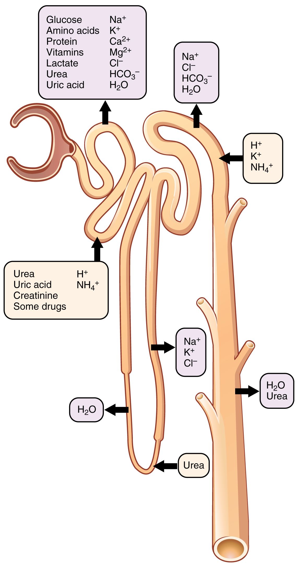
Each kidney holds roughly a million nephrons, and each nephron is a factory that does the same four jobs: filtration, reabsorption, secretion, and excretion. Blood arrives at the glomerulus, a tuft of capillaries wedged inside Bowman's capsule, and pressure squeezes water and small solutes out into the tubule. That filtrate is enormous — about 180 liters a day. Since nobody urinates 180 liters, the obvious conclusion is the important one: the nephron's real work is not making urine, it is taking almost all of it back. Roughly 99 percent of the filtered water is reabsorbed. Urine is the leftover 1 percent.
This is the single insight that makes diuretics make sense. A diuretic does not squeeze more fluid out of the blood. It interferes with reabsorption. Water follows sodium, so any drug that blocks sodium from being pulled back out of the tubule leaves that sodium in the filtrate, and the water stays with it and leaves as urine. Every diuretic in this chapter is a variation on that one theme: block sodium reabsorption somewhere, and water comes along.
The segments, in order
Filtrate leaves Bowman's capsule and enters the proximal convoluted tubule (PCT), the workhorse, where about 65 percent of filtered sodium and water — plus glucose, amino acids, and bicarbonate — are reclaimed. Next comes the loop of Henle, which dives into the salty medulla; its thick ascending limb pumps out another 20–25 percent of the sodium using the Na⁺-K⁺-2Cl⁻ cotransporter. Then the distal convoluted tubule (DCT) reclaims perhaps 5–10 percent through a sodium-chloride cotransporter. Finally the collecting duct fine-tunes the last 2–3 percent under hormonal control: aldosterone tells it to keep sodium and dump potassium, and antidiuretic hormone (ADH) tells it to keep water by inserting aquaporins.
The percentages tell you the potency ranking before you learn a single drug name. Block the loop, where a quarter of the sodium is handled, and you get a powerful diuresis. Block the DCT, where under a tenth is handled, and you get a moderate one. Block the collecting duct, which is only fine-tuning, and you get a weak diuresis — but exquisite control over potassium.
| Segment | Sodium reclaimed | Transporter / control | Diuretic that acts here |
|---|---|---|---|
| Proximal tubule | ~65% | Na⁺/H⁺ exchange; carbonic anhydrase; osmotic gradient | Carbonic anhydrase inhibitors; osmotic diuretics |
| Thick ascending limb | ~20–25% | Na⁺-K⁺-2Cl⁻ cotransporter | Loop diuretics — most potent |
| Distal convoluted tubule | ~5–10% | Na⁺-Cl⁻ cotransporter | Thiazides — moderate |
| Collecting duct | ~2–3% | Aldosterone-driven Na⁺ channels (ENaC); ADH/aquaporins | Potassium-sparing — weak, but K⁺-protective |
Why potassium is always in the conversation
Look at the collecting duct again. Aldosterone reabsorbs sodium there and trades it for potassium, which goes out into the urine. Now imagine a loop or thiazide diuretic upstream, dumping a flood of sodium into the collecting duct. More sodium arriving means more sodium–potassium trading, which means more potassium lost. That is why loop and thiazide diuretics cause hypokalemia — it is not a random side effect, it is arithmetic. And drugs that block that trade at the collecting duct do the reverse: they hold potassium in, which is why they are called potassium-sparing and why their signature danger is hyperkalemia.
- The nephron filters ~180 L/day and reabsorbs about 99% of it; urine is the remainder.
- Diuretics work by blocking sodium reabsorption — water follows sodium out.
- Potency tracks the segment: loop > thiazide > potassium-sparing, because that is the order of how much sodium each segment handles.
- The collecting duct trades sodium for potassium under aldosterone — the reason most diuretics wreck potassium in one direction or the other.
Section 2Diuretic classes
Loop diuretics — the fire hose
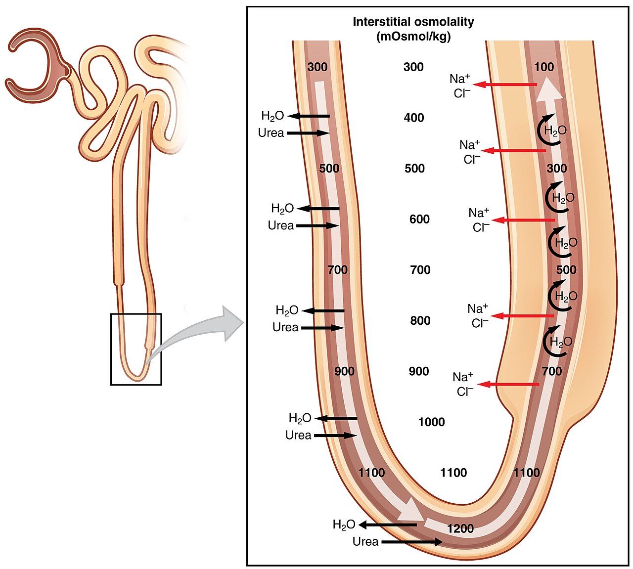
Furosemide is the prototype, joined by bumetanide, torsemide, and ethacrynic acid. They block the Na⁺-K⁺-2Cl⁻ cotransporter in the thick ascending limb, and the result is the most powerful diuresis in pharmacology — up to 20–25 percent of filtered sodium out the door. IV furosemide begins working within minutes, which is exactly what you want in acute pulmonary edema, when a patient is drowning in their own fluid and cannot wait. Loops also keep working when kidney function is poor, unlike thiazides, so they are the diuretic of chronic kidney disease. Their classic uses: heart failure with fluid overload, pulmonary edema, edema of renal or hepatic origin.
The trade-off is that a fire hose flushes out everything. Loops waste potassium, magnesium, calcium, sodium, and chloride and can cause dehydration, orthostatic hypotension, and metabolic alkalosis. They are also ototoxic — tinnitus and hearing loss, made far more likely by rapid IV push or by pairing with an aminoglycoside. Push IV furosemide slowly, no faster than about 20 mg/minute in an adult, and slower still in the elderly. Because furosemide is a sulfonamide derivative, ask about sulfa allergy; ethacrynic acid is the non-sulfa alternative.
Thiazides — the everyday drug
Hydrochlorothiazide is the prototype; chlorthalidone (longer-acting, generally preferred for blood pressure) and metolazone round out the group. They block the Na⁺-Cl⁻ cotransporter in the distal tubule — a smaller job, so a gentler diuresis, but the mild sustained vasodilation that comes with it makes thiazides a cornerstone of hypertension therapy. They are also, delightfully, the only diuretic that retains calcium, which makes them useful in calcium-based kidney stones and mildly protective of bone. Note the ceiling: thiazides lose effectiveness once the glomerular filtration rate falls below roughly 30 mL/min. That is when the prescriber switches to a loop. Metolazone is the exception worth knowing — it keeps working at a low GFR, which is why it is deliberately added to a loop diuretic when a patient stops responding to the loop alone. That combination is potent enough to be dangerous: expect a brisk diuresis and watch potassium and volume status closely.
Teach patients the metabolic mischief: thiazides raise glucose, uric acid (watch for gout flares), lipids, and calcium, while lowering potassium, sodium, and magnesium. Thiazide-induced hyponatremia is a real and underappreciated cause of confusion and falls in older adults.
Potassium-sparing — the counterweight
Two mechanisms, one result. Spironolactone and eplerenone are aldosterone antagonists: they block aldosterone's receptor in the collecting duct. Amiloride and triamterene block the sodium channel directly without touching the receptor. Either way, sodium leaves and potassium stays. The diuresis is weak, so these are rarely used alone for fluid — they are used with a loop or thiazide to blunt potassium loss, and, in the case of spironolactone and eplerenone, because they independently reduce mortality in heart failure with reduced ejection fraction by blocking aldosterone-driven cardiac remodeling. Spironolactone is also standard for the ascites of cirrhosis.
The danger is hyperkalemia — and it is a lethal one. Teach patients to avoid salt substitutes, which are largely potassium chloride, and never to add a potassium supplement without being told to. Combining a potassium-sparing diuretic with an ACE inhibitor or ARB stacks the risk. Spironolactone is also non-selective at steroid receptors, so it causes gynecomastia, breast tenderness, and menstrual irregularity; eplerenone is selective and largely avoids this.
Osmotic and carbonic anhydrase inhibitors — the specialists
Mannitol is an osmotic diuretic: a sugar alcohol that is filtered but not reabsorbed, so it drags water with it osmotically all the way down the tubule. Its real job is usually not the urine — it is pulling water out of swollen tissue. Mannitol is used for increased intracranial pressure, cerebral edema, and acutely raised intraocular pressure. Because it expands plasma volume before it diureses, it can precipitate heart failure and pulmonary edema in a vulnerable patient. Mannitol crystallizes in cold solution, so always use an in-line filter and inspect the bag; warm it if crystals are present.
Acetazolamide blocks carbonic anhydrase in the proximal tubule, dumping bicarbonate. Weak as a diuretic, but useful in glaucoma, altitude sickness, and to alkalinize urine. It predictably causes a metabolic acidosis — which is sometimes the point.
| Class | Prototype | Site & key effect | Nursing alert |
|---|---|---|---|
| Loop | Furosemide (bumetanide, torsemide, ethacrynic acid) | Blocks Na⁺-K⁺-2Cl⁻ in thick ascending limb; most potent; works in renal impairment | Hypokalemia, hypomagnesemia, hypocalcemia, dehydration, orthostasis; ototoxic — push IV slowly (≤20 mg/min), avoid with aminoglycosides; daily weight & I&O; sulfa cross-sensitivity |
| Thiazide | Hydrochlorothiazide (chlorthalidone, metolazone) | Blocks Na⁺-Cl⁻ in distal tubule; moderate; first-line for HTN; spares calcium | Hypokalemia, hyponatremia, hyperglycemia, hyperuricemia (gout), hyperlipidemia; ineffective if GFR <30 mL/min (metolazone excepted — still works, and is added to a loop for diuretic resistance); give in the morning |
| Potassium-sparing (aldosterone antagonist) | Spironolactone (eplerenone) | Blocks aldosterone receptor in collecting duct; weak diuresis; mortality benefit in HFrEF; ascites | Hyperkalemia — no salt substitutes, no K⁺ supplements; caution with ACE inhibitors/ARBs; gynecomastia (spironolactone) |
| Potassium-sparing (channel blocker) | Triamterene (amiloride) | Blocks collecting-duct Na⁺ channels directly; weak | Hyperkalemia; often combined with HCTZ to balance potassium; triamterene can tint urine blue-green |
| Osmotic | Mannitol | Filtered, not reabsorbed; osmotically drags water; reduces ICP and cerebral/ocular edema | In-line filter required (crystallizes); transient volume expansion may cause pulmonary edema/HF; monitor serum osmolality, neuro status, hourly urine output |
| Carbonic anhydrase inhibitor | Acetazolamide | Blocks proximal carbonic anhydrase; dumps bicarbonate; glaucoma, altitude sickness | Predictable metabolic acidosis; hypokalemia; paresthesias; sulfa derivative |
- Loops (furosemide) are the most potent, work in renal failure, and are ototoxic if pushed fast.
- Thiazides (hydrochlorothiazide) are first-line for hypertension, spare calcium, and fail below a GFR of ~30.
- Potassium-sparing agents cause hyperkalemia; spironolactone adds a survival benefit in heart failure and causes gynecomastia.
- Mannitol treats increased ICP, needs an in-line filter, and can transiently worsen heart failure.
- Loops and thiazides drop potassium; potassium-sparing agents raise it — and they are frequently paired for exactly that reason.
Section 3IV fluids
Tonicity is a comparison, not a property
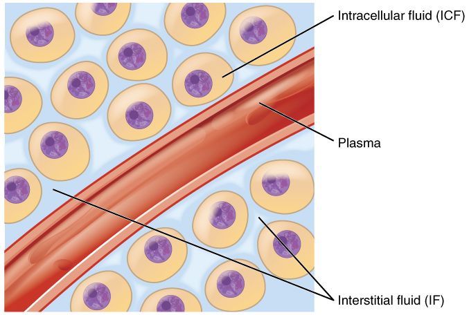
Tonicity describes how a solution's solute concentration compares to that inside the patient's cells. Normal serum osmolality is roughly 275–295 mOsm/kg, and that is the reference point for everything below. Water always moves toward the higher concentration of solute, so the tonicity of the bag decides which way water travels once it is in the vein. That is the whole lesson. An isotonic fluid matches serum, so no net water movement occurs and the fluid simply stays where you put it — expanding the extracellular space. A hypotonic fluid is more dilute than serum, so water leaves the vessel and moves into the cells, swelling them. A hypertonic fluid is more concentrated, so it pulls water out of the cells and into the vessel.
Remember it physically: isotonic stays, hypotonic enters the cell, hypertonic exits the cell. Now attach the danger to each. Too much isotonic fluid overloads the circulation → pulmonary edema. Too much hypotonic fluid swells cells → cerebral edema. Too much hypertonic fluid drags water into an already-full vasculature → fluid overload and shriveled cells.
The bags you will actually hang
0.9% sodium chloride (normal saline) and lactated Ringer's (LR) are the isotonic workhorses: resuscitation, hypovolemia, hemorrhage, dehydration, surgery, and a keep-vein-open line. Normal saline is the only fluid compatible with blood products. Large volumes of it, though, produce a hyperchloremic metabolic acidosis, because you are pouring in far more chloride than plasma normally carries. Lactated Ringer's is more physiologic — it carries potassium, calcium, and lactate that the liver converts to bicarbonate — but avoid it in liver failure (lactate is not cleared) and in significant hyperkalemia, and never run it in the same line as blood, because its calcium can trigger clotting in the tubing. D5W is a special case: isotonic in the bag, but the dextrose is metabolized within minutes and what remains is free water, so it behaves as hypotonic once infused. Do not use it for resuscitation, and do not use it in a patient at risk of cerebral edema.
0.45% sodium chloride (half-normal saline) is the everyday hypotonic fluid, used for cellular dehydration and as maintenance in a patient with hypernatremia or diabetic ketoacidosis after initial resuscitation. Hypotonic fluids are contraindicated in head injury, stroke, and any raised ICP — the last thing a swollen brain needs is more water inside its cells. 3% sodium chloride is the hypertonic fluid you will see in an ICU for symptomatic hyponatremia or cerebral edema; it is a high-alert drug, usually needs a central line and a pump, and demands frequent sodium checks. Correcting sodium too fast is its own catastrophe — generally not more than 8–10 mEq/L in 24 hours — because rapid correction of chronic hyponatremia can cause osmotic demyelination syndrome, a devastating and irreversible brain injury. D5½NS and D5LR are hypertonic maintenance fluids that provide some calories.
| Fluid | Tonicity | Where the water goes / when to use | Nursing alert |
|---|---|---|---|
| 0.9% NaCl (normal saline) | Isotonic | Stays extracellular; resuscitation, hypovolemia, blood administration, KVO | Only fluid compatible with blood products; large volumes → hyperchloremic metabolic acidosis; overload risk in HF/renal disease |
| Lactated Ringer's | Isotonic | Stays extracellular; surgery, burns, trauma, GI losses; lactate → bicarbonate | Avoid in liver failure and hyperkalemia; never with blood (calcium clots the line) |
| D5W | Isotonic in the bag, hypotonic in the body | Dextrose burns off → free water; free-water replacement, drug diluent, hypernatremia | Not for resuscitation; contraindicated in raised ICP; monitor glucose |
| 0.45% NaCl (½NS) | Hypotonic | Water enters cells; cellular dehydration, hypernatremia, DKA maintenance | Cerebral edema risk — contraindicated in head injury/stroke/raised ICP; can cause hypotension & vascular collapse |
| 3% NaCl | Hypertonic | Pulls water out of cells; symptomatic hyponatremia, cerebral edema | High-alert; pump + frequent Na⁺; correct ≤8–10 mEq/L per 24 h to avoid osmotic demyelination; watch for overload |
| D5½NS / D5LR / D5NS | Hypertonic | Maintenance with calories; post-op fluid | Fluid overload; monitor glucose; not for a patient already fluid-overloaded |
- Isotonic stays, hypotonic enters the cell, hypertonic exits the cell — every fluid decision starts here.
- Normal saline is the only fluid to hang with blood; lactated Ringer's is avoided in liver failure and never runs with blood.
- D5W is isotonic in the bag but functionally hypotonic in the patient — never for resuscitation, never in raised ICP.
- Hypotonic fluids risk cerebral edema; hypertonic fluids risk fluid overload and, if sodium is corrected faster than ~8–10 mEq/L/day, osmotic demyelination.
Section 4Electrolyte replacement
Potassium — the one that will kill you fastest
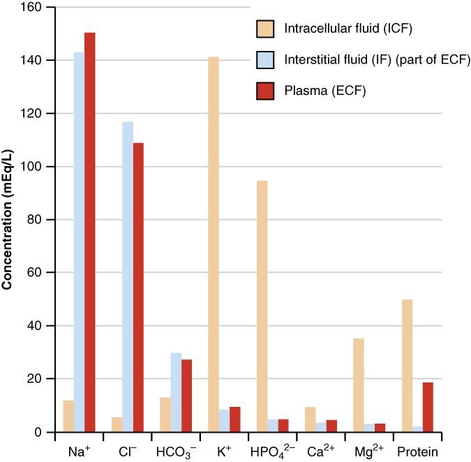
Normal serum potassium is 3.5–5.0 mEq/L, and that narrow window is not an accident: almost all the body's potassium lives inside cells, so the small amount in serum is a razor-thin margin between a beating heart and a stopped one. Both directions are dangerous. Hypokalemia flattens T waves, produces U waves, and invites ventricular dysrhythmias — and it dramatically potentiates digoxin toxicity, so a patient on both a loop diuretic and digoxin deserves a hard look at every potassium result. Hyperkalemia gives peaked T waves, a widening QRS, and eventually cardiac arrest.
The replacement rules exist because people have died from breaking them, and they are worth memorizing exactly:
- Never give potassium IV push. Never. Not ever. IV push potassium causes fatal cardiac arrest. It is always diluted and always infused.
- Always on a pump, never by gravity, and never as a bolus.
- Peripheral infusion generally does not exceed 10 mEq/hour; higher rates require central access and continuous cardiac monitoring per institutional policy.
- Never exceed the facility's maximum concentration — commonly 10 mEq per 100 mL peripherally.
- Verify urine output (a common threshold is at least 30 mL/hour) before infusing — potassium is renally excreted, and giving it to an anuric patient stacks it up.
- Potassium burns. Phlebitis and infiltration are common; assess the site frequently and stop for pain.
- Oral potassium irritates the GI tract — give with food and a full glass of water, and teach that extended-release tablets are not crushed.
For hyperkalemia, the order of operations matters. IV calcium gluconate comes first — it does not lower potassium at all, it stabilizes the cardiac membrane so the heart survives the next few minutes. Then insulin with dextrose (and sometimes albuterol or sodium bicarbonate) shifts potassium into cells temporarily. Only then do you actually remove it, with a binder such as sodium zirconium cyclosilicate or patiromer, a loop diuretic, or dialysis. Stabilize, shift, eliminate.
Calcium — the muscle and the vein
Normal total serum calcium is roughly 8.5–10.5 mg/dL, and remember that about half of it is bound to albumin — a low albumin makes total calcium look falsely low, so check an ionized calcium when it matters. Hypocalcemia produces the classic neuromuscular irritability: tingling around the mouth and fingertips, muscle cramps, Trousseau's sign (carpal spasm when the blood-pressure cuff inflates) and Chvostek's sign (facial twitch when the cheek is tapped), and, at the extreme, laryngospasm and tetany. Hypercalcemia does the opposite — the muscles and mind go slack: weakness, lethargy, constipation, kidney stones, confusion.
Two IV forms exist and they are not interchangeable. Calcium chloride carries about three times the elemental calcium of calcium gluconate and is far more damaging if it extravasates, so gluconate is preferred peripherally and chloride is generally reserved for central access in an emergency. Either way, push slowly — a fast push causes bradycardia and hypotension — and keep the patient on a cardiac monitor. Do not mix calcium with sodium bicarbonate or phosphate, which precipitate in the line. Calcium also potentiates digoxin and can precipitate toxicity. Teach oral calcium with vitamin D for absorption, spaced away from certain antibiotics it chelates.
Magnesium — check the reflexes
Normal magnesium runs about 1.5–2.5 mEq/L. It is quietly essential: magnesium is a cofactor for hundreds of enzymes, and — critically for the bedside — you cannot correct a potassium until you have corrected the magnesium. Refractory hypokalemia is almost always undiagnosed hypomagnesemia, because low magnesium drives renal potassium wasting no matter how much potassium you give. Hypomagnesemia causes hyperreflexia, tremor, positive Chvostek and Trousseau signs, and torsades de pointes — for which IV magnesium is itself the treatment. It is common in chronic alcohol use, malnutrition, and loop diuretic therapy.
Hypermagnesemia is the mirror image and is the risk you manage during any magnesium infusion. Watch the sequence: loss of deep tendon reflexes comes first, then respiratory depression, then hypotension and bradycardia, then cardiac arrest. That is why the reflex check is not busywork — a diminished patellar reflex is the early warning that buys you time. During IV magnesium (for eclampsia, for example) monitor reflexes, respiratory rate, blood pressure, and urine output, and keep calcium gluconate at the bedside as the antidote.
| Electrolyte | Normal range | Deficiency & excess | Nursing alert |
|---|---|---|---|
| Potassium | 3.5–5.0 mEq/L | Low: flat T waves, U waves, dysrhythmias, digoxin toxicity. High: peaked T waves, wide QRS, arrest | NEVER IV push; dilute, pump, ≤10 mEq/h peripherally; confirm urine output; watch the site (burns); do not crush ER tablets |
| Calcium | 8.5–10.5 mg/dL total | Low: perioral tingling, cramps, Trousseau & Chvostek, tetany, laryngospasm. High: weakness, lethargy, constipation, stones | Gluconate peripherally, chloride centrally; push slowly on a monitor; never mix with bicarbonate or phosphate; potentiates digoxin; check ionized Ca if albumin is low |
| Magnesium | 1.5–2.5 mEq/L | Low: hyperreflexia, tremor, torsades, refractory hypokalemia. High: reflexes lost → respiratory depression → arrest | Check deep tendon reflexes — first sign of toxicity; monitor RR, BP, urine output; calcium gluconate is the antidote; fix Mg before you can fix K⁺ |
- Potassium is never given IV push — always diluted, always on a pump, generally ≤10 mEq/h peripherally, and only with adequate urine output.
- Hyperkalemia is treated stabilize (calcium) → shift (insulin + dextrose) → eliminate (binders, diuretic, dialysis).
- Calcium gluconate is safer peripherally than calcium chloride; push slowly, never with bicarbonate or phosphate.
- Correct magnesium before potassium; during magnesium infusion, loss of deep tendon reflexes is the first warning and calcium gluconate is the antidote.
Section 5Poisoning & antidotes
The patient comes before the poison
The first instinct in a poisoning is to hunt for the antidote. Resist it. Most poisonings have no antidote at all, and the patients who survive do so because of supportive care: airway, breathing, circulation, glucose, and time. So the sequence is ABCs first, decontamination second, antidote third if one exists, and enhanced elimination last. Along the way, call Poison Control — in the United States, 1-800-222-1222 — early rather than late. They do this all day.
For decontamination, activated charcoal is the mainstay, and it works by adsorbing the poison in the gut before it is absorbed. It is most useful within about an hour of ingestion, and it is contraindicated in a patient with a depressed level of consciousness or an unprotected airway, because aspirated charcoal causes a severe pneumonitis. It also does not bind everything: the classic misses are iron, lithium, alcohols, and the strong acids and alkalis — and corrosives should never be given charcoal anyway, since it obscures endoscopy. Do not induce vomiting. Syrup of ipecac is obsolete; it causes harm and does not improve outcomes.
The antidotes worth knowing cold
Learn these as pairs, because they are the ones that show up on exams and in real emergencies. Acetaminophen overdose is the leading cause of acute liver failure in the United States, and it is deceptively quiet — the patient may look fine for a day while a toxic metabolite is destroying hepatocytes. Acetylcysteine replenishes glutathione and is dramatically effective when started early, ideally within 8–10 hours; use the Rumack-Matthew nomogram to decide after a single acute ingestion. Warn patients that the oral form smells like rotten eggs; the IV form can cause an anaphylactoid reaction.
Opioid overdose is diagnosed at the bedside by the triad of respiratory depression, pinpoint pupils, and a depressed level of consciousness. Naloxone reverses it within minutes — but its duration is shorter than most opioids, so the patient can re-sedate, and repeat doses or an infusion may be needed. Never leave a reversed patient unmonitored. In an opioid-dependent patient, naloxone precipitates acute withdrawal, which is miserable but not fatal; respiratory arrest is. Benzodiazepine overdose has an antidote too, flumazenil, but it is used cautiously and rarely: in a chronic benzodiazepine user or a mixed overdose it can precipitate intractable seizures.
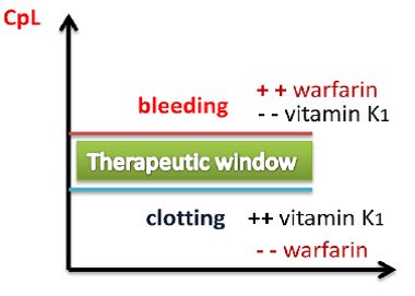
The anticoagulants each have a partner. Warfarin is reversed by vitamin K (phytonadione), and, when the bleeding is life-threatening, by prothrombin complex concentrate. Heparin is reversed by protamine sulfate. Dabigatran has idarucizumab; the factor Xa inhibitors apixaban and rivaroxaban have andexanet alfa. Digoxin toxicity — nausea, visual halos or yellow-green vision, bradycardia, confusion — is treated with digoxin immune Fab, and remember that hypokalemia makes digoxin toxicity far more likely, which loops right back to Section 2. Organophosphate or cholinergic poisoning presents as the SLUDGE picture (salivation, lacrimation, urination, defecation, GI upset, emesis) and is treated with atropine plus pralidoxime. And iron — historically a leading cause of pediatric poisoning death, though fatalities fell sharply after unit-dose blister packaging, and it remains a serious pediatric ingestion — is chelated with deferoxamine.
| Poison / drug | Antidote | Key toxic signs | Nursing alert |
|---|---|---|---|
| Acetaminophen | Acetylcysteine | Quiet early phase, then RUQ pain, rising AST/ALT, hepatic failure | Most effective within 8–10 h; oral form smells of sulfur (dilute, use a straw); IV can cause anaphylactoid reaction; use the Rumack-Matthew nomogram |
| Opioids | Naloxone | Respiratory depression + pinpoint pupils + ↓LOC | Shorter duration than the opioid — re-sedation risk, repeat doses/infusion; precipitates withdrawal in dependence; keep monitoring |
| Benzodiazepines | Flumazenil | Sedation, ataxia, slurred speech; respiratory depression if mixed | Use cautiously — may precipitate seizures in chronic users or mixed (e.g., TCA) overdose |
| Warfarin | Vitamin K (phytonadione) | Bleeding, elevated INR, bruising, hematuria | Onset takes hours — PCC/FFP for life-threatening bleed; monitor INR; teach consistent vitamin K intake |
| Heparin | Protamine sulfate | Bleeding, elevated aPTT, thrombocytopenia (HIT) | Give slowly — rapid administration causes hypotension & anaphylaxis; only partially reverses LMWH |
| Digoxin | Digoxin immune Fab | Nausea, anorexia, yellow-green/halo vision, bradycardia, confusion | Hold for apical pulse <60; hypokalemia potentiates toxicity; narrow therapeutic range (0.5–2 ng/mL) |
| Organophosphates / cholinergics | Atropine + pralidoxime | SLUDGE, bronchorrhea, bradycardia, miosis | Decontaminate the skin — protect yourself with PPE; large repeated atropine doses may be needed |
| Iron | Deferoxamine | Vomiting, GI bleeding, shock, later hepatic failure; historically a leading cause of pediatric poisoning death (fatalities fell sharply after unit-dose packaging, but still a serious ingestion) | Charcoal does not bind iron; urine may turn reddish-brown ("vin rosé"); teach childproof storage |
| Dabigatran | Idarucizumab | Bleeding | Factor Xa inhibitors (apixaban, rivaroxaban) use andexanet alfa instead |
| Most oral ingestions | Activated charcoal (adsorbent, not an antidote) | — | Within ~1 h; contraindicated with ↓LOC or unprotected airway; does not bind iron, lithium, alcohols, corrosives; never induce vomiting |
- Supportive care first — ABCs, then decontamination, then antidote; call Poison Control early.
- Activated charcoal works within about an hour, is contraindicated with a depressed LOC, and misses iron, lithium, alcohols, and corrosives; never induce vomiting.
- Learn the pairs: acetaminophen–acetylcysteine, opioids–naloxone, warfarin–vitamin K, heparin–protamine, digoxin–immune Fab, organophosphates–atropine + pralidoxime, iron–deferoxamine.
- Naloxone's duration is shorter than the opioid's — expect re-sedation and keep monitoring.
The chapter in one breath
- The nephron reabsorbs ~99% of what it filters; every diuretic works by blocking sodium reabsorption at one segment, and water follows sodium.
- Potency follows the segment: loop > thiazide > potassium-sparing, because that is the order of how much sodium each segment handles.
- Furosemide is the fire hose (ototoxic if pushed fast, wastes K⁺); hydrochlorothiazide is the everyday antihypertensive (spares calcium, fails below GFR 30); spironolactone spares K⁺ and saves lives in heart failure; mannitol pulls water off a swollen brain.
- Isotonic stays, hypotonic enters the cell, hypertonic exits the cell — and the danger of each is overload, cerebral edema, and overload again, respectively.
- Normal saline is the only fluid to hang with blood; D5W acts hypotonic once the dextrose burns off; 3% NaCl is high-alert and sodium must not rise faster than ~8–10 mEq/L per day.
- Potassium is never IV push — always diluted, pumped, rate-limited, and given only with adequate urine output.
- Hyperkalemia goes stabilize → shift → eliminate; fix magnesium before potassium; during magnesium infusion, lost reflexes are the first warning and calcium gluconate is the antidote.
- In poisoning, supportive care beats antidote-hunting — but know the pairs cold, and remember that naloxone wears off before the opioid does.
ReferenceWords worth knowing
A quick reference for the terms in this chapter — skim it now, or circle back whenever a word trips you up.
- Activated charcoal
- An adsorbent given orally to bind an ingested poison before absorption; useful within about an hour, contraindicated with a depressed level of consciousness, and ineffective against iron, lithium, alcohols, and corrosives.
- Aldosterone antagonist
- A potassium-sparing diuretic (spironolactone, eplerenone) that blocks aldosterone's receptor in the collecting duct, retaining potassium and reducing mortality in heart failure with reduced ejection fraction.
- Chvostek's sign
- A facial muscle twitch elicited by tapping over the facial nerve; a sign of hypocalcemia.
- Collecting duct
- The final nephron segment, where aldosterone drives sodium reabsorption in exchange for potassium and ADH controls water reabsorption.
- Diuretic
- A drug that increases urine output by blocking sodium reabsorption somewhere along the nephron.
- Glomerulus
- The capillary tuft inside Bowman's capsule where blood is filtered to form filtrate.
- High-alert medication
- A drug carrying a heightened risk of significant harm when used in error — concentrated potassium, hypertonic saline, insulin, and anticoagulants among them.
- Hyperkalemia
- Serum potassium above about 5.0 mEq/L; produces peaked T waves, a widening QRS, and cardiac arrest; the signature risk of potassium-sparing diuretics.
- Hypertonic
- A solution more concentrated than serum; it pulls water out of cells and into the vascular space.
- Hypokalemia
- Serum potassium below about 3.5 mEq/L; causes flattened T waves, U waves, dysrhythmias, and potentiates digoxin toxicity; the signature risk of loop and thiazide diuretics.
- Hypotonic
- A solution more dilute than serum; water moves into cells, swelling them — the reason these fluids are contraindicated in raised intracranial pressure.
- Isotonic
- A solution matching serum osmolality; it stays in the extracellular space and causes no net water shift across cell membranes.
- Loop diuretic
- The most potent diuretic class (furosemide, bumetanide, torsemide); works even in renal impairment and is ototoxic if infused rapidly.
- Loop of Henle
- The nephron segment whose thick ascending limb reabsorbs 20–25% of filtered sodium via the Na⁺-K⁺-2Cl⁻ cotransporter; the target of loop diuretics.
- Nephron
- The functional unit of the kidney, performing filtration, reabsorption, secretion, and excretion; each kidney holds roughly a million.
- Osmotic demyelination syndrome
- Irreversible brain injury caused by correcting chronic hyponatremia too rapidly; prevented by limiting sodium correction to roughly 8–10 mEq/L in 24 hours.
- Osmotic diuretic
- A filtered, non-reabsorbed solute (mannitol) that osmotically drags water along the tubule; used to reduce intracranial and intraocular pressure.
- Ototoxicity
- Damage to hearing or balance; a loop diuretic risk that rises with rapid IV administration and with concurrent aminoglycosides.
- Potassium-sparing diuretic
- A weak diuretic acting at the collecting duct that retains potassium; the class whose defining danger is hyperkalemia.
- Thiazide diuretic
- A moderate diuretic (hydrochlorothiazide, chlorthalidone) acting on the distal tubule's Na⁺-Cl⁻ cotransporter; first-line for hypertension, spares calcium, ineffective below a GFR of about 30 mL/min.
- Tonicity
- A solution's solute concentration compared with that inside the body's cells; it determines which direction water moves after infusion.
- Trousseau's sign
- Carpal spasm provoked by inflating a blood-pressure cuff on the upper arm; a sign of hypocalcemia.