The Urinary System
Two fist-sized organs filter your entire blood volume roughly every half hour, and the vocabulary built around them shows up on nearly every chart you will ever read. This chapter gives you the urinary word parts in the order the fluid actually travels — kidney, renal pelvis, ureter, bladder, urethra, meatus — then layers on the symptom suffixes, the pathology terms, the imaging studies, the surgeries and the abbreviations. You will learn why the same organ carries two roots (ren/o and nephr/o), how a single letter separates ureter/o from urethr/o, and why pyel/o and py/o are the most expensive two-letter difference in the chapter. By the end you should be able to open a urinalysis report, a nephrology consult or an operative note and take most of it apart without a dictionary — because you know the pieces, not because you memorized the whole term.
By the end of this chapter you can
- Trace urine from glomerulus to urinary meatus, naming each structure and its combining form.
- Explain when English medicine uses ren/o and when it uses nephr/o, and predict which one a new term will carry.
- Distinguish ur/o, ureter/o and urethr/o, and pyel/o from py/o, in unfamiliar terms.
- Build and decode symptom terms using the suffixes -uria, -uresis and -lithiasis (dysuria, oliguria, nocturia, enuresis, nephrolithiasis).
- Read a urinalysis line by line, translating hematuria, pyuria, proteinuria, glycosuria, ketonuria and bacteriuria.
- Separate the major kidney diseases — glomerulonephritis, pyelonephritis, nephrotic syndrome, hydronephrosis, polycystic kidney disease, AKI and CKD.
- Classify lower-tract problems including cystitis, urethral stricture, urinary retention and the four patterns of incontinence.
- Match urinary procedures to their suffixes, contrasting nephrectomy, nephrostomy, nephrotomy, lithotripsy and cystoscopy.
- Interpret the abbreviations a real chart uses: UA, UTI, BUN, eGFR, I&O, HD, TURP, ESWL.
Section 1The Tract, End to End
One organ, two roots
The kidney is the first structure in medicine that forces students to accept an uncomfortable truth: the same organ can have two perfectly correct combining forms. Ren/o comes from Latin, nephr/o from Greek, and English medicine kept both. They are not interchangeable in practice. Latin roots gravitate toward anatomical adjectives and toward terms that entered through anatomy: renal artery, renal pelvis, renal cortex, renal failure, renal colic. Greek roots gravitate toward pathology, procedures and specialties: nephritis, nephrology, nephrectomy, nephrostomy, nephropathy.
That pattern is not a rule you can bet a licence on, but it is a reliable instinct. When you meet an unfamiliar term and have to guess, ask whether it names a structure (lean Latin) or names a disease, a study or a surgery (lean Greek). Decode nephr/o/megaly (nef-roh-MEG-ah-lee) as kidney + enlargement, and ren/al as pertaining to the kidney. Both point at the same organ; only the register differs.
Two more facts about location earn their keep. The kidneys sit retroperitoneal (ret-roh-per-ih-toh-NEE-al) — behind (retro-) the peritoneum — which is why a surgeon can reach a kidney through the flank without entering the abdominal cavity, and why kidney bleeding can hide for a long time. And they sit against the posterior body wall around the twelfth rib, which is why a nurse assesses kidney tenderness at the costovertebral angle (CVA), the corner between the last rib and the spine.
| Word part | Meaning | Example term | What the example means |
|---|---|---|---|
| ren/o | kidney (Latin) | renal | pertaining to the kidney |
| nephr/o | kidney (Greek) | nephrology | study of the kidney |
| cortic/o | cortex, outer region | renal cortical cyst | fluid sac in the outer kidney tissue |
| medull/o | medulla, inner region | medullary sponge kidney | malformation of the inner kidney tubules |
| glomerul/o | glomerulus (little ball of capillaries) | glomerulopathy | disease of the filtering tufts |
| pyel/o | renal pelvis (the funnel) | pyelogram | image of the renal pelvis |
| cali/o, calic/o | calyx (cup) | caliectasis | dilation of the calices |
| tubul/o | tubule, small tube | acute tubular necrosis | death of the tubule cells inside the nephron |
Inside the kidney: cortex to calyx
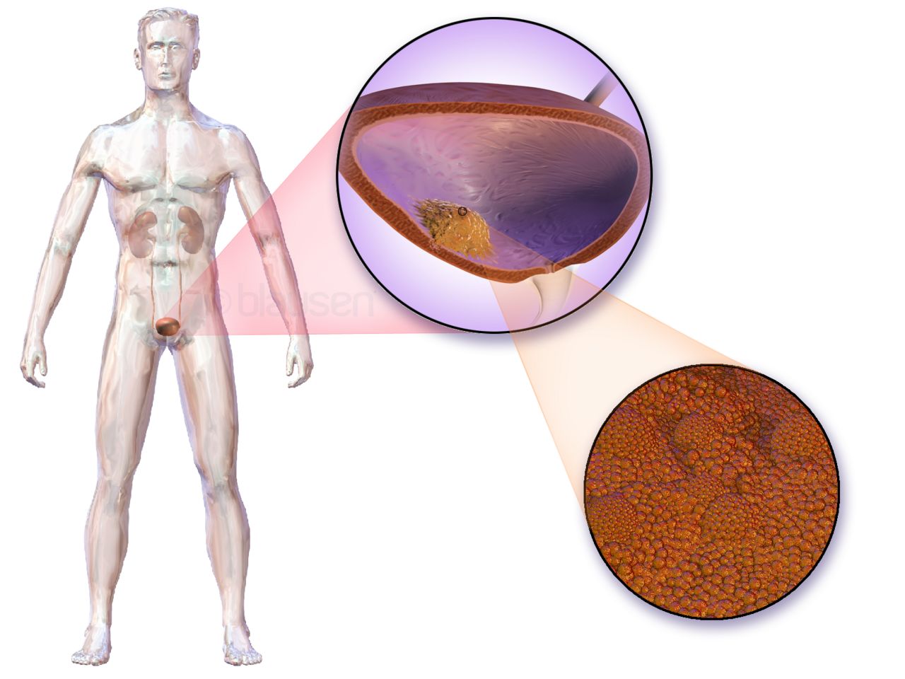
Cut a kidney lengthwise and you see three zones. The outer cortex holds the glomeruli. The inner medulla is arranged in cone-shaped renal pyramids whose tips (papillae) drip urine into cup-shaped calices. The calices merge into the renal pelvis (pel-vis), the funnel that hands urine to the ureter. Notice how much of urinary imaging vocabulary lives in that funnel: a pyelogram shows it, pyelonephritis inflames it along with the kidney tissue, a pyeloplasty rebuilds it when it is obstructed.
The microscopic unit is the nephron (NEF-ron), about a million per kidney. Blood enters a knot of capillaries called the glomerulus (gloh-MER-yoo-lus), which behaves like a sieve: water, electrolytes, glucose, urea and small wastes are pushed out into the tubule, while blood cells and large proteins are meant to stay in. Everything downstream in the tubule is a negotiation — reabsorb what the body needs, secrete what it does not. That single sentence explains a surprising share of the chapter’s pathology. When the sieve leaks, you get proteinuria and hematuria. When the tubule is injured, you lose the ability to concentrate urine.
Decode glomerul/o/nephr/itis (gloh-mer-yoo-loh-neh-FRY-tis) = glomerulus + kidney + inflammation = inflammation of the kidney centered on the glomeruli. Decode pyel/o/nephr/itis (pye-eh-loh-neh-FRY-tis) = renal pelvis + kidney + inflammation = infection that has climbed into the kidney and its funnel. Two diseases, two very different causes, and the roots tell you where each one lives.
The drainage line: ureter, bladder, urethra, meatus
Each kidney sends urine down one ureter (YOO-reh-ter), a muscular tube about 25 to 30 cm long that moves urine by peristalsis, not gravity. Both ureters enter the back of the urinary bladder at an angle, forming a flap-valve that normally prevents backflow. The triangular region marked by the two ureteral openings and the urethral outlet is the trigone (TRY-gohn), and it matters clinically because it is a common site of infection and tumor and because it is exquisitely sensitive to catheter balloons.
From the bladder, urine leaves through the single urethra (yoo-REE-thrah) and exits at the urinary meatus (mee-AY-tus), the external opening. Length is not trivia here: the female urethra is roughly 4 cm, the male urethra roughly 20 cm and it passes through the prostate. Those two anatomical facts explain most of the epidemiology in Section 4 — why women get far more bladder infections, and why men over sixty get far more obstruction.
The act of emptying is micturition (mik-choo-RISH-un), also called voiding or urination. You will see all three words used interchangeably in charting, though void dominates in nursing documentation because it is short and unambiguous.
| Word part | Meaning | Example term | What the example means |
|---|---|---|---|
| ureter/o | ureter | ureterostomy | surgical opening of a ureter to the surface |
| cyst/o | urinary bladder (Greek) | cystitis | inflammation of the bladder |
| vesic/o | urinary bladder (Latin) | vesicoureteral reflux | backflow from bladder into ureter |
| urethr/o | urethra | urethritis | inflammation of the urethra |
| meat/o | meatus, opening | meatotomy | incision to enlarge the urinary opening |
| trigon/o | trigone of the bladder | trigonitis | inflammation of the bladder trigone |
| ur/o | urine, urinary tract | urology | study of the urinary tract |
| urin/o | urine | urinary | pertaining to urine |
Singular and plural in the urinary tract
Urinary anatomy is unusually rich in Latin and Greek plurals, and they appear in radiology reports constantly. A report reading “dilated calices bilaterally” is describing more than one calyx. “Multiple calculi” means more than one calculus — more than one stone. Learn the endings once and they carry into every other chapter.
| Singular | Plural | Rule | Seen in |
|---|---|---|---|
| glomerulus | glomeruli | -us to -i | “sclerosis of 40% of glomeruli” |
| calculus | calculi | -us to -i | “two obstructing calculi” |
| calyx (calix) | calices (calyces) | -yx/-ix to -ices | “blunted calices” |
| pelvis | pelves | -is to -es | “bilateral renal pelves dilated” |
| papilla | papillae | -a to -ae | “papillae are necrotic” |
| hilum | hila | -um to -a | “vessels enter at the hila” |
| cortex | cortices | -ex to -ices | “thinned renal cortices” |
| diverticulum | diverticula | -um to -a | “bladder diverticula” |
Learn the tract in flow order — glomerulus, tubule, calyx, renal pelvis, ureter, bladder, urethra, meatus — and attach one combining form to each stop, because nearly every urinary term you meet names a place on that line.
Section 2Reading the Urine: The -uria Family
One suffix, an entire vocabulary
The suffix -uria means “urine condition” or “a condition of the urine.” Put almost any root in front of it and you have named a finding. This is the single highest-yield pattern in the chapter, because it converts a long list of unrelated-looking clinical words into one rule you can apply to words you have never seen.
Decode hemat/uria (hee-mah-TOO-ree-ah) = blood + urine condition = blood in the urine. Decode dys/uria (dis-YOO-ree-ah) = painful or difficult + urine condition = painful urination. Decode olig/uria (ol-ih-GYOO-ree-ah) = scanty + urine condition = markedly decreased urine output. Decode an/uria (an-YOO-ree-ah) = without + urine condition = essentially no urine produced. Notice that the suffix does not commit to a mechanism — it only tells you that something about the urine is abnormal. The root supplies the specific.
Two related endings deserve separate shelf space. -uresis means the act of urinating: diuresis (dye-yoo-REE-sis) is increased urine output, and a diuretic is a drug that causes it. Enuresis (en-yoo-REE-sis) is involuntary urination, most often used for bedwetting in a child old enough to have control (nocturnal enuresis). Do not confuse diuresis (making a lot of urine) with dysuria (it hurts to urinate) — they sound similar and mean nothing alike.
| Word part | Meaning | Example term | What the example means |
|---|---|---|---|
| -uria | urine condition | hematuria | blood in the urine |
| -uresis | urination | diuresis | increased output of urine |
| olig/o | scanty, few | oliguria | low urine output (under about 400 mL/day, or under 0.5 mL/kg/hr, in an adult) |
| an- | without | anuria | virtually no urine output (under about 100 mL/day) |
| poly- | many, much | polyuria | excessive volume of urine |
| dys- | painful, difficult | dysuria | painful or burning urination |
| noct/i | night | nocturia | waking at night to urinate |
| -dipsia | thirst | polydipsia | excessive thirst |
| py/o | pus | pyuria | pus (white cells) in the urine |
| bacteri/o | bacteria | bacteriuria | bacteria in the urine |
What the specimen contains, and the words for it
A urinalysis (yoo-rih-NAL-ih-sis), abbreviated UA, has three layers: how the urine looks, what the chemical dipstick detects, and what the microscope sees. Learn the vocabulary in that order and the report reads top to bottom.
Appearance terms are plain but precise. Normal urine is clear and straw to amber. Turbid or cloudy urine suggests cells, crystals or infection. Hematuria may be gross (visible to the eye) or microscopic (found only under the lens); the distinction changes how urgently a patient is worked up. Specific gravity (SG) compares urine density to water and tells you whether the kidney is concentrating: a dehydrated patient makes concentrated, high-SG urine, while a patient with damaged tubules or diabetes insipidus makes dilute, low-SG urine no matter how dry they are.
The chemical strip contributes most of the -uria words. Proteinuria (proh-tee-NOO-ree-ah) and its more specific cousin albuminuria (al-byoo-mih-NOO-ree-ah) signal a leaking glomerular filter. Glycosuria (gly-koh-SOO-ree-ah), sugar in the urine, appears when blood glucose exceeds what the tubules can reabsorb — the classic first clue in undiagnosed diabetes mellitus. Ketonuria (kee-toh-NOO-ree-ah) means the body is burning fat for fuel, seen in starvation, low-carbohydrate states and diabetic ketoacidosis. A positive nitrite or leukocyte esterase pad points toward infection, which the microscope then confirms as pyuria and bacteriuria.
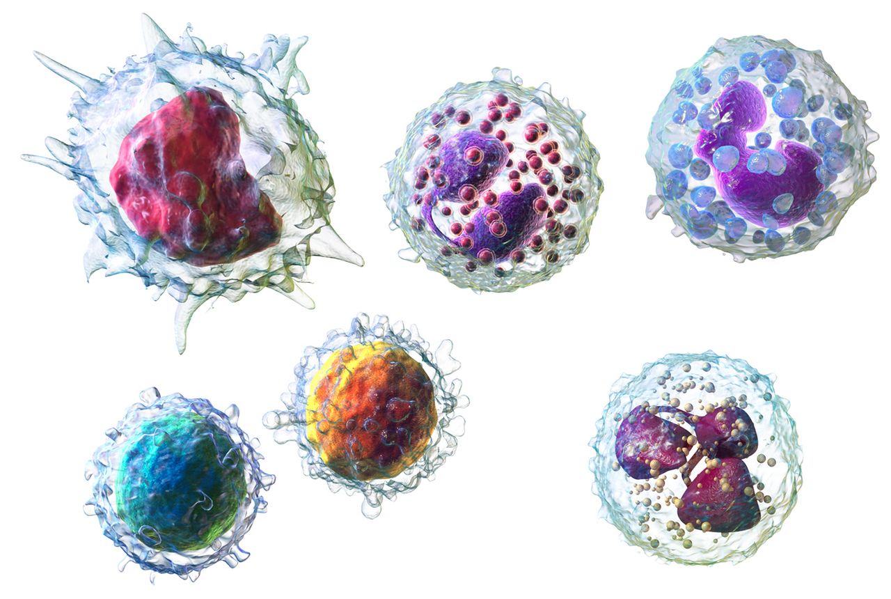
Under the microscope you also meet casts — cylindrical molds of the renal tubule made of protein and whatever cells were passing through. Red cell casts are close to proof that bleeding came from the glomerulus rather than from a stone or a tumor downstream, which is a beautiful example of a single microscopic finding localizing disease along the tract you learned in Section 1. Crystalluria is crystals in the urine, often a hint about what a stone is made of.
| Word part | Meaning | Example term | What the example means |
|---|---|---|---|
| albumin/o | albumin (a blood protein) | albuminuria | albumin leaking into the urine |
| protein/o | protein | proteinuria | abnormal protein in the urine |
| glycos/o, glyc/o | sugar, glucose | glycosuria | glucose in the urine |
| keton/o | ketone bodies | ketonuria | ketones in the urine |
| hemat/o, hem/o | blood | hemoglobinuria | free hemoglobin in the urine |
| lith/o | stone | urolithiasis | abnormal condition of stones in the urinary tract |
| crystall/o | crystal | crystalluria | crystals in the urine |
| azot/o | nitrogen (urea) | azotemia | nitrogen wastes accumulating in the blood |
| -emia | blood condition | uremia | urine wastes in the blood, with symptoms |
When the wastes back up: azotemia and uremia
The kidney’s job is to move nitrogen wastes out of the blood. When it stops doing that, the wastes rise, and medicine has two words for the situation. Azot/emia (az-oh-TEE-mee-ah) = nitrogen + blood condition, and it is a laboratory statement: the blood urea nitrogen (BUN) and creatinine are elevated. Ur/emia (yoo-REE-mee-ah) = urine + blood condition, and it is a clinical statement: the patient has symptoms from those wastes — nausea, itching, confusion, a metallic taste, pericardial rub, uremic frost on the skin in extreme cases.
Put plainly, every uremic patient is azotemic, but not every azotemic patient is uremic. A mildly dehydrated post-operative patient can be azotemic on Tuesday and normal on Wednesday after fluids. Uremia describes a body being poisoned by what it cannot excrete, and it is one of the indications for starting dialysis.
The two numbers behind those words are worth knowing before you meet them on a chart. BUN reflects urea and is sensitive to hydration, diet and gastrointestinal bleeding. Creatinine (kree-AT-ih-nin) is a muscle breakdown product filtered fairly steadily, so it is a better mirror of kidney function — and it is the number plugged into the eGFR (estimated glomerular filtration rate), the single figure that stages chronic kidney disease.
One practical note about where these words come from. I&O — intake and output — is the chart section where oliguria, polyuria and anuria are actually detected. A 70 kg patient charted at 25 mL/hr for four consecutive hours is oliguric — that is below the 0.5 mL/kg/hr threshold — whether or not anyone has written the word yet. Learn the terms and the numbers behind them together; either one alone leaves you unable to act.
Master the suffix -uria and its relatives and you can decode almost every urinary symptom and urinalysis finding on sight, because the root in front simply names what is wrong with the urine.
Section 3Disorders of the Kidney
Inflammation, infection and the two great syndromes
Nephritis (neh-FRY-tis) is the umbrella word for inflammation of the kidney, and the more useful terms narrow it by location. Glomerulonephritis attacks the filtering tufts, usually through an immune mechanism — classically days to weeks after a streptococcal throat or skin infection in children. Its signature is a nephritic picture: hematuria with red cell casts, mild to moderate proteinuria, hypertension and edema. Pyelonephritis is bacterial, almost always arriving from below after a bladder infection climbs the ureter, and its signature is fever, chills, flank pain and CVA tenderness on top of the burning and frequency of a bladder infection.
Set against nephritic disease is nephrotic syndrome (neh-FROT-ik), which is not one disease but a pattern: massive proteinuria, low blood albumin (hypoalbuminemia), widespread edema and high cholesterol. Decode the difference this way — in the nephritic pattern the filter is inflamed and leaks cells; in the nephrotic pattern the filter is porous and leaks protein. Both are glomerular. Both may end in the same place if untreated. But the urinalysis distinguishes them in one glance.
Interstitial nephritis names inflammation of the tissue between the tubules, most often a drug reaction. And glomerulosclerosis (gloh-mer-yoo-loh-skleh-ROH-sis) = glomerulus + hardening + abnormal condition, the scarring endpoint that diabetes and hypertension eventually produce — which is why those two chronic diseases, not primary kidney disease, are the leading causes of kidney failure in the United States.
| Word part | Meaning | Example term | What the example means |
|---|---|---|---|
| -itis | inflammation | nephritis | inflammation of the kidney |
| -osis | abnormal condition | hydronephrosis | abnormal accumulation of urine in the kidney |
| -pathy | disease | nephropathy | any disease of the kidney |
| -megaly | enlargement | nephromegaly | enlarged kidney |
| -sclerosis | hardening | nephrosclerosis | hardening of the kidney’s small arteries |
| -ptosis | drooping, downward displacement | nephroptosis | a dropped or “floating” kidney |
| -ectasis | dilation, stretching | ureterectasis | dilated ureter |
| -lithiasis | condition of stones | nephrolithiasis | kidney stones |
| hydr/o | water, fluid | hydroureter | ureter distended with backed-up urine |
| -cele | hernia, pouching | ureterocele | ballooning of the ureter into the bladder |
Stones, obstruction and backpressure
Nephrolithiasis (nef-roh-lih-THIGH-ah-sis) = kidney + stone + abnormal condition. The broader term urolithiasis covers a stone anywhere in the tract, and ureterolithiasis pins it to the ureter, which is where stones cause the most pain. A stone lodged in a ureter produces renal colic: waves of excruciating flank pain radiating toward the groin, often with nausea, restlessness and gross or microscopic hematuria. The word calculus (plural calculi) is the formal term for the stone itself.
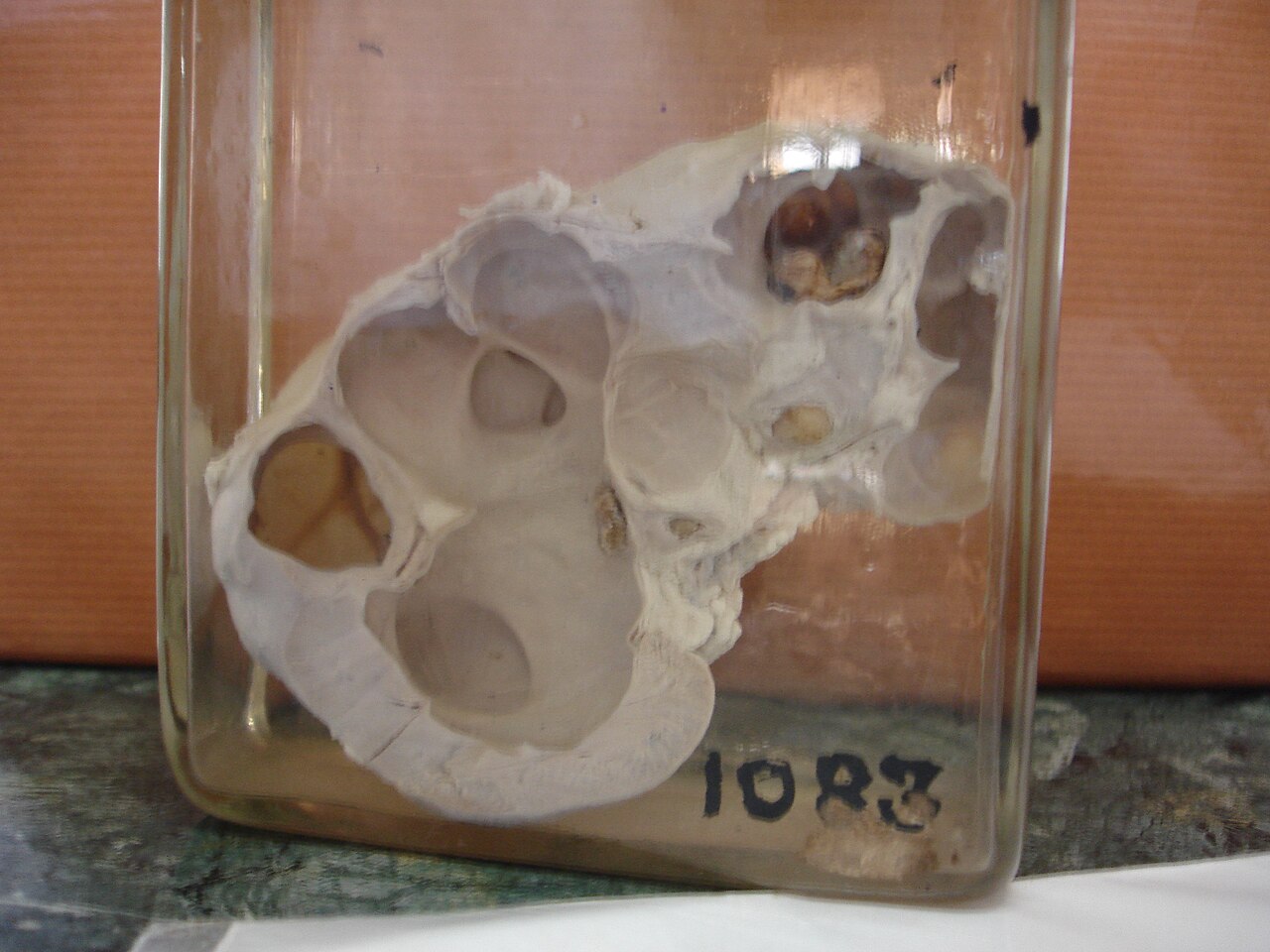
Anything that blocks the tract — a stone, a stricture, a tumor, an enlarged prostate, a pregnancy — causes urine to back up. That backpressure has its own vocabulary. Hydro/ureter is a ureter swollen with trapped urine. Hydro/nephr/osis (high-droh-neh-FROH-sis) is the kidney’s collecting system dilated by the same trapped urine, and if it persists it flattens and destroys the cortex. Pyo/nephr/osis is the same dilated system filled with pus instead, and it demands emergent drainage.
Two more structural terms show up often enough to learn now. Nephroptosis (nef-rop-TOH-sis) is a kidney that drops out of its normal position when the patient stands, occasionally kinking the ureter; it is repaired by nephropexy, surgical fixation. Polycystic kidney disease (PKD) is an inherited disorder in which both kidneys fill with fluid-filled cysts that gradually replace working tissue — enormous kidneys, hypertension, hematuria and, in roughly half of autosomal dominant cases, kidney failure by about age sixty.
Failure: acute, chronic and the numbers behind them
Acute kidney injury (AKI) is an abrupt drop in kidney function over hours to days, marked by rising creatinine and often by oliguria. It is classified by where the problem sits relative to the kidney — a distinction that is pure directional terminology. Prerenal means before the kidney: poor perfusion from hemorrhage, dehydration, heart failure or sepsis. Intrarenal (or intrinsic) means inside the kidney itself: acute tubular necrosis, glomerulonephritis, nephrotoxic drugs. Postrenal means after the kidney: obstruction anywhere from renal pelvis to meatus. Decode the prefixes and you have the differential.
Chronic kidney disease (CKD) is irreversible loss of nephrons over months to years, staged 1 through 5 by eGFR. Stage 5, an eGFR below 15, is end-stage kidney disease (ESKD, still widely charted as ESRD), the point at which survival requires dialysis or transplant. Along the way the patient accumulates the predictable consequences of nephrons that no longer regulate: anemia (less erythropoietin), bone disease (less activated vitamin D), hyperkalemia, metabolic acidosis and fluid overload.
Tumors round out the section. Renal cell carcinoma (RCC) is the common adult kidney cancer, notorious for being silent until it produces the late triad of hematuria, flank pain and a palpable mass. Wilms tumor, also called nephroblastoma (nef-roh-blas-TOH-mah), is the childhood counterpart — the suffix -blastoma flags a tumor of immature, embryonic cells, which is exactly why it occurs in toddlers rather than adults.
Wilms tumor is also a lesson in eponyms. Named for the German surgeon Max Wilms, it survives in daily speech alongside its descriptive twin nephroblastoma. Current style drops the possessive apostrophe — write Wilms tumor, not Wilms’ tumor — and the same convention now governs Down syndrome and Bright disease, the nineteenth-century eponym for what we now call glomerulonephritis. Where a descriptive alternative exists, prefer it in writing; the eponym is what you will hear spoken on rounds.
Kidney disease terms sort themselves by mechanism — inflamed filter, blocked plumbing, scarred tissue or malignant growth — and the suffix on each term (-itis, -osis, -sclerosis, -oma) tells you which mechanism you are reading about.
Section 4The Bladder, the Urethra and the Problem of Control
Infection from below
Urinary tract infection (UTI) is the umbrella; the useful terms specify a floor of the building. Cystitis (sis-TIE-tis) is bladder infection — burning, urgency, frequency, suprapubic discomfort, cloudy urine, usually no fever. Urethritis (yoo-ree-THRY-tis) is inflammation of the urethra, often sexually transmitted in young adults. Pyelonephritis, from Section 3, is the same organisms having reached the kidney, and the arrival of fever and flank pain is what separates a nuisance from an admission. Clinicians also divide UTIs into lower (bladder and urethra) and upper (ureters and kidneys), which is simply the same anatomy in plainer words.
Why women get most of them is anatomy, not hygiene: a 4 cm urethra opening close to the perineum gives gut bacteria a short commute. Why hospitalized patients get them is hardware. A catheter-associated urinary tract infection (CAUTI) is one of the most tracked, most preventable hospital-acquired infections in the country, and the single most effective prevention is removing the catheter as soon as it is no longer needed.
Not every positive culture is a disease. Asymptomatic bacteriuria means bacteria are present without symptoms, and outside of pregnancy and certain urologic procedures it is generally not treated — treating it breeds resistance without helping the patient. That single distinction, buried in one word ending in -uria, drives a large share of antibiotic stewardship policy.
| Term | Built from | What it means |
|---|---|---|
| cystitis | cyst/o + -itis | inflammation of the urinary bladder |
| urethritis | urethr/o + -itis | inflammation of the urethra |
| trigonitis | trigon/o + -itis | inflammation of the bladder trigone |
| cystocele | cyst/o + -cele | bladder herniating into the vaginal wall |
| vesicovaginal fistula | vesic/o + vagin/o | abnormal opening between bladder and vagina |
| vesicoureteral reflux | vesic/o + ureter/o | urine flowing backward from bladder to ureter |
| urethral stricture | urethr/o + strictura (narrowing) | scarred narrowing of the urethra |
| cystalgia | cyst/o + -algia | pain in the urinary bladder |
| urethrostenosis | urethr/o + -stenosis | narrowing of the urethra |
Storage problems and emptying problems
Every lower-tract complaint is ultimately one of two failures: the bladder will not hold, or the bladder will not empty. Sorting a patient’s symptoms into those two buckets is the first thing a urology clinic does, and each bucket has its own vocabulary.
Storage failure produces urgency (a sudden compelling need), frequency (voiding often, in small amounts), nocturia and incontinence — the involuntary loss of urine. Incontinence itself comes in four named patterns worth memorizing. Stress incontinence leaks with coughing, laughing or lifting, because abdominal pressure overwhelms a weakened outlet. Urge incontinence leaks because the detrusor muscle contracts without permission, the hallmark of overactive bladder. Overflow incontinence dribbles from a bladder so full it spills, which is really an emptying problem wearing a storage disguise. Functional incontinence means the urinary tract works but the person cannot reach a toilet in time — immobility, dementia, restraints, a bed rail.
Emptying failure produces hesitancy (a delay in starting), a weak or intermittent stream, straining, a sense of incomplete emptying, and finally urinary retention — urine the bladder cannot expel. Acute retention is agonizing and requires immediate catheterization. Chronic retention may be nearly painless, which makes it dangerous: a bladder that never empties develops high pressures, then reflux, then hydronephrosis, then kidney damage. The volume left behind after voiding is the post-void residual (PVR), measured by bladder ultrasound or catheter.
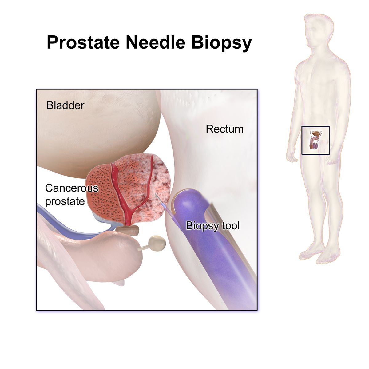
Two causes dominate emptying failure. In men over fifty it is usually benign prostatic hyperplasia (BPH), a non-cancerous enlargement of the prostate squeezing the urethra it surrounds. In patients with spinal cord injury, multiple sclerosis, stroke or long-standing diabetes it is neurogenic bladder — loss of the nerve signaling that coordinates the detrusor with the sphincter. The nerve version can fail in either direction, spastic or flaccid, which is why it appears in both buckets.
Bladder tumors, stones and the chronically irritated bladder
Bladder cancer is most often urothelial carcinoma, arising from the transitional epithelium that lines the entire tract from renal pelvis to urethra. Its cardinal presentation is painless gross hematuria in an older adult, frequently a current or former smoker — and painless is the operative word, because the absence of pain is what distinguishes it from a stone or an infection. Any adult with unexplained hematuria earns an evaluation.
Stones that formed in the kidney often finish their journey in the bladder, where cystolithiasis causes intermittent obstruction, pain and infection. Bladder diverticula are outpouchings of the bladder wall forced out by chronic high pressure, and because they empty poorly they collect stagnant urine, stones and infection.
Finally, interstitial cystitis, also called bladder pain syndrome, is chronic bladder pain with urgency and frequency in the absence of infection. It is the diagnosis reached after cultures keep returning negative, and it reminds you of something important about terminology: -itis promises inflammation, but a term that has been in use for a century may keep its suffix long after the science changes. Read the suffix as a strong hint, not a guarantee.
One last word pair to keep straight. Continence is the ability to hold urine; in/continence adds the negating prefix in-, meaning not. It is not the same as retention, which is the opposite failure — and a patient who leaks may still be retaining. Overflow incontinence is exactly that paradox: a full, non-emptying bladder that dribbles at the top. Draining it relieves both problems at once.
Sort every lower-tract term into storage failure or emptying failure, because that one question separates urgency, frequency and incontinence from hesitancy, weak stream and retention — and points to entirely different causes and treatments.
Section 5Tests, Procedures, Dialysis and the Chart
Looking, imaging and measuring
Diagnostic terms are built from the same three procedural suffixes you have used since Chapter 2. -scopy means visual examination with an instrument, -graphy means the process of recording an image, and -gram means the image itself. Attach them to the urinary roots and the studies name themselves.
Cyst/o/scopy (sis-TOS-koh-pee) is visual examination of the bladder through a scope passed up the urethra — the definitive test for hematuria and bladder tumors. Ureter/o/scopy (yoo-ree-ter-OS-koh-pee) advances a thinner scope into the ureter, usually to find and remove a stone. Nephr/o/scopy looks inside the kidney through a tract made in the flank. Cyst/o/urethr/o/gram (sis-toh-yoo-REE-throh-gram) is a picture of bladder and urethra; add “voiding” and the abbreviation VCUG and you have the pediatric study that detects vesicoureteral reflux by imaging a child in the act of urinating.
Imaging vocabulary also carries a bit of history. The intravenous pyelogram (IVP) injected contrast into a vein and photographed it as the kidneys excreted it; it defined urology for fifty years and has now been largely replaced by CT urography, which sees the same anatomy and much more. A retrograde pyelogram runs the contrast the other direction — up from the bladder through the ureters — when the kidney cannot excrete contrast itself. KUB is simply a plain abdominal film centered on kidneys, ureters and bladder, and it remains a fast way to spot a calcium stone.
The laboratory side is short and consequential: BUN, creatinine, eGFR, creatinine clearance (a timed urine collection that measures filtration directly), urinalysis, and urine culture and sensitivity (C&S), which identifies the organism and tells you which antibiotic will kill it. Urodynamic studies measure bladder pressures and flow rates to distinguish the storage failures from the emptying failures of Section 4. And a renal biopsy — bi/o (life) + -opsy (process of viewing), a sample of living tissue examined under the microscope — is what finally names the specific glomerular disease when the urinalysis can only describe a pattern.
| Word part | Meaning | Example term | What the example means |
|---|---|---|---|
| -scopy | visual examination | cystoscopy | viewing the inside of the bladder |
| -graphy | process of recording | urography | imaging of the urinary tract |
| -gram | the record or image | pyelogram | image of the renal pelvis |
| son/o | sound | renal sonography | ultrasound imaging of the kidney |
| retro- | backward, behind | retrograde pyelogram | contrast injected upward from the bladder |
| -meter, -metry | measure, measurement | cystometry | measurement of bladder pressure and capacity |
| -lysis | separation, breakdown | urinalysis | analysis of the urine |
| trans- | across, through | transurethral | performed through the urethra |
Operations: the suffix decides everything
Three suffixes account for most urinary surgery, and confusing them is the classic error of the first clinical semester. -ectomy is excision — take it out. -otomy is incision — cut into it, then close it. -ostomy is the creation of a permanent or semi-permanent opening — a new mouth to the outside or to another structure. Applied to one root, they describe three completely different operations with three completely different recoveries.
Nephr/ectomy (neh-FREK-toh-mee) removes a kidney, typically for cancer or a non-functioning organ. Nephr/otomy cuts into the kidney. Nephr/o/lith/otomy (nef-roh-lih-THOT-oh-mee) cuts into the kidney to remove a stone; add “percutaneous” and the abbreviation PCNL and you have the operation done through a small flank tract for large stones. Nephr/ostomy (neh-FROS-toh-mee) creates an opening from the kidney to the skin so a tube can drain an obstructed kidney immediately, before anything is definitively repaired.
The same three suffixes ripple down the tract. Cystectomy removes the bladder, usually for invasive cancer, and forces the surgeon to build a new drainage route — commonly an ileal conduit, in which a short segment of ileum carries urine to a stoma on the abdominal wall. That new opening is a urostomy, and its cousin the cystostomy (or suprapubic catheter) drains the bladder directly through the lower abdominal wall when the urethra cannot be used.
Repair and destruction suffixes complete the set. -plasty is surgical repair: urethroplasty (yoo-REE-throh-plas-tee) rebuilds a strictured urethra, pyeloplasty repairs an obstructed renal pelvis. -pexy is surgical fixation: nephropexy anchors a dropped kidney. -rrhaphy is suture: cystorrhaphy sews a torn bladder. And -tripsy is crushing: lithotripsy (LITH-oh-trip-see) breaks stones, most familiarly as extracorporeal shock wave lithotripsy (ESWL), where the shock waves are generated outside (extra-) the body (corpor/o) and focused on the stone.
Catheters, dialysis and transplant
Urinary catheterization (kath-eh-ter-ih-ZAY-shun) is passing a tube into the bladder to drain it. An indwelling (Foley) catheter stays in place, held by a small inflated balloon. A straight or intermittent catheter drains and comes out immediately, which is safer for long-term use in neurogenic bladder because it does not leave a permanent highway for bacteria. Continuous bladder irrigation (CBI) flushes sterile fluid through a three-way catheter to keep clots from obstructing after prostate or bladder surgery.
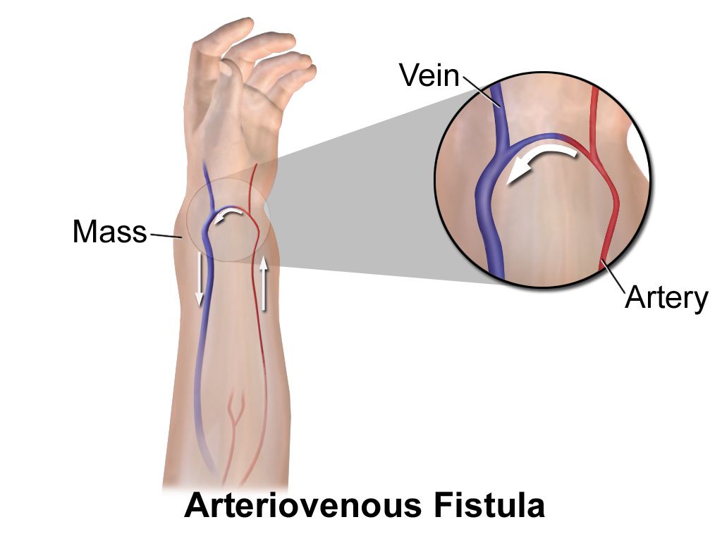
When kidney function is gone, filtration must be done artificially. Dia/lysis = through + separation: wastes pass through a semipermeable membrane out of the blood. In hemodialysis (HD) the membrane is a machine’s dialyzer, and the patient needs reliable high-flow vascular access, usually an arteriovenous fistula (AVF) surgically created in the arm. In peritoneal dialysis (PD) the membrane is the patient’s own peritoneum, and dialysate is cycled into and out of the abdomen through an implanted catheter. Both are renal replacement therapy (RRT); neither restores the kidney’s hormonal work, which is why dialysis patients still need erythropoietin and vitamin D.
Kidney transplant is the definitive treatment for end-stage kidney disease. The graft is usually placed in the iliac fossa of the pelvis rather than in the native kidney bed, and the failed kidneys are commonly left in place. The vocabulary of rejection — hyperacute, acute, chronic — and of the immunosuppression that prevents it belongs to Chapter 8, but the surgical terms belong here: nephrectomy for the donor, ureteroneocystostomy for the new connection of donor ureter to recipient bladder. Decode that last monster in pieces: ureter/o + ne/o (new) + cyst/o + -ostomy = a new opening between ureter and bladder. Long terms are not hard terms; they are just several short ones standing in a line.
| Abbreviation | Stands for | Why you will see it |
|---|---|---|
| UA | urinalysis | first-line urine test |
| C&S | culture and sensitivity | identifies organism and antibiotic |
| UTI / CAUTI | urinary tract infection / catheter-associated UTI | diagnosis and quality metric |
| BUN | blood urea nitrogen | waste-clearance lab |
| Cr, eGFR | creatinine, estimated glomerular filtration rate | stages kidney function |
| AKI / CKD / ESKD | acute kidney injury / chronic kidney disease / end-stage kidney disease | the failure spectrum |
| I&O | intake and output | fluid balance charting |
| PVR | post-void residual | detects incomplete emptying |
| KUB | kidneys, ureters, bladder (plain film) | quick stone film |
| IVP / VCUG | intravenous pyelogram / voiding cystourethrogram | contrast imaging studies |
| HD / PD / RRT | hemodialysis / peritoneal dialysis / renal replacement therapy | dialysis modality |
| ESWL / PCNL | extracorporeal shock wave lithotripsy / percutaneous nephrolithotomy | stone procedures |
| BPH / TURP | benign prostatic hyperplasia / transurethral resection of the prostate | obstruction and its surgery |
| SG | specific gravity | urine concentration |
Urinary procedures are named by suffix — look, image, cut out, cut into, open, repair, fix, crush — so identifying the suffix tells you what was done before you have even read the root.
The chapter in one breath
The urinary system runs one direction, and its vocabulary follows the flow. Blood is filtered at the glomerulus, refined along the tubule, collected in the calices and renal pelvis, carried down the ureter, stored in the bladder and released through the urethra and meatus. The kidney carries two roots, Latin ren/o for anatomy and Greek nephr/o for disease and surgery. Symptoms cluster under one suffix: -uria converts any root into a urinary finding, from dysuria and hematuria to oliguria, polyuria and nocturia. Kidney disease sorts by mechanism — inflamed filter (glomerulonephritis), climbing infection (pyelonephritis), leaking filter (nephrotic syndrome), blocked plumbing (hydronephrosis, nephrolithiasis), scarred tissue (nephrosclerosis), and tumor. Lower-tract disease sorts into failure to store and failure to empty. Procedures are named by suffix, and the difference between -ectomy, -otomy and -ostomy is the difference between an organ removed, an organ opened and closed, and an organ now draining into a bag. Know the parts and the long terms stop being long.
Words worth knowing
| Term | What it means |
|---|---|
| nephron | the microscopic filtering unit of the kidney; about a million per kidney |
| glomerulus (gloh-MER-yoo-lus) | the capillary tuft where blood is filtered; plural glomeruli |
| renal pelvis | funnel-shaped basin that collects urine and hands it to the ureter; plural pelves |
| trigone (TRY-gohn) | triangular bladder floor bounded by the two ureteral openings and the urethra |
| micturition (mik-choo-RISH-un) | the act of urinating; voiding |
| dysuria (dis-YOO-ree-ah) | painful or difficult urination |
| oliguria (ol-ih-GYOO-ree-ah) | markedly decreased urine output |
| nocturia (nok-TYOO-ree-ah) | waking from sleep to urinate |
| enuresis (en-yoo-REE-sis) | involuntary urination, classically bedwetting |
| diuresis (dye-yoo-REE-sis) | increased production and output of urine |
| azotemia (az-oh-TEE-mee-ah) | elevated nitrogen wastes in the blood, a laboratory finding |
| uremia (yoo-REE-mee-ah) | symptomatic illness caused by those retained wastes |
| glomerulonephritis (gloh-mer-yoo-loh-neh-FRY-tis) | inflammation of the kidney centered on the glomeruli |
| pyelonephritis (pye-eh-loh-neh-FRY-tis) | bacterial infection of the kidney and renal pelvis |
| nephrotic syndrome (neh-FROT-ik) | heavy proteinuria, low albumin, edema and hyperlipidemia |
| hydronephrosis (high-droh-neh-FROH-sis) | dilation of the renal collecting system from obstruction |
| nephrolithiasis (nef-roh-lih-THIGH-ah-sis) | stones in the kidney; a stone is a calculus, plural calculi |
| vesicoureteral reflux (ves-ih-koh-yoo-REE-ter-al) | backward flow of urine from bladder into ureter |
| cystoscopy (sis-TOS-koh-pee) | visual examination of the bladder through the urethra |
| lithotripsy (LITH-oh-trip-see) | crushing of a stone, often by focused external shock waves |