Medical-Surgical Nursing II
Endocrine Disorders: Diabetes Mellitus
Diabetes is the disease you will meet on every unit you ever work, attached to every other diagnosis you have already studied — the heart failure patient, the post-op abdomen, the pneumonia, the pressure injury that will not close. It is also the disease in which nursing judgement is measured in minutes: a capillary glucose of 48 mg/dL, a patient breathing deeply and smelling faintly of acetone, an insulin drip running while the potassium is 3.1 mEq/L. This chapter builds the reasoning from the inside out. First the mechanism — why absent insulin produces ketones and why resistant insulin produces dehydration. Then the assessment and the numbers that define the disease. Then the drugs, organised by what they actually do, because that is what tells you when they cause hypoglycaemia and when they do not. Then the two hyperglycaemic emergencies side by side, with the treatment sequence you will be expected to defend: fluids first, potassium before insulin, insulin last. Then the slow complications and the teaching that prevents them.
By the end of this chapter you can
- Explain how absolute versus relative insulin deficiency produces the differing presentations of type 1 and type 2 diabetes mellitus, including the role of insulin resistance, lipolysis and ketogenesis.
- Apply the four diagnostic criteria for diabetes (A1C, fasting plasma glucose, 2-hour OGTT, random glucose with symptoms) and identify prediabetes from the same panel.
- Perform a focused assessment for hyperglycaemia, hypoglycaemia and the complication surveillance schedule, including monofilament testing and urine albumin-to-creatinine ratio.
- Compare insulins by onset, peak and duration and predict the hypoglycaemia window for each; demonstrate safe mixing, storage and high-alert administration practice.
- Classify non-insulin agents by mechanism — biguanide, sulfonylurea, DPP-4 inhibitor, GLP-1 receptor agonist, SGLT2 inhibitor, thiazolidinedione — and state the monitoring that matters for each.
- Differentiate diabetic ketoacidosis from hyperosmolar hyperglycaemic state by laboratory profile and sequence the treatment: fluids, then potassium, then insulin.
- Recognise and treat hypoglycaemia using the rule of 15, IV dextrose or glucagon, and identify hypoglycaemia unawareness.
- Teach sick-day rules, foot care, injection-site rotation and when to call, and construct an SBAR escalation for a deteriorating patient.
- Modify assessment and glycaemic targets for the older adult, and delegate appropriately within the RN scope.
Section 1Two Ways the Pancreas Fails
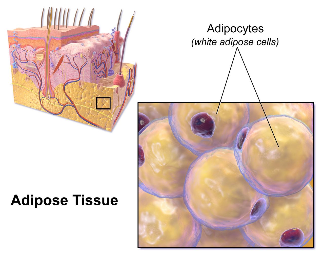
Insulin is the storage signal, and everything follows from that
Beta cells in the islets of Langerhans release insulin when blood glucose rises after a meal. Insulin does three things worth memorising because every sign of diabetes traces back to one of them. It opens GLUT4 transporters in muscle and adipose tissue so glucose leaves the blood and enters cells. It tells the liver to stop making new glucose — to suppress glycogenolysis and gluconeogenesis. And it suppresses lipolysis, keeping fatty acids locked inside adipocytes. Alpha cells in the same islet release glucagon, which does the opposite, and the fed-fasted balance between the two normally keeps plasma glucose in a remarkably narrow band of roughly 70 to 140 mg/dL across a whole day of eating and sleeping.
Remove insulin and all three brakes release at once. Glucose cannot enter muscle, so it accumulates in plasma. The liver, seeing no insulin signal, interprets the situation as starvation and pours out still more glucose. Adipose tissue releases free fatty acids, which the liver converts to ketone bodies — acetoacetate, beta-hydroxybutyrate and acetone — as an alternative fuel. That is the whole of diabetic ketoacidosis in one sentence: the cells are starving in a sea of sugar, and the liver keeps setting the table.
Once plasma glucose exceeds the renal threshold of roughly 180 mg/dL, the proximal tubule cannot reabsorb it all and glucose spills into urine. Glucose is osmotically active, so it drags water and electrolytes with it. This osmotic diuresis is why the classic presentation is polyuria and polydipsia, why the patient is volume depleted before anyone thinks to check a set of orthostatics, and why sodium, potassium, magnesium and phosphate all leave the body through the bladder while the serum values may still look deceptively normal.
Type 1: the beta cells are gone
Type 1 diabetes is autoimmune destruction of beta cells — a T-cell mediated insulitis that unfolds over months to years in a genetically susceptible person, often after a viral trigger. By the time hyperglycaemia appears, roughly 80 to 90 percent of beta-cell mass is already lost. Autoantibodies to glutamic acid decarboxylase 65 (GAD65), islet antigen 2, insulin and zinc transporter 8 are typically detectable, and C-peptide — the fragment cleaved from proinsulin when the body makes its own insulin — is low or undetectable. That C-peptide value is how a laboratory distinguishes a patient making no insulin from one making plenty but using it badly.
Presentation is fast and dramatic: days to a few weeks of polyuria, polydipsia, polyphagia and weight loss, often in a child, adolescent or young adult, and roughly a third of new cases arrive already in ketoacidosis. Because there is no endogenous insulin at all, these patients are ketosis prone and require exogenous insulin from diagnosis, permanently. There is no such thing as skipping the basal dose. A brief “honeymoon phase” of partial remission after starting insulin can fool families into thinking the disease resolved — it has not, and teaching should say so plainly.
Do not assume type 1 means young. Latent autoimmune diabetes in adults (LADA) is a slower autoimmune process presenting in the thirties, forties or later, frequently misclassified as type 2 until oral agents fail unusually quickly. A lean adult whose glucose control deteriorates within a year or two of diagnosis, or who arrives with ketones, deserves antibody and C-peptide testing.
Type 2: the insulin is there and nobody is listening
Type 2 diabetes begins with insulin resistance in muscle, liver and fat, usually driven by visceral adiposity, inactivity and genetics. The pancreas compensates by making more insulin, and for years it succeeds — the patient is hyperinsulinaemic and euglycaemic and has no idea. Over time the beta cells cannot sustain the output, secretion declines, and glucose rises first after meals, then fasting. Because there is still some circulating insulin, lipolysis stays largely suppressed and ketones usually do not accumulate. That single fact is why type 2 patients decompensate into a hyperosmolar, non-ketotic state rather than into ketoacidosis.
Onset is insidious, and the diagnosis is frequently made by a complication rather than a symptom. A patient presents with a non-healing foot ulcer, recurrent vaginal candidiasis, blurred vision, an unexpected myocardial infarction or a routine A1C drawn before surgery. Look for acanthosis nigricans — velvety hyperpigmentation in the axillae and posterior neck — and for the rest of the metabolic syndrome: central obesity, hypertension, elevated triglycerides, low HDL.
Two other categories matter on a medical-surgical floor. Secondary diabetes follows pancreatitis, pancreatectomy, Cushing syndrome, acromegaly, or drugs — glucocorticoids above all, plus thiazides, atypical antipsychotics, tacrolimus and some antiretrovirals. Steroid-induced hyperglycaemia is a daily reality: prednisone given each morning produces a predictable afternoon and evening glucose surge with a relatively normal fasting value, so the insulin adjustment belongs in the middle of the day, not at bedtime. Gestational diabetes is hyperglycaemia first recognised in pregnancy; it usually resolves at delivery but confers a lifetime type 2 risk that warrants screening every one to three years afterwards.
Making the diagnosis
Four tests establish diabetes, and any one of them will do — but in an asymptomatic patient the abnormal result must be confirmed, either by repeating the same test on a new sample or by a second different test from the same sample. A patient with unequivocal hyperglycaemic symptoms and a random glucose of 200 mg/dL or more needs no confirmation. Note that the A1C reflects average glycaemia over roughly the preceding three months, the lifespan of a red cell, and is therefore unreliable in haemoglobinopathies, recent transfusion, haemolysis, iron-deficiency anaemia, pregnancy and advanced chronic kidney disease.
| Test | Normal | Prediabetes | Diabetes |
|---|---|---|---|
| A1C | Below 5.7% | 5.7–6.4% | 6.5% or higher |
| Fasting plasma glucose (no caloric intake 8 h) | Below 100 mg/dL | 100–125 mg/dL (impaired fasting glucose) | 126 mg/dL or higher |
| 2-hour plasma glucose during 75-g OGTT | Below 140 mg/dL | 140–199 mg/dL (impaired glucose tolerance) | 200 mg/dL or higher |
| Random plasma glucose | — | — | 200 mg/dL or higher with classic symptoms or hyperglycaemic crisis |
Screen asymptomatic adults beginning at age 35, and earlier at any age with a BMI of 25 kg/m² or more (23 in Asian Americans) plus one risk factor — first-degree relative with diabetes, high-risk race or ethnicity, history of cardiovascular disease, hypertension, HDL below 35 mg/dL or triglycerides above 250 mg/dL, polycystic ovary syndrome, physical inactivity, acanthosis nigricans, or prior gestational diabetes. Repeat at least every three years if normal, annually in prediabetes. Prediabetes is not a benign label: intensive lifestyle intervention aiming at seven percent weight loss and 150 minutes of weekly activity reduces progression more effectively than metformin does, and it is the single highest-yield teaching conversation in primary care.
Absent insulin means the liver makes ketones and the patient becomes acidotic; deficient-but-present insulin means the patient becomes profoundly dehydrated and hyperosmolar instead — the same hyperglycaemia takes two different exits.
Section 2Assessment, Diagnostics and Surveillance
The history and physical that actually changes management
Start with the glycaemic history, not the diagnosis list. What is the regimen, in units and in times of day — and what does the patient actually take at home, which is frequently a different answer? When was the last dose? Has anyone been checking glucose, how often, and what have the numbers looked like? Any hypoglycaemic episodes, and did the patient feel them coming? Any hospitalisations for high or low sugars? Who fills the prescriptions, and can they afford them? Insulin rationing is a real and lethal cause of ketoacidosis, and patients rarely volunteer it unless asked without judgement.
Symptoms of hyperglycaemia are the three P’s plus fatigue: polyuria, polydipsia, polyphagia, blurred vision from osmotic lens swelling, weight loss, slow wound healing, recurrent yeast or urinary infections. Symptoms of hypoglycaemia divide into two groups you should keep separate in your head because they appear at different thresholds. Autonomic or adrenergic symptoms come first, usually below 70 mg/dL — tremor, palpitations, anxiety, diaphoresis, hunger, pallor. Neuroglycopenic symptoms come as the brain runs out of substrate, usually below 54 mg/dL — confusion, slurred speech, blurred or double vision, incoordination, bizarre behaviour, seizure, coma.
The physical examination is largely a search for complications. Take a supine and standing blood pressure. Inspect the mouth for candidiasis and periodontal disease. Auscultate for carotid bruits. Palpate dorsalis pedis and posterior tibial pulses and note capillary refill and hair distribution on the lower legs. Take both shoes and both socks off — every time, on every diabetic patient, no exceptions — and inspect the plantar surfaces, the heels and between every toe. Examine injection sites for lipohypertrophy, the rubbery subcutaneous thickening that develops where a patient injects repeatedly into the same spot, and which makes insulin absorption erratic and unpredictable.
Glycaemic monitoring: A1C, fingersticks and continuous sensors
The A1C measures the fraction of haemoglobin that has been non-enzymatically glycated, and it correlates to an estimated average glucose: an A1C of 6% corresponds to roughly 126 mg/dL, 7% to about 154, 8% to about 183, 9% to about 212, and 10% to about 240. Check it every three months when therapy has changed or the target is not met, and every six months when the patient is stable at goal. The general adult target is below 7%, but a target is a clinical judgement, not a reflex — below 6.5% for a young patient with long life expectancy and no hypoglycaemia risk, and a looser ceiling of below 8% for a frail elder with limited life expectancy, cognitive impairment, advanced complications or hypoglycaemia unawareness, where the harm of a low sugar plainly outweighs the benefit of a tight one. Read that looser figure as an upper limit to stay under, never as a number to climb to; and in very poor health or at end of life, stop managing to an A1C at all and manage instead to glucose values, avoiding both hypoglycaemia and symptomatic hyperglycaemia.
Capillary glucose targets for most non-pregnant adults are 80 to 130 mg/dL before meals and below 180 mg/dL at the peak, one to two hours after the start of a meal. Continuous glucose monitoring adds a metric worth knowing by name: time in range, the percentage of readings between 70 and 180 mg/dL, with a goal above 70 percent, less than 4 percent of time below 70 mg/dL and less than 1 percent below 54 mg/dL. Sensors report interstitial glucose, which lags plasma by five to fifteen minutes, so when a sensor value and the patient’s symptoms disagree — or when the glucose is falling rapidly — confirm with a fingerstick before you act.
Two patterns of morning hyperglycaemia are commonly confused and are distinguished by a single 0200–0300 glucose check. The dawn phenomenon is a physiological pre-waking surge of growth hormone and cortisol; the 3 a.m. glucose is normal or high and rises further by morning, and the fix is more or later-timed basal insulin. The Somogyi effect is rebound hyperglycaemia after unrecognised nocturnal hypoglycaemia; the 3 a.m. glucose is low, and the fix is less evening insulin or a bedtime snack. Adding insulin to a Somogyi pattern makes the patient worse, which is exactly why the 3 a.m. check is worth the interrupted sleep.
Complication surveillance is a schedule, not an impulse
The chronic complications of diabetes are silent until they are not, so the surveillance calendar is itself a nursing intervention. Ask about it at every encounter and document the gaps — discharge teaching that includes “you are due for your eye exam” is worth more than another lecture on carbohydrates.
| Surveillance | Frequency | Target or action threshold |
|---|---|---|
| A1C | Every 6 months at goal; every 3 months if not | Individualised; below 7% for most adults |
| Dilated retinal examination | Type 2 at diagnosis; type 1 within 5 years of onset; then annually | Refer any retinopathy to ophthalmology |
| Urine albumin-to-creatinine ratio and eGFR | Annually (type 2 from diagnosis; type 1 after 5 years) | Albuminuria at 30 mg/g or higher with hypertension → ACE inhibitor or ARB (monitor potassium and creatinine) |
| Comprehensive foot examination with 10-g monofilament | Annually; visual inspection every visit | Loss of protective sensation → podiatry, protective footwear |
| Blood pressure | Every visit | Below 130/80 mmHg for most |
| Lipid panel | Annually | Moderate-intensity statin ages 40–75; high-intensity with added risk |
| Immunisations | Influenza annually; pneumococcal, hepatitis B, COVID-19 per schedule | Diabetes is a standing indication for all of these |
The 10-gram Semmes-Weinstein monofilament test deserves a specific description because students are often asked to perform it. With the patient’s eyes closed, touch the filament perpendicular to the plantar skin at standard sites until it buckles into a C shape, hold about one second, and ask the patient to say when and where it is felt. Avoid callus, ulcer and scar. Inability to feel the buckling filament at any tested site means loss of protective sensation — that patient can stand on a tack, or wear a shoe with a pebble in it all day, and never know. Everything about their footwear teaching changes from that moment.
Assessment in diabetes is two jobs at once: find today’s glucose problem, and audit the surveillance schedule that prevents the complication you will otherwise meet in five years.
Section 3Insulin and the Non-Insulin Agents
Insulin by onset, peak and duration
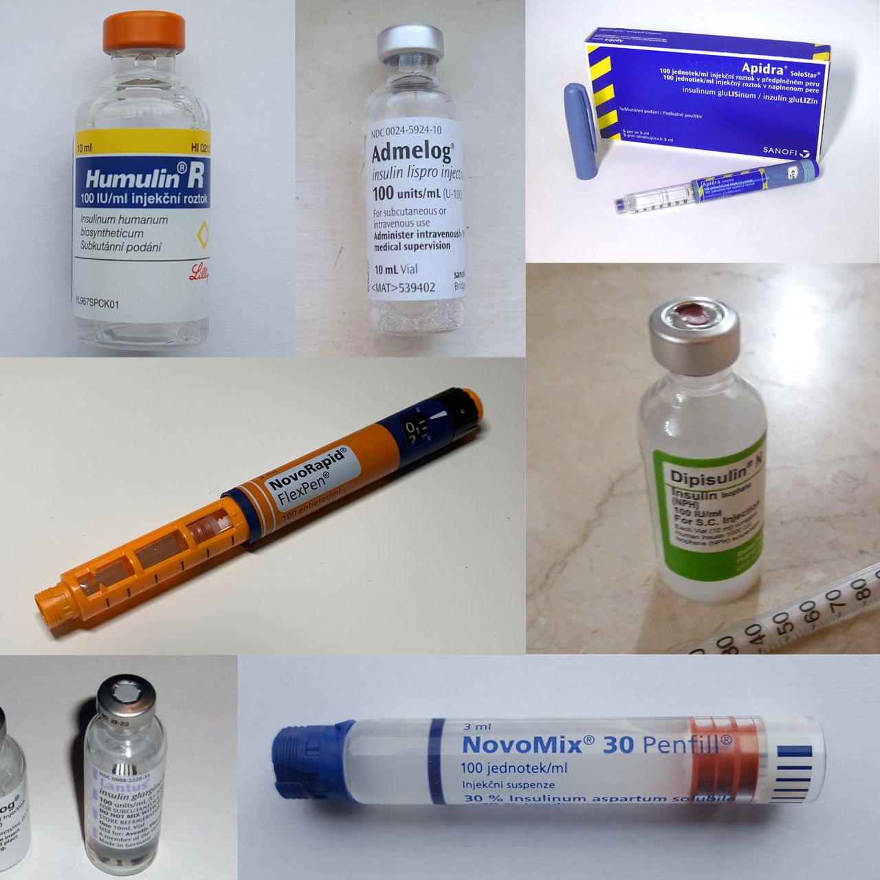
Every insulin question you will ever be asked reduces to a timeline. If you know when the drug peaks, you know when the patient is most likely to go low, when the tray must be in front of them, and when to schedule the fingerstick. Memorise the table below as a shape, not as trivia.
| Class and examples | Onset | Peak | Duration | Nursing timing |
|---|---|---|---|---|
| Rapid-acting: lispro, aspart, glulisine | 10–30 min | 30 min–3 h | 3–5 h | Give 0–15 min before eating; tray must be present. Never give if the meal may not arrive. |
| Ultra-rapid: faster aspart, inhaled human insulin | Within 15 min (inhaled about 12 min) | About 30–60 min | About 2.5–3 h | Inhaled form is contraindicated in asthma and COPD (bronchospasm). |
| Short-acting: regular (U-100) | 30 min–1 h | 2–4 h | 5–8 h | Give 30 min before the meal. The only insulin given IV. |
| Intermediate: NPH (isophane) | 1–2 h | 4–12 h | 12–18 h | Cloudy; roll gently to resuspend. The 4–12 h peak is the classic nocturnal hypoglycaemia window. |
| Long-acting: glargine U-100, detemir | 2–4 h (detemir 1–3 h) | No pronounced peak | Up to 24 h (detemir often twice daily) | Same time daily. Do not mix with any other insulin in a syringe. |
| Ultra-long: degludec, glargine U-300 | About 1–6 h | No peak | Beyond 24 h (degludec beyond 42 h) | Flexible dosing time; steady state takes 3–4 days, so do not chase numbers daily. |
Mixing rules are narrow and absolute. Only NPH and regular are mixed in the same syringe: inject air into the NPH (cloudy) vial, inject air into the regular (clear) vial, then draw up the regular first — clear before cloudy, so that no cloudy NPH contaminates the clear vial. Administer within five minutes of mixing. Glargine, detemir and degludec are never mixed with anything and never diluted. Premixed products such as 70/30 NPH-regular or 75/25 lispro protamine-lispro carry the peak profile of their intermediate component and demand a fixed, reliable meal schedule.
Insulin is a high-alert drug and should be handled like one
Insulin appears near the top of every list of medications most frequently involved in fatal error. Use an insulin syringe only — never a tuberculin or standard syringe — and match the syringe to the concentration. Never write or accept the abbreviation “U” for units; “10U” has been read as 100. Perform an independent double check of the drug, concentration, dose and pump settings with a second nurse per facility policy, particularly for any insulin infusion, any paediatric dose and any concentrated product (U-200 lispro, U-300 glargine, U-500 regular).
Insulin pens are single-patient devices, period. Even with the needle changed, biological material can be aspirated back into the cartridge, and pens have transmitted bloodborne infection between patients. Label the pen with the patient name and keep it in that patient’s drawer. Prime with two units before each injection to clear air, inject at a 90-degree angle, and count to ten before withdrawing the needle so the full dose is delivered rather than tracking back out.
Absorption varies by site — abdomen is fastest, then upper arm, then thigh, then buttock — so teach the patient to use one anatomic region consistently for a given injection and to rotate systematically within it, about an inch apart, avoiding a two-inch radius around the umbilicus. Exercise of a limb, heat, massage and hot showers all accelerate absorption from that site. Unopened vials and pens are refrigerated until the printed expiry; once in use, most products are kept at room temperature and discarded after 28 days (some, including detemir and degludec, allow longer — check the package). Never freeze insulin, never use it if it is discoloured, clumped or frosted-looking, and do not inject cold insulin, which stings.
Non-insulin agents, grouped by what they do
Whether a drug causes hypoglycaemia on its own is entirely a function of mechanism. Agents that force insulin release regardless of glucose — sulfonylureas and meglitinides — do. Agents that improve sensitivity, slow absorption, promote glucose loss in urine or act only in the presence of a rising glucose generally do not, unless combined with insulin or a secretagogue.
| Class (examples) | Mechanism | Key adverse effects | Nursing implications |
|---|---|---|---|
| Biguanide: metformin | Reduces hepatic glucose output; improves insulin sensitivity | GI upset, metallic taste, vitamin B12 deficiency, rare lactic acidosis | First-line in type 2. Take with food; extended release at night. Contraindicated if eGFR below 30 mL/min/1.73 m². Withhold at the time of iodinated contrast when eGFR is 30–60 or with acute kidney injury and restart after 48 h with stable renal function. No hypoglycaemia alone. |
| Sulfonylureas: glipizide, glimepiride, glyburide | Close beta-cell potassium channels, forcing insulin release | Hypoglycaemia, weight gain, disulfiram-like reaction with alcohol | Give about 30 min before a meal; never on an empty, skipped-meal day. Long duration means prolonged hypoglycaemia requiring admission. Avoid glyburide in older adults (Beers criteria). |
| Meglitinides: repaglinide, nateglinide | Same channel, rapid on and off | Hypoglycaemia (shorter) | Take with each meal; skip the dose if the meal is skipped. |
| Thiazolidinedione: pioglitazone | PPAR-gamma agonist; increases peripheral insulin sensitivity | Fluid retention, weight gain, heart failure exacerbation, fractures, macular oedema | Contraindicated in NYHA class III–IV heart failure. Daily weights; report a gain above 3 lb in a day or oedema and dyspnoea. Full effect takes 4–12 weeks. |
| DPP-4 inhibitors: sitagliptin, linagliptin, saxagliptin | Block degradation of endogenous incretins | Arthralgia, pancreatitis, urticaria; heart failure hospitalisation (saxagliptin, alogliptin) | Weight neutral, oral, modest A1C effect. Renal dose adjustment for most except linagliptin. |
| GLP-1 receptor agonists: semaglutide, liraglutide, dulaglutide, exenatide (tirzepatide is dual GIP/GLP-1) | Glucose-dependent insulin release, suppressed glucagon, delayed gastric emptying, satiety | Nausea, vomiting, pancreatitis, gallstones; thyroid C-cell tumours in rodents | Weight loss and cardiovascular benefit. Contraindicated with personal or family history of medullary thyroid carcinoma or MEN 2. Titrate slowly; small low-fat meals. Delayed emptying raises aspiration risk — anaesthesia must know before any sedation or surgery. |
| SGLT2 inhibitors: empagliflozin, dapagliflozin, canagliflozin | Block renal glucose reabsorption; glucose exits in urine | Genital mycotic and urinary infection, volume depletion and orthostasis, euglycaemic DKA, rare Fournier gangrene | Cardiovascular and kidney protection independent of glucose. Teach perineal hygiene and fluid intake. Hold 3–4 days before surgery or during acute illness. Check ketones for DKA symptoms even when glucose is near normal. |
| Alpha-glucosidase inhibitors: acarbose, miglitol | Delay intestinal carbohydrate breakdown | Flatulence, bloating, diarrhoea | Take with the first bite of each meal. If hypoglycaemia occurs on combined therapy, treat with glucose or dextrose, not sucrose — table sugar cannot be split. |
| Amylin analogue: pramlintide | Slows gastric emptying, suppresses glucagon, increases satiety | Severe hypoglycaemia with insulin, nausea | Separate syringe from insulin, separate site. Mealtime insulin is reduced by 50% when started. |
Glycaemic management in the hospitalised patient
Inpatient targets are deliberately looser than outpatient ones because the hospitalised patient eats unpredictably, receives steroids, goes NPO for procedures and has less physiological reserve for a low. Aim for 140 to 180 mg/dL for most critically ill and non-critically ill inpatients; a tighter 110 to 140 band is reasonable only for selected patients when it can be achieved without hypoglycaemia. Below 100 mg/dL, reassess the regimen; below 70, treat and change something.
A basal-bolus-correction regimen — long-acting insulin for background needs, rapid-acting insulin with meals, and a correction scale for the gap — is superior to sliding-scale insulin used alone. Sliding scale alone is reactive: it treats a high number after it has already happened, produces rollercoaster control, and is specifically inappropriate as sole therapy in type 1 diabetes. Time your fingersticks to the regimen: before meals and at bedtime for the eating patient, every four to six hours for the NPO patient, and hourly for anyone on an insulin infusion.
Watch the situations that predictably destabilise glucose: enteral feeding started or interrupted (an abrupt tube-feed hold with prandial coverage already given is a classic hypoglycaemic event), parenteral nutrition, glucocorticoids, octreotide, vasopressors, dialysis and any acute illness. And know that hypoglycaemia in a hospitalised patient is a reportable safety event in most facilities, not a private nursing footnote.
Know when each insulin peaks and you know when your patient will crash; know each oral agent’s mechanism and you know whether it can cause a low at all.
Section 4The Acute Crises: Hypoglycaemia, DKA and HHS
Hypoglycaemia is the emergency that kills first
Between a glucose of 40 and a glucose of 400, treat the 40. Hyperglycaemia damages the patient over hours to years; hypoglycaemia damages the brain in minutes. Level 1 hypoglycaemia is a glucose below 70 mg/dL, level 2 is below 54 mg/dL (clinically significant neuroglycopenia), and level 3 is severe hypoglycaemia of any value causing cognitive impairment requiring another person’s assistance.
For the conscious patient who can swallow safely, use the rule of 15: give 15 grams of fast-acting carbohydrate, wait 15 minutes, recheck. Fifteen grams is four glucose tablets, 4 ounces (half a cup) of juice or regular soda, one tablespoon of honey or sugar, or 8 ounces of milk. Repeat the cycle until the glucose is above 70 mg/dL, then give a snack containing protein and complex carbohydrate if the next meal is more than an hour away. Avoid chocolate, ice cream and peanut butter as the initial treatment — fat slows gastric emptying and delays the rise. Never put food or fluid in the mouth of a patient who is not fully awake and able to protect the airway.
For the unresponsive patient, the patient having a seizure, or anyone NPO or unable to swallow: if IV access exists, give 25 to 50 mL of 50% dextrose (12.5 to 25 g) by slow IV push per protocol, then start or resume a dextrose-containing infusion, because a D50 push wears off quickly. If there is no IV access, give glucagon 1 mg intramuscularly or subcutaneously, or 3 mg intranasally, and turn the patient onto their side — glucagon frequently causes vomiting. Glucagon mobilises hepatic glycogen, so it works poorly in the malnourished, the alcohol-intoxicated and anyone with depleted stores; those patients need dextrose. Recheck in 15 minutes, feed as soon as the patient is alert and safe, then investigate the cause. A sulfonylurea-induced or long-acting-insulin-induced low will relapse: that patient stays, gets a dextrose infusion and serial checks, and does not go home from the emergency department.
Hypoglycaemia unawareness is the loss of adrenergic warning symptoms, seen with autonomic neuropathy, long-duration diabetes, repeated lows and beta-blocker therapy. Beta blockers blunt tremor and tachycardia but not diaphoresis — so in a beta-blocked patient, sudden unexplained sweating is your warning sign. Treatment for unawareness is deliberately relaxing targets and scrupulously avoiding lows for several weeks, which restores symptom perception.
DKA and HHS side by side
Both are hyperglycaemic emergencies, both are usually precipitated by infection, non-adherence or a new stressor such as myocardial infarction, stroke, pancreatitis, trauma, surgery or a new drug. The difference is whether enough insulin remains to suppress lipolysis.
| Feature | Diabetic ketoacidosis | Hyperosmolar hyperglycaemic state |
|---|---|---|
| Typical patient | Type 1 (also type 2 under severe stress); often younger | Type 2, often older, often with limited access to water |
| Onset | Hours to 1–2 days | Days to weeks |
| Plasma glucose | Usually above 250 mg/dL (may be near normal in euglycaemic DKA) | Usually above 600 mg/dL, often above 1000 |
| Arterial pH | Below 7.30 (severe below 7.00) | Above 7.30 |
| Serum bicarbonate | Below 18 mEq/L (severe below 10) | Above 18 mEq/L |
| Ketones (serum beta-hydroxybutyrate) | Positive; 3.0 mmol/L or higher | Absent or small |
| Anion gap | Elevated, above 10–12 mEq/L | Normal or mildly elevated |
| Effective serum osmolality | Variable, often below 320 mOsm/kg | Above 320 mOsm/kg |
| Fluid deficit | About 6 L (roughly 100 mL/kg) | About 9–12 L (roughly 100–200 mL/kg) |
| Distinctive findings | Kussmaul respirations, fruity acetone breath, abdominal pain, nausea and vomiting | Profound dehydration, altered mental status, focal neurologic deficits, seizures; no Kussmaul, no acetone breath |
| Mortality | Roughly 1–5% | Roughly 5–20% |
Two calculations belong in your pocket. Anion gap equals sodium minus the sum of chloride and bicarbonate; it is the metric of ketoacid accumulation and its closure — not the glucose — tells you the DKA is resolving. Effective serum osmolality equals two times the sodium plus glucose divided by 18; above 320 mOsm/kg it correlates with the obtundation of HHS, and a patient who is not fully alert with an osmolality below 320 needs a hunt for another cause of altered mentation. Remember also that hyperglycaemia dilutes serum sodium: add roughly 1.6 mEq/L to the measured sodium for every 100 mg/dL of glucose above 100 to get the corrected value that guides fluid choice.
The treatment sequence — fluids, potassium, insulin, and dextrose
Fluids first. Start isotonic 0.9% sodium chloride at roughly 15 to 20 mL/kg in the first hour (commonly 1 to 1.5 L in an adult), guided by haemodynamics and cardiac and renal status. After the initial resuscitation, the fluid is chosen by the corrected sodium: if it is normal or high, switch to 0.45% sodium chloride at 250 to 500 mL/h; if it is low, continue 0.9%. Volume alone lowers glucose substantially by dilution and restored renal perfusion, and it is the intervention that reverses the shock physiology. Monitor intake and output hourly, and in the elderly or anyone with heart failure or chronic kidney disease, watch for the crackles and jugular distension of overshoot.
Potassium before insulin. This is the sentence to carry out of this chapter. Total body potassium is severely depleted in both crises — on the order of 3 to 5 mEq/kg — even though the serum value on arrival is often normal or high, because acidosis and insulin deficiency have shifted potassium out of cells. Insulin drives potassium straight back in, and if you start the drip on a hypokalaemic patient you can precipitate lethal arrhythmia and respiratory muscle weakness. The rule: if serum potassium is below 3.3 mEq/L, hold the insulin and replace potassium (about 20 to 30 mEq/h) until it exceeds 3.3. If potassium is 3.3 to 5.2, run insulin and add 20 to 30 mEq of potassium to each litre of fluid to keep it in the 4 to 5 range. If it is above 5.2, withhold potassium, start insulin, and recheck in two hours. Confirm urine output before giving potassium, and keep the patient on continuous cardiac monitoring throughout.
Then insulin. Regular insulin is the only insulin given intravenously: a continuous infusion at 0.1 unit/kg/h, with or without an initial 0.1 unit/kg bolus (0.14 unit/kg/h if no bolus is used). Prime the tubing with the insulin solution before connecting it, because insulin adsorbs to plastic and the first millilitres are effectively wasted. Aim for a glucose fall of 50 to 75 mg/dL per hour; a faster drop risks cerebral oedema, particularly in children and adolescents, and a plateau usually means the fluid or insulin rate is inadequate. In HHS, insulin doses are lower and fluid resuscitation is proportionally more important.
Then dextrose. When the glucose reaches about 200 mg/dL in DKA, or about 250 to 300 mg/dL in HHS, change the fluid to one containing 5% dextrose and reduce the insulin infusion to roughly 0.02 to 0.05 unit/kg/h. This looks paradoxical to students — giving sugar to a hyperglycaemic patient — but the insulin must keep running to close the anion gap and stop ketogenesis, and dextrose is what makes that safe. DKA is resolved when the glucose is below 200 mg/dL plus two of: bicarbonate 15 mEq/L or higher, venous pH above 7.30, anion gap 12 mEq/L or less. Bicarbonate therapy is reserved for a pH below 6.9. Phosphate is replaced only for a level below 1.0 mg/dL with cardiac dysfunction, respiratory depression or haemolytic anaemia.
What you monitor and what makes you call
During active treatment expect hourly capillary glucose, basic metabolic panel every two to four hours, continuous cardiac monitoring, strict hourly intake and output, and neurologic checks. Falling mental status during treatment — especially in a young patient whose numbers are improving — is cerebral oedema until proven otherwise: headache, vomiting, bradycardia with rising blood pressure, then deterioration. Stop and escalate immediately.
Do not forget euglycaemic ketoacidosis. Patients taking an SGLT2 inhibitor, pregnant patients, and those who have been vomiting or eating nothing can be fully ketoacidotic with a glucose under 250 mg/dL. If the patient is nauseated, tachypnoeic and acidotic, check ketones and a gas regardless of how reassuring the glucose looks. And in every case, find the precipitant: culture, chest film, ECG, medication review. Treating the DKA without treating the pyelonephritis that caused it simply buys you a second admission.
Here is the escalation you should be able to give without notes. S: “This is Priya, RN, on 5-West with Mr. Alvarez in 512, a 24-year-old with type 1 diabetes admitted with vomiting.” B: “He was admitted this morning; he ran out of insulin four days ago.” A: “In the last hour his respirations have gone from 18 to 32 and deep, he is drowsy, his glucose is 486, his breath smells fruity and his blood pressure has dropped from 118/70 to 92/54 with a heart rate of 128.” R: “I think he is in DKA. I need you to see him now. I would like a stat basic metabolic panel, serum beta-hydroxybutyrate, venous blood gas, magnesium and phosphorus, a large-bore IV with isotonic fluid, and orders for an insulin infusion and cardiac monitoring.”
Fluids first, potassium before insulin, insulin next, dextrose when the glucose reaches 200 — and the anion gap, not the glucose, tells you when the ketoacidosis is actually over.
Section 5Chronic Complications, Teaching and Discharge
Microvascular disease: eyes, kidneys, nerves
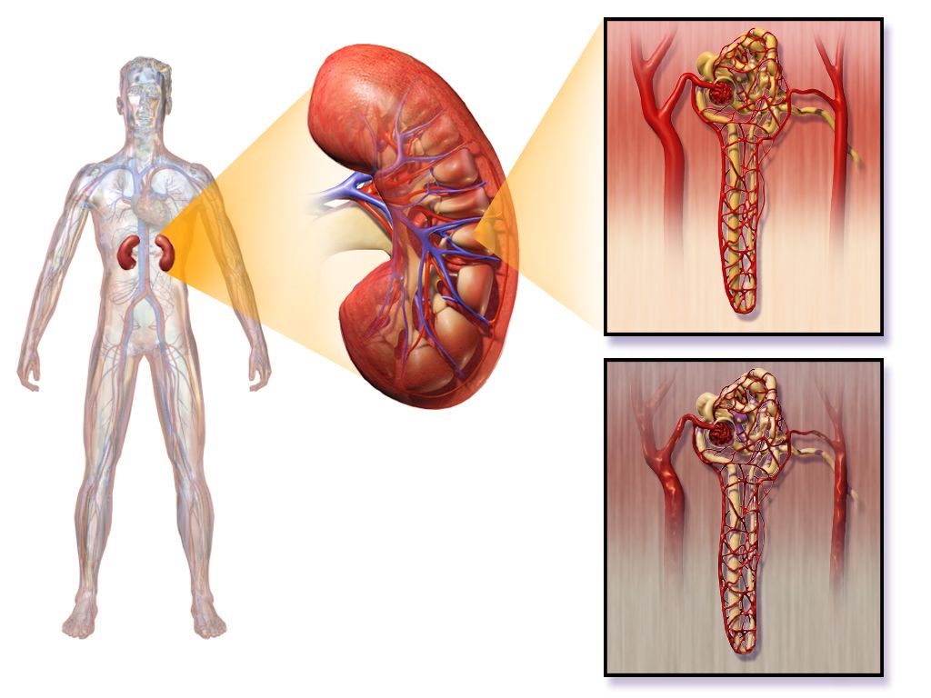
Chronic hyperglycaemia glycates proteins and thickens basement membranes throughout the microcirculation. In the retina this produces diabetic retinopathy — non-proliferative changes of microaneurysms, dot-and-blot haemorrhages and hard exudates, progressing to proliferative retinopathy with fragile neovascular vessels that bleed into the vitreous or drag the retina off. It is asymptomatic until it is severe, which is the entire argument for the annual dilated examination. Sudden painless vision loss, a shower of new floaters or a “curtain” across the field is a same-day ophthalmology emergency. Diabetic macular oedema is the more common cause of gradual central vision loss and is treated with intravitreal anti-VEGF injections.
Diabetic kidney disease begins with hyperfiltration, then persistent albuminuria at a urine albumin-to-creatinine ratio of 30 mg/g or higher, then declining eGFR. An ACE inhibitor or ARB is indicated for albuminuria with hypertension and slows progression by reducing intraglomerular pressure; monitor potassium and creatinine after starting or increasing the dose, and expect a modest creatinine rise that is acceptable. SGLT2 inhibitors and, increasingly, non-steroidal mineralocorticoid antagonists add further renal protection. Blood pressure control below 130/80 mmHg matters at least as much as glucose control here. As eGFR falls, insulin clearance falls with it and insulin requirements paradoxically decrease — a patient with worsening kidney function who suddenly starts running low is telling you something about their kidneys, not their diet.
Diabetic neuropathy is most often a distal symmetric polyneuropathy in a stocking-glove distribution: numbness, tingling, burning and lancinating pain that is worse at night, with loss of vibratory sense and ankle reflexes early. Pharmacological options include pregabalin, gabapentin, duloxetine and amitriptyline; opioids are not first-line. Autonomic neuropathy is the quieter and more dangerous form — orthostatic hypotension, resting tachycardia, silent myocardial ischaemia, gastroparesis, neurogenic bladder with retention and overflow, erectile dysfunction, and hypoglycaemia unawareness. Gastroparesis produces early satiety, bloating and wildly erratic post-meal glucose; manage with small, frequent, low-fat, low-fibre meals, and note that metoclopramide carries a boxed warning for tardive dyskinesia and is limited to under 12 weeks.
Macrovascular disease and the diabetic foot
Atherosclerotic cardiovascular disease is the leading cause of death in diabetes, and diabetes is a coronary risk equivalent. Because of autonomic neuropathy, myocardial infarction may present without chest pain — as dyspnoea, fatigue, nausea, confusion or simply unexplained hyperglycaemia. Take “I just feel off” in a diabetic patient as seriously as crushing substernal pain in anyone else and obtain an ECG. Peripheral arterial disease produces claudication, diminished pulses, hair loss, dependent rubor and cool, shiny, thin skin; cerebrovascular disease produces stroke, which Chapter 3 takes up in detail.
The diabetic foot ulcer is where neuropathy, ischaemia and immune dysfunction meet, and it precedes the majority of non-traumatic lower-limb amputations. Neuropathy removes the pain that would otherwise stop the patient from walking on a wound; motor neuropathy deforms the arch and toes so pressure concentrates under the metatarsal heads; ischaemia starves the healing; hyperglycaemia impairs neutrophil function. Assessment of any ulcer records site, size in centimetres, depth, undermining, wound bed appearance, exudate, odour, surrounding callus or cellulitis, and whether bone is palpable on gentle probing — a positive probe-to-bone strongly suggests osteomyelitis. Management combines pressure offloading, debridement, moist wound care, targeted antibiotics for infection and vascular assessment. Note that a warm, red, swollen, relatively painless foot in a neuropathic patient may be Charcot neuroarthropathy rather than infection, and it requires immediate immobilisation to prevent midfoot collapse.
Foot teaching is concrete and non-negotiable. Inspect both feet daily, using a mirror or a family member for the soles. Wash daily in lukewarm water tested with a thermometer or the elbow — never the foot — and dry carefully between the toes. Moisturise the tops and bottoms but not between the toes. Cut nails straight across, or have podiatry do it. Never use over-the-counter corn removers, razor blades or heating pads. Never go barefoot, indoors or out. Shake out every shoe before putting it on. Break in new shoes 1 to 2 hours a day. Wear seamless, non-constricting socks. Report any blister, cut, redness, callus, ingrown nail or colour change the day it appears — not at the next scheduled visit.
Self-management teaching and sick-day rules
Discharge teaching is prioritised by what will kill the patient soonest if missed: hypoglycaemia recognition and treatment first, insulin administration technique second, sick-day rules third, then foot care, monitoring, nutrition and follow-up. Use teach-back for every psychomotor skill. Watch the patient draw up and inject their own dose before they leave, and watch the caregiver do it too. Ask directly about cost, refrigeration, vision, dexterity, literacy and transport, because the most elegant regimen fails against a broken refrigerator or a $300 copay.
Nutrition teaching centres on consistent carbohydrate intake, portion awareness (the plate method: half non-starchy vegetables, one quarter lean protein, one quarter starch) and carbohydrate counting for those on prandial insulin, where a typical starting ratio is one unit per 15 grams. Emphasise fibre, whole grains and limited added sugar and saturated fat. Alcohol inhibits hepatic gluconeogenesis and can cause delayed hypoglycaemia many hours later — teach patients to drink only with food, in moderation, and to check glucose before bed. Physical activity of 150 minutes per week improves insulin sensitivity for up to 48 hours; teach patients on insulin or secretagogues to check before, sometimes during and after exercise, to carry fast carbohydrate, and to avoid injecting into a limb about to be exercised. Do not exercise if the glucose is above 250 mg/dL with ketones present — exercise will worsen ketosis.
Sick-day rules prevent most avoidable hyperglycaemic crises, and they are counterintuitive enough that they must be taught explicitly and rehearsed. Illness raises counter-regulatory hormones, so glucose and insulin needs go up even when the patient is eating less.
| Sick day | What the patient does |
|---|---|
| Insulin | Never stop taking insulin, even when not eating. Basal continues. Doses often need to increase. |
| Glucose checks | Every 3–4 hours, day and night. |
| Ketone checks | Every 3–4 hours in type 1, and any time glucose exceeds 240 mg/dL or vomiting occurs. |
| Fluids | About 8 oz of fluid every hour while awake — sugar-free if glucose is high; carbohydrate-containing (broth, juice, regular soda, about 15 g per hour) if unable to eat solid food. |
| Food | Try to maintain usual carbohydrate intake in small, soft, frequent amounts. |
| Call the provider | Vomiting or diarrhoea persisting beyond 4 hours or unable to keep fluids down; glucose above 240 mg/dL on two consecutive checks with moderate or large ketones; fever above 101.5 °F not settling; rapid breathing, chest pain, weight loss, drowsiness or confusion; illness lasting beyond 1–2 days. |
The older adult, delegation and the red flags
Older adults present differently and are more fragile in both directions. Hypoglycaemia in an elder rarely looks like tremor and diaphoresis — it looks like a fall, a new confusion, slurred speech mistaken for stroke, or a family member saying “she is just not herself.” Check a glucose on every older adult with altered mental status, every time. Hyperglycaemia likewise presents atypically as new incontinence, delirium, weakness or dehydration, and because the thirst mechanism blunts with age and mobility may prevent reaching a glass of water, HHS develops quietly over days. Renal clearance declines, so sulfonylureas and renally cleared insulins accumulate; glyburide is on the Beers list and should be avoided. Glycaemic targets loosen deliberately — an A1C below 7.5% for a healthy older adult and below 8.0% with multiple comorbidities or cognitive impairment — upper limits, not goals to drift up to — while in very poor health or at end of life there is no A1C target at all, only glucose values managed to avoid hypoglycaemia and symptomatic hyperglycaemia. Polypharmacy, vision loss, arthritic hands and cost all bear directly on whether a regimen can be executed at home.
Delegation follows one principle: the nursing process cannot be delegated. Assessment, teaching, evaluation, clinical judgement and any unstable patient remain with the RN. Assistive personnel may, within facility policy and training, obtain point-of-care capillary glucose readings, record intake and output, assist with meals and positioning, obtain vital signs on stable patients, and report observations such as a reddened heel or an untouched tray — and they must be told explicitly which values to report immediately and to whom. Insulin administration, insulin infusion titration, evaluating whether a hypoglycaemic episode has resolved, initial teaching of injection technique, and the assessment of a new foot ulcer are RN responsibilities. A licensed practical or vocational nurse may administer subcutaneous insulin and reinforce previously taught content within state scope and policy, but the initial assessment, the initial teaching and the unstable patient stay with the registered nurse.
| Red flag | What it may mean | First action |
|---|---|---|
| Glucose below 54 mg/dL, or any low with confusion | Severe hypoglycaemia | Treat immediately — oral carbohydrate if alert and swallowing, otherwise IV dextrose or glucagon; recheck in 15 min; stay with the patient |
| Deep rapid respirations with fruity breath, vomiting, abdominal pain | DKA | Glucose, ketones, gas and metabolic panel; isotonic fluid; cardiac monitoring; escalate now |
| Glucose above 600 mg/dL with drowsiness or focal deficits in an older adult | HHS | Airway and neuro check, large-bore access, isotonic fluid, escalate; look for the infection that caused it |
| Falling level of consciousness during DKA treatment, with headache and vomiting | Cerebral oedema | Stop, escalate immediately; slow the correction; neurologic checks |
| Nausea, dyspnoea, acidosis with a near-normal glucose on an SGLT2 inhibitor | Euglycaemic DKA | Check ketones and a gas anyway; hold the agent; escalate |
| Fever, erythema, purulent or malodorous foot wound, probe-to-bone | Deep infection or osteomyelitis | Cultures, vascular and wound assessment, offloading, urgent provider notification |
| Sudden painless vision loss or a shower of floaters | Vitreous haemorrhage or retinal detachment | Same-day ophthalmology referral |
| Unexplained hyperglycaemia, fatigue or dyspnoea in a long-standing diabetic | Silent myocardial infarction | ECG and troponin; do not wait for chest pain |
The complications of diabetes are prevented at the bedside by three unglamorous habits: taking the socks off, checking a glucose on every confused patient, and making sure the patient can actually perform at home the regimen you documented as taught.
The chapter in one breath
Diabetes is one hyperglycaemia with two mechanisms. Type 1 is autoimmune destruction of beta cells — absolute deficiency, low C-peptide, ketosis prone, insulin required forever. Type 2 is insulin resistance followed by beta-cell exhaustion — insidious onset, enough residual insulin to suppress ketogenesis, so decompensation runs toward hyperosmolarity instead. Diagnosis rests on an A1C of 6.5% or higher, a fasting glucose of 126 mg/dL or higher, a 2-hour OGTT value of 200 mg/dL or higher, or a random 200 with symptoms; prediabetes occupies the band just below each. Management is glucose targets individualised to the patient, insulin chosen by its onset-peak-duration profile, and non-insulin agents understood by mechanism so you know which ones can drop a sugar. Acutely, hypoglycaemia outranks hyperglycaemia every time: treat the 40 before the 400. DKA brings acidosis, ketones, an anion gap and Kussmaul breathing; HHS brings glucose above 600, osmolality above 320 and profound obtundation without ketones. Both are treated in the same order — isotonic fluid first, potassium replaced before insulin is started if it is below 3.3 mEq/L, then a regular insulin infusion, then dextrose added when the glucose reaches about 200 so the drip can keep closing the gap. Chronically, the work is surveillance, foot care, sick-day rules and teaching that survives discharge.
Words worth knowing
| Term | What it means |
|---|---|
| Absolute insulin deficiency | No endogenous insulin production, as in type 1 diabetes after beta-cell destruction; obligates exogenous insulin and predisposes to ketoacidosis. |
| Insulin resistance | Reduced tissue response to circulating insulin, the initiating defect in type 2 diabetes; compensated by hyperinsulinaemia until beta cells fail. |
| C-peptide | Fragment cleaved from proinsulin during endogenous insulin production; low or absent in type 1, normal or high in early type 2. |
| A1C (glycated haemoglobin) | Percentage of haemoglobin glycated over the red cell lifespan; reflects roughly 3 months of average glycaemia. Diabetes at 6.5% or higher. |
| Osmotic diuresis | Water and electrolyte loss driven by glucose exceeding the renal reabsorption threshold near 180 mg/dL; cause of polyuria and volume depletion. |
| Ketogenesis | Hepatic conversion of free fatty acids to acetoacetate, beta-hydroxybutyrate and acetone when insulin is absent; the source of DKA acidosis. |
| Anion gap | Sodium minus (chloride plus bicarbonate); elevated above 10–12 mEq/L in DKA and the marker whose closure signals resolution. |
| Effective serum osmolality | Two times sodium plus glucose divided by 18; above 320 mOsm/kg correlates with the obtundation of HHS. |
| Kussmaul respirations | Deep, rapid, laboured breathing that blows off carbon dioxide to compensate for metabolic acidosis; characteristic of DKA, absent in HHS. |
| Euglycaemic DKA | Ketoacidosis with glucose below about 250 mg/dL, seen with SGLT2 inhibitors, pregnancy, starvation and vomiting; missed if you screen on glucose alone. |
| Rule of 15 | Treatment of conscious hypoglycaemia: 15 g of fast carbohydrate, recheck in 15 minutes, repeat until glucose exceeds 70 mg/dL. |
| Hypoglycaemia unawareness | Loss of adrenergic warning symptoms from autonomic neuropathy, repeated lows or beta blockade; the first sign may be neuroglycopenia. |
| Dawn phenomenon | Pre-waking hyperglycaemia from growth hormone and cortisol surge; 3 a.m. glucose is normal or high. |
| Somogyi effect | Rebound morning hyperglycaemia following unrecognised nocturnal hypoglycaemia; 3 a.m. glucose is low and evening insulin must be reduced. |
| Lipohypertrophy | Rubbery subcutaneous thickening at repeatedly used injection sites causing erratic insulin absorption; prevented by systematic site rotation. |
| Basal-bolus regimen | Long-acting insulin for background needs plus rapid-acting insulin at meals with a correction component; preferred over sliding scale alone. |
| Time in range | Percentage of continuous glucose monitor readings between 70 and 180 mg/dL; goal above 70% with less than 4% below 70 mg/dL. |
| Loss of protective sensation | Inability to perceive the 10-g monofilament; the patient cannot feel injury and requires intensified foot protection. |
| Charcot neuroarthropathy | Progressive destruction of bones and joints in a neuropathic foot, presenting warm, swollen and red with little pain; needs immobilisation and offloading. |
| Sick-day rules | Illness protocol: never stop insulin, check glucose and ketones every 3–4 hours, maintain fluids and carbohydrate, and call for persistent vomiting or ketones. |
Check yourself
Endocrine Disorders: Thyroid, Parathyroid, Adrenal and Pituitary
Every hormone in this chapter is a message, and every disorder is either too much message, too little message, or a message that never left the building. Learn the loop and the diseases stop being a list to memorise: the pituitary shouts at a gland, the gland answers with hormone, and the hormone tells the pituitary to be quiet. That single sentence explains why a high TSH means a lazy thyroid, why a suppressed TSH with a high free T4 means the gland has gone rogue, and why a patient on long-term prednisone cannot make cortisol on the day she gets pneumonia. Endocrine emergencies are quiet until they are not. Myxedema coma, thyroid storm, hypocalcaemic tetany, adrenal crisis and severe hyponatraemia all present first as vital-sign drift and a change in mentation, and all of them kill through airway, circulation or seizure. Your job on a busy floor is to notice the drift, name the pattern and escalate before the crash.
By the end of this chapter you can
- Explain negative feedback in the hypothalamic—pituitary—target axis and use paired hormone levels to distinguish primary from secondary (central) endocrine disease.
- Interpret TSH, free T4, serum calcium, PTH, cortisol, serum and urine osmolality against real reference ranges and state what the nurse does with each result.
- Prioritise nursing care in myxedema coma and thyroid storm, including why a thionamide must precede iodine and why aspirin is withheld.
- Deliver post-thyroidectomy care that anticipates airway obstruction from haematoma, recurrent laryngeal nerve injury and hypocalcaemia.
- Distinguish hyperparathyroidism from hypoparathyroidism by symptom cluster, ECG change and emergency drug, and demonstrate Chvostek and Trousseau signs.
- Compare Cushing syndrome and adrenal insufficiency, and manage adrenal crisis with fluids and IV hydrocortisone while teaching sick-day rules.
- Differentiate SIADH from diabetes insipidus using volume status, urine specific gravity and osmolality, and correct sodium at a safe rate.
- Recognise older-adult presentations such as apathetic thyrotoxicosis and drug-induced hyponatraemia, and state which tasks may and may not be delegated to assistive personnel.
- Compose an SBAR escalation for each endocrine red flag in this chapter.
Section 1Reading the Axes: The Logic That Makes Endocrine Make Sense
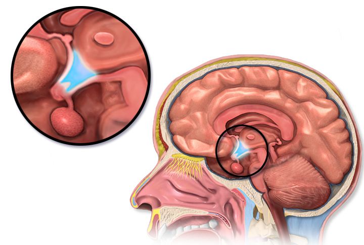
Feedback loops, and why the trophic hormone tells you where the lesion is
The hypothalamus secretes releasing hormones into a private portal circulation that drains straight into the anterior pituitary. The pituitary answers with trophic hormones — thyroid-stimulating hormone (TSH), adrenocorticotropic hormone (ACTH), growth hormone (GH), follicle-stimulating and luteinising hormone, prolactin. Those trophic hormones travel in the systemic blood to a target gland, and the target gland releases the hormone that actually does the work: thyroxine, cortisol, oestrogen, testosterone. The finished hormone then circles back and tells both the hypothalamus and the pituitary to stop shouting. That is negative feedback, and it is the whole game.
Here is the payoff for the bedside. If the target gland is the broken part — a thyroid destroyed by autoimmune thyroiditis, an adrenal cortex wrecked by autoimmune adrenalitis — then the target hormone is low and the pituitary, hearing no feedback, screams louder. So you see a low target hormone with a high trophic hormone. That is primary disease. If the pituitary or hypothalamus is the broken part, it cannot shout at all, so both hormones are low: low target hormone with a low or inappropriately normal trophic hormone. That is secondary, or central, disease. The same two-line rule works for excess. A rogue gland making too much hormone will suppress the trophic hormone to nearly nothing; a pituitary tumour making too much trophic hormone will drive the target hormone up while the trophic hormone stays high, which is biochemically impossible unless something upstream is autonomous.
Students lose points on this because they memorise diseases instead of patterns. Read the pair. A TSH of 18 mIU/L with a free T4 of 0.4 ng/dL is a failing thyroid. A TSH of 0.9 mIU/L with a free T4 of 0.4 ng/dL is a failing pituitary and a much more dangerous patient, because a pituitary that cannot make TSH usually cannot make ACTH either, and cortisol deficiency kills faster than hypothyroidism.
| Pattern | Trophic hormone | Target hormone | Where the lesion is | Classic example |
|---|---|---|---|---|
| Primary deficiency | High | Low | Target gland | Hashimoto hypothyroidism; Addison disease |
| Secondary (central) deficiency | Low or normal | Low | Pituitary or hypothalamus | Pituitary apoplexy; long-term exogenous steroids |
| Primary excess | Suppressed | High | Target gland | Graves disease; adrenal cortisol-secreting adenoma |
| Secondary excess | High or normal | High | Pituitary or ectopic tumour | Cushing disease; ectopic ACTH from small-cell lung cancer |
Stimulate when you suspect too little, suppress when you suspect too much
Random hormone levels are noisy. Cortisol swings with the clock, GH is secreted in bursts, and a single value can be normal in a patient who has almost no reserve. So endocrinology provokes the gland. When you suspect deficiency, you stimulate: give synthetic ACTH (cosyntropin 250 mcg IV or IM) and measure cortisol at 30 and 60 minutes. A healthy adrenal cortex jumps above roughly 18 to 20 mcg/dL. A failing one does not move. When you suspect excess, you suppress: give 1 mg of dexamethasone at 11 pm and draw cortisol at 8 am. A normal axis obediently shuts down to below 1.8 mcg/dL; an autonomous cortisol source ignores the instruction. The same grammar governs the water-deprivation test for diabetes insipidus and the oral glucose tolerance test for acromegaly.
Your nursing role in provocative testing is not glamorous and it is entirely about the validity of the result. Confirm the exact draw times and write them on the tubes; a cortisol drawn at 45 minutes instead of 30 is uninterpretable. Hold or document interfering drugs. Biotin supplements, which patients take for hair and nails and never think to report, distort many immunoassays including TSH and free T4 and should be stopped several days before. Keep the patient supine and calm if the test involves catecholamines. And for a water-deprivation test, the patient must not sneak fluids — which means the RN, not the assistive personnel, owns the observation, because the test is a live risk of hypernatraemia.
The labs that lie, and the assessment that does not
Acute illness perturbs every axis. In non-thyroidal illness syndrome (formerly euthyroid sick syndrome) a critically ill patient shows a low T3, sometimes a low free T4 and a TSH that is low during illness and rebounds high during recovery — none of it meaning thyroid disease. Steroids, dopamine, amiodarone, lithium, opioids and heparin all bend results. This is why a good endocrine assessment leans on the physical exam and the trend: weight over months, resting heart rate, skin and hair texture, deep-tendon-reflex relaxation phase, orthostatic vital signs, and the family’s report of how the person has changed. A daughter saying “she used to do the crossword” is data.
Older adults deserve a standing warning. Endocrine disease in a person over seventy-five rarely announces itself with textbook signs. Hypothyroidism looks like dementia or depression. Hyperthyroidism looks like new atrial fibrillation and weight loss with no tremor or agitation at all — apathetic thyrotoxicosis. Hypercalcaemia looks like constipation and confusion. Adrenal insufficiency looks like recurrent falls. If an older patient has an unexplained change in cognition, function or sodium, an endocrine cause belongs on your differential.
Interpret every endocrine result as a pair — trophic hormone with target hormone — because the pair, not the single value, tells you whether the gland, the pituitary or a drug is the problem.
Section 2The Thyroid: From Sluggish to Storm
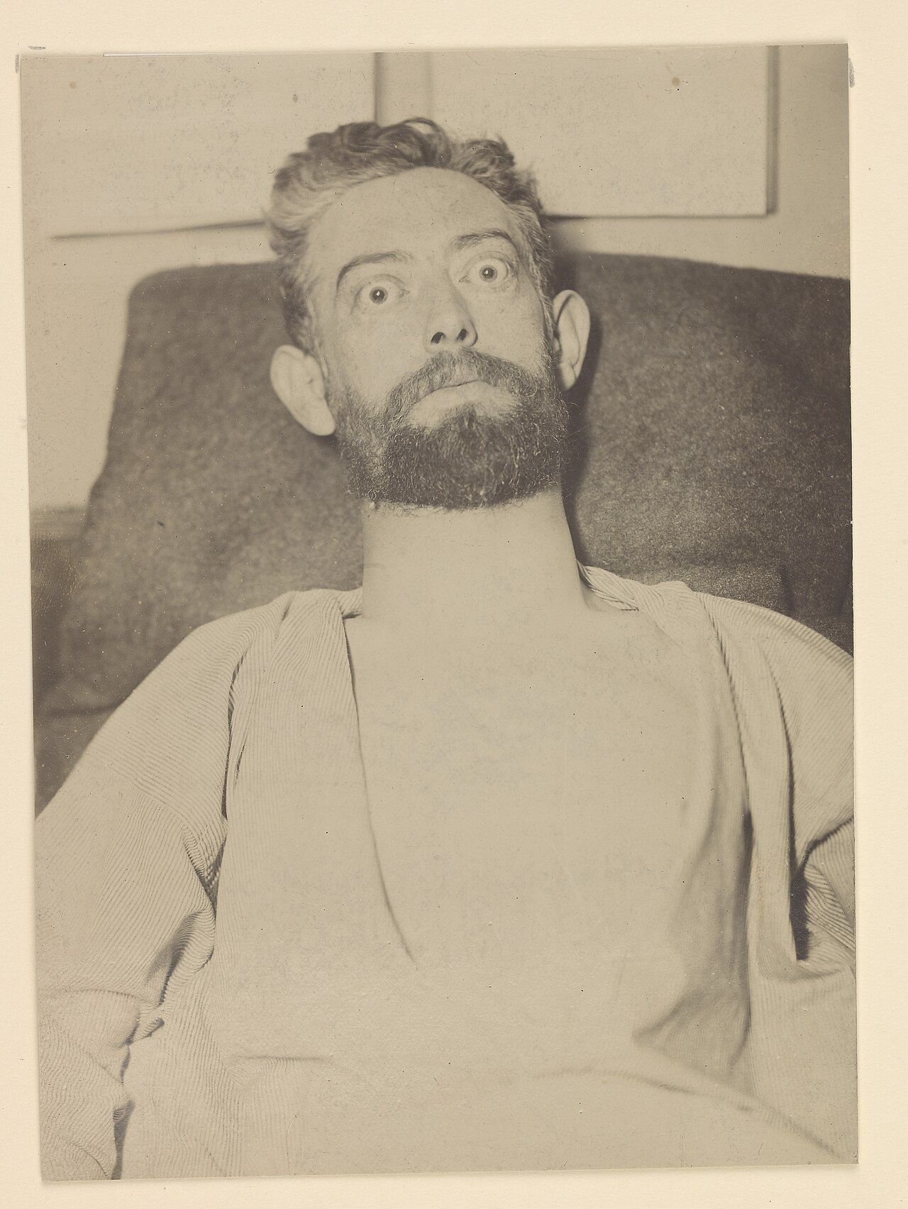
Hypothyroidism: everything slows, including the diaphragm
Thyroid hormone sets the basal metabolic rate of essentially every cell, so deficiency slows everything at once. Heat production falls, giving cold intolerance and a low-normal temperature. Gut motility falls, giving constipation. Cardiac output falls through bradycardia and reduced contractility, and diastolic hypertension appears because vessels stiffen. Cholesterol clearance falls, so the lipid panel worsens. Glycosaminoglycans accumulate in the dermis and draw water in, producing the non-pitting periorbital and facial puffiness called myxedema, along with generalised non-pitting swelling of the hands and feet, the coarse skin, the hoarse voice and the enlarged tongue. The separate sign called pretibial myxedema is not part of this picture — it is Graves dermopathy over the shins and belongs to the hyperthyroid patient. Reflexes show a characteristically slow relaxation phase. The most common cause in iodine-sufficient countries is Hashimoto thyroiditis, an autoimmune destruction marked by anti-thyroid peroxidase antibodies; other causes include prior radioactive iodine, thyroidectomy, amiodarone, lithium and, in the newborn, congenital absence — which is why every state screens.
Diagnosis is a TSH first. Reference range is roughly 0.4 to 4.0 mIU/L. An elevated TSH with a low free T4 (normal roughly 0.8 to 1.8 ng/dL) is overt primary hypothyroidism. An elevated TSH with a normal free T4 is subclinical disease, which may be treated if the TSH exceeds 10 mIU/L or the patient is pregnant or symptomatic. Treatment is levothyroxine, a synthetic T4 that the body converts to active T3. A healthy adult starts near 1.6 mcg/kg per day. An adult over sixty, or anyone with coronary disease, starts at 12.5 to 25 mcg daily and titrates slowly, because thyroid hormone raises myocardial oxygen demand and a full replacement dose can precipitate angina, infarction or a tachyarrhythmia. Recheck TSH six to eight weeks after any dose change — not sooner, because the axis takes that long to re-equilibrate.
Teaching levothyroxine is a nursing skill in itself. Take it on an empty stomach with water, thirty to sixty minutes before breakfast, at the same time daily. Separate it by at least four hours from calcium carbonate, iron, magnesium, aluminium antacids, sucralfate, cholestyramine and proton-pump inhibitors, all of which block absorption. Do not switch brands casually. It is lifelong; feeling well is a sign the dose is right, not a reason to stop. Report palpitations, chest pain, insomnia, tremor or unintended weight loss — those are over-replacement, and in an older adult chronic over-replacement causes atrial fibrillation and accelerates osteoporosis.
Myxedema coma: the hypothermic, hypercapnic, hyponatraemic patient
Myxedema coma is decompensated hypothyroidism, usually in an older woman in winter, precipitated by infection, cold exposure, surgery, sedatives, opioids or stopping levothyroxine. The picture is hypothermia often below 35 degrees Celsius, bradycardia, hypotension, hypoventilation with carbon dioxide retention, non-pitting oedema, hyponatraemia, hypoglycaemia and a depressed level of consciousness that progresses to coma. Mortality remains substantial even when treated, and the killer is respiratory failure.
Priorities are airway and ventilation first — anticipate intubation, monitor capnography and arterial blood gases, and hold every sedative and opioid you reasonably can, since these patients clear drugs at a crawl. Then IV levothyroxine, typically a loading dose of 200 to 400 mcg followed by 50 to 100 mcg daily, with liothyronine added in some protocols. Critically, give IV hydrocortisone 100 mg every 8 hours before or with the thyroid hormone, because coexisting adrenal insufficiency is common and giving thyroid hormone first accelerates cortisol metabolism and can tip the patient into adrenal crisis. Rewarm passively with blankets; active external rewarming vasodilates a patient with no cardiac reserve and drops the blood pressure. Treat hyponatraemia with fluid restriction rather than aggressive saline unless the sodium is dangerously low, monitor glucose, and hunt the precipitant — culture, chest film, urinalysis.
Hyperthyroidism, Graves disease and thyroid storm
Excess thyroid hormone speeds everything and up-regulates beta-adrenergic receptors, which is why the patient looks adrenergic: tachycardia, palpitations, atrial fibrillation, systolic hypertension with a widened pulse pressure, fine tremor, warm moist skin, heat intolerance, weight loss with a ravenous appetite, frequent stools, anxiety, insomnia and proximal muscle weakness. Graves disease, an autoimmune condition driven by thyroid-stimulating immunoglobulin, adds a diffuse goitre, ophthalmopathy with exophthalmos and lid lag, and rarely pretibial myxedema. Toxic multinodular goitre and a toxic adenoma cause hyperthyroidism without eye disease. Subacute thyroiditis causes a painful gland with transient hormone leak and a low radioactive iodine uptake — an important distinction, because thionamides do not help a gland that is leaking rather than manufacturing.
Labs show a suppressed TSH, often below 0.1 mIU/L, with a high free T4 or, in about five percent, an isolated high T3. A radioactive iodine uptake and scan sorts overproduction (high uptake) from leak (low uptake). Medical treatment has three arms. Beta blockers — propranolol, atenolol, or esmolol in the unstable patient — control symptoms within hours and are the first thing the patient actually feels. Thionamides block hormone synthesis: methimazole is preferred for most adults because it is once daily and less hepatotoxic; propylthiouracil (PTU) is reserved for the first trimester of pregnancy and for thyroid storm, because it also blocks peripheral conversion of T4 to T3. Radioactive iodine ablation is the common definitive therapy in the United States and usually leads to permanent hypothyroidism, which is an accepted trade. It is absolutely contraindicated in pregnancy and breastfeeding, because iodine-131 crosses the placenta and ablates the fetal thyroid and is excreted in breast milk: document a negative pregnancy test before dosing, confirm lactation has stopped, and teach that conception is deferred for roughly six months afterwards. Teach the radiation precautions for the first several days as well — sleep alone, limit close contact with pregnant women and young children, sit to void and flush the toilet twice, wash hands thoroughly, and keep laundry, towels and eating utensils separate. Surgery is chosen for large compressive goitres, suspected malignancy, pregnancy when drugs fail, or severe ophthalmopathy.
| Drug | Mechanism | Onset | Nursing implications |
|---|---|---|---|
| Propranolol | Non-selective beta blockade; high doses also reduce T4-to-T3 conversion | Hours | Hold and call for heart rate below 60 or systolic below 90; caution in asthma and decompensated heart failure; masks hypoglycaemia in diabetes |
| Methimazole | Blocks thyroid peroxidase, halting hormone synthesis | 2 to 6 weeks for full effect | Teach the agranulocytosis warning: sore throat, mouth ulcers or fever means stop and get a CBC with differential the same day; teratogenic in the first trimester |
| Propylthiouracil | Blocks synthesis and peripheral T4-to-T3 conversion | Days for conversion effect | Preferred in storm and in the first trimester; monitor for hepatotoxicity — dark urine, jaundice, right-upper-quadrant pain; check LFTs |
| Iodine (SSKI, Lugol solution) | Acutely blocks hormone release and, by the Wolff–Chaikoff effect, blocks iodide organification and new synthesis | Hours | Give at least one hour AFTER a thionamide; dilute in juice and use a straw to protect enamel; effect wanes after 10 to 14 days |
| Hydrocortisone | Reduces T4-to-T3 conversion; treats relative adrenal insufficiency | Hours | Monitor glucose and potassium; do not omit in storm |
| Cholestyramine | Binds hormone in the enterohepatic circulation | Days | Adjunct only; separate from all other oral drugs by four hours |
Thyroid storm is hyperthyroidism plus decompensation, usually triggered by infection, surgery, trauma, iodinated contrast, childbirth or abrupt withdrawal of antithyroid drugs. Expect fever above 38.5 and often above 40 degrees Celsius, heart rate above 140 with atrial fibrillation, agitation progressing to delirium and coma, vomiting and diarrhoea with dehydration, and high-output heart failure. Treat in an order that matters: beta blocker first for the cardiovascular emergency, thionamide (PTU) second to stop synthesis, iodine at least one hour after the thionamide — give iodine first and you simply hand the gland raw material — then hydrocortisone, cooling blankets and acetaminophen. Never give aspirin; salicylates displace thyroid hormone from thyroxine-binding globulin and raise the free fraction. Continuous cardiac monitoring, cooling, aggressive IV fluid with glucose, and a relentless search for the trigger complete the picture.
Nodules, cancer, and the neck after thyroidectomy
Thyroid nodules are common and mostly benign. Ultrasound characterises them and fine-needle aspiration samples the suspicious ones. Papillary carcinoma is the most frequent and has an excellent prognosis; follicular, medullary (associated with multiple endocrine neoplasia type 2 and elevated calcitonin) and anaplastic follow in decreasing frequency and increasing aggression. Treatment is usually total thyroidectomy, often with radioactive iodine ablation, then lifelong levothyroxine at a dose deliberately high enough to suppress TSH in higher-risk disease.
Post-thyroidectomy nursing is a masterclass in anticipating three complications. First, airway. A haematoma in a closed neck compartment compresses the trachea within hours; so does laryngeal oedema. Keep the head of bed elevated 30 to 45 degrees, support the head and neck with pillows when the patient moves, and check the dressing — including behind the neck, where blood tracks by gravity — every fifteen minutes initially. A complaint of neck tightness, pressure, or difficulty swallowing is an emergency, not a comfort issue. Keep suction, oxygen and an emergency tracheostomy tray at the bedside. Second, the recurrent laryngeal nerve. Ask the patient to speak every hour or two; a weak or hoarse voice can be transient oedema, but bilateral injury produces stridor and complete airway obstruction. Third, hypocalcaemia from parathyroid injury or removal, which typically appears 24 to 72 hours postoperatively — covered next section. Also watch for the rarest complication, thyroid storm, in a patient who was not adequately prepared preoperatively.
Delegation lines are sharp here. The nursing assistant may take and report vital signs, assist with positioning, record intake and output, and provide oral care. The RN performs the airway assessment, the voice check, the neuromuscular irritability check, the dressing evaluation, the teaching and every judgement about escalation. An unstable postoperative thyroidectomy patient is never handed off to unlicensed personnel.
Hypothyroid emergencies kill through hypoventilation and hypothermia, hyperthyroid emergencies kill through fever and arrhythmia, and the neck after thyroidectomy kills through airway compression — so airway and cardiac monitoring outrank every other task.
Section 3Parathyroid Glands and the Calcium Emergency
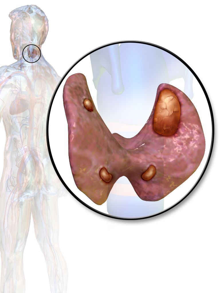
Parathyroid hormone in one paragraph
Parathyroid hormone (PTH) exists to defend the serum ionised calcium. When calcium falls, PTH rises and does three things: it pulls calcium out of bone by activating osteoclasts, it makes the kidney reabsorb calcium while dumping phosphate in the urine, and it activates vitamin D to 1,25-dihydroxycholecalciferol so the gut absorbs more calcium. Remember the reciprocal relationship: PTH drives calcium up and phosphate down. That is why primary hyperparathyroidism shows a high calcium with a low phosphate, and hypoparathyroidism shows a low calcium with a high phosphate.
Reference values you must know cold: total serum calcium roughly 8.5 to 10.5 mg/dL, ionised calcium roughly 4.5 to 5.6 mg/dL (1.12 to 1.4 mmol/L), phosphate 2.5 to 4.5 mg/dL, magnesium 1.8 to 2.4 mg/dL, and intact PTH about 10 to 65 pg/mL. Because roughly 40 percent of serum calcium is bound to albumin and only the ionised fraction is physiologically active, a hypoalbuminaemic patient can look hypocalcaemic while the physiologically active fraction is fine. Correct it: add 0.8 mg/dL to the measured calcium for every 1 g/dL that albumin sits below 4.0 g/dL. Better still, in the critically ill, ask for an ionised calcium.
Hyperparathyroidism and hypercalcaemia: stones, bones, groans and moans
Primary hyperparathyroidism is usually a single benign adenoma secreting PTH autonomously; a minority is hyperplasia, often part of multiple endocrine neoplasia. Secondary hyperparathyroidism is the appropriate response of healthy glands to chronic kidney disease, where phosphate retention and failed vitamin D activation drive calcium down; here calcium is low or low-normal while PTH is high. Tertiary disease is what happens when chronically stimulated glands become autonomous. The other big cause of hypercalcaemia on a med-surg floor is malignancy — PTH-related peptide from squamous tumours, or direct bone lysis in myeloma and breast cancer — and there the PTH is appropriately suppressed.
The classic mnemonic still earns its keep. Stones: nephrolithiasis, polyuria, nephrogenic diabetes insipidus from calcium’s effect on the collecting duct. Bones: bone pain, osteoporosis, pathological fracture. Abdominal groans: anorexia, nausea, constipation, peptic ulcer disease, pancreatitis. Psychic moans: fatigue, depression, confusion, and at high levels stupor and coma. Add muscle weakness with diminished deep tendon reflexes and, on the monitor, a shortened QT interval with bradyarrhythmias. Hypercalcaemia potentiates digoxin, so a patient on digoxin with a rising calcium is at real risk of toxicity.
A hypercalcaemic crisis — calcium above roughly 14 mg/dL with altered mentation — is managed in a fixed order. Volume first: these patients are profoundly dehydrated from calcium-induced polyuria and vomiting, so give isotonic saline at 200 to 300 mL per hour with careful attention to the older heart. Loop diuretics come after the patient is volume replete, and only if fluid overload threatens; giving furosemide to a dry patient worsens everything. Calcitonin 4 units/kg subcutaneously or intramuscularly every 12 hours works within hours but tachyphylaxes in 48. Intravenous bisphosphonates — zoledronic acid 4 mg over 15 minutes, or pamidronate — take two to four days but hold the calcium down; check renal function and dental status first, since osteonecrosis of the jaw is a real if uncommon risk. Denosumab is used when bisphosphonates cannot be. Cinacalcet, a calcimimetic, tricks the parathyroid’s calcium-sensing receptor into reading calcium as high and turns PTH off. Dialysis is the backstop.
Nursing care for chronic hypercalcaemia is mundane and effective: encourage 3 to 4 litres of fluid daily unless contraindicated, mobilise the patient because immobility resorbs bone, add fibre for constipation, screen every medication for thiazides, lithium and calcium-containing antacids, and institute fall precautions. Parathyroidectomy cures primary disease, and the postoperative watch is the mirror image — a sudden drop into hypocalcaemia, sometimes dramatic in “hungry bone syndrome” where demineralised bone soaks up calcium for days.
Hypoparathyroidism and tetany: the emergency you can see at the bedside
The commonest cause of hypoparathyroidism is surgical — the glands removed, devascularised or bruised during thyroidectomy, radical neck dissection or parathyroidectomy. Autoimmune destruction, congenital absence in DiGeorge syndrome, and severe hypomagnesaemia (which blocks PTH secretion and PTH action) are the others. Falling ionised calcium destabilises nerve membranes and lowers the threshold for depolarisation, so the presentation is neuromuscular irritability that escalates.
Early: perioral and fingertip numbness and tingling, muscle cramps, anxiety. Then hyperactive reflexes, carpopedal spasm, and the two named signs — Chvostek sign, a twitch of the ipsilateral facial muscles when you tap the facial nerve just anterior to the earlobe, and Trousseau sign, carpal spasm after inflating a blood-pressure cuff 20 mmHg above systolic for three minutes. Trousseau is the more specific of the two. Late and lethal: laryngospasm with stridor, bronchospasm, seizures, a prolonged QT interval that can degenerate into torsades de pointes, and hypotension with decreased contractility. On the postoperative thyroid patient, a complaint of tingling around the mouth at hour 20 is not anxiety — it is the first rung of a ladder that ends in laryngospasm.
Acute symptomatic hypocalcaemia is treated with IV calcium gluconate, typically 1 to 2 grams diluted in 50 to 100 mL of D5W infused over 10 to 20 minutes, followed by a continuous infusion if symptoms persist, with the patient on continuous cardiac monitoring throughout because rapid administration causes bradycardia and hypotension. Calcium gluconate is preferred peripherally; calcium chloride contains three times the elemental calcium but is a severe vesicant and should go through a central line. Check and replace magnesium — hypocalcaemia that will not correct is hypomagnesaemia until proven otherwise. Never mix calcium with sodium bicarbonate or phosphate in the same line; it precipitates. Long term, patients take oral calcium carbonate or citrate with calcitriol (activated vitamin D, because a patient without PTH cannot activate their own), and increasingly recombinant PTH. Teach seizure precautions, a high-calcium and low-phosphate diet, and the tingling-means-call rule.
| Hypercalcaemia (high PTH or malignancy) | Hypocalcaemia (low PTH) | |
|---|---|---|
| Calcium / phosphate | Calcium high, phosphate low | Calcium low, phosphate high |
| Neuromuscular | Weakness, hyporeflexia, lethargy, confusion | Tingling, cramps, tetany, hyperreflexia, seizures |
| Gastrointestinal | Constipation, nausea, ulcer, pancreatitis | Diarrhoea, abdominal cramping |
| ECG | Shortened QT, bradyarrhythmia, digoxin sensitivity | Prolonged QT, risk of torsades de pointes |
| Airway risk | Low | High — laryngospasm and stridor |
| Emergency drug | Isotonic saline, then calcitonin and a bisphosphonate | IV calcium gluconate; correct magnesium |
Falling calcium is an airway and rhythm emergency — perioral tingling after neck surgery buys the patient a cardiac monitor, an ionised calcium, a magnesium level and calcium gluconate at the ready, not reassurance.
Section 4The Adrenal Glands: Too Much Cortisol, Too Little Cortisol, and the Medulla
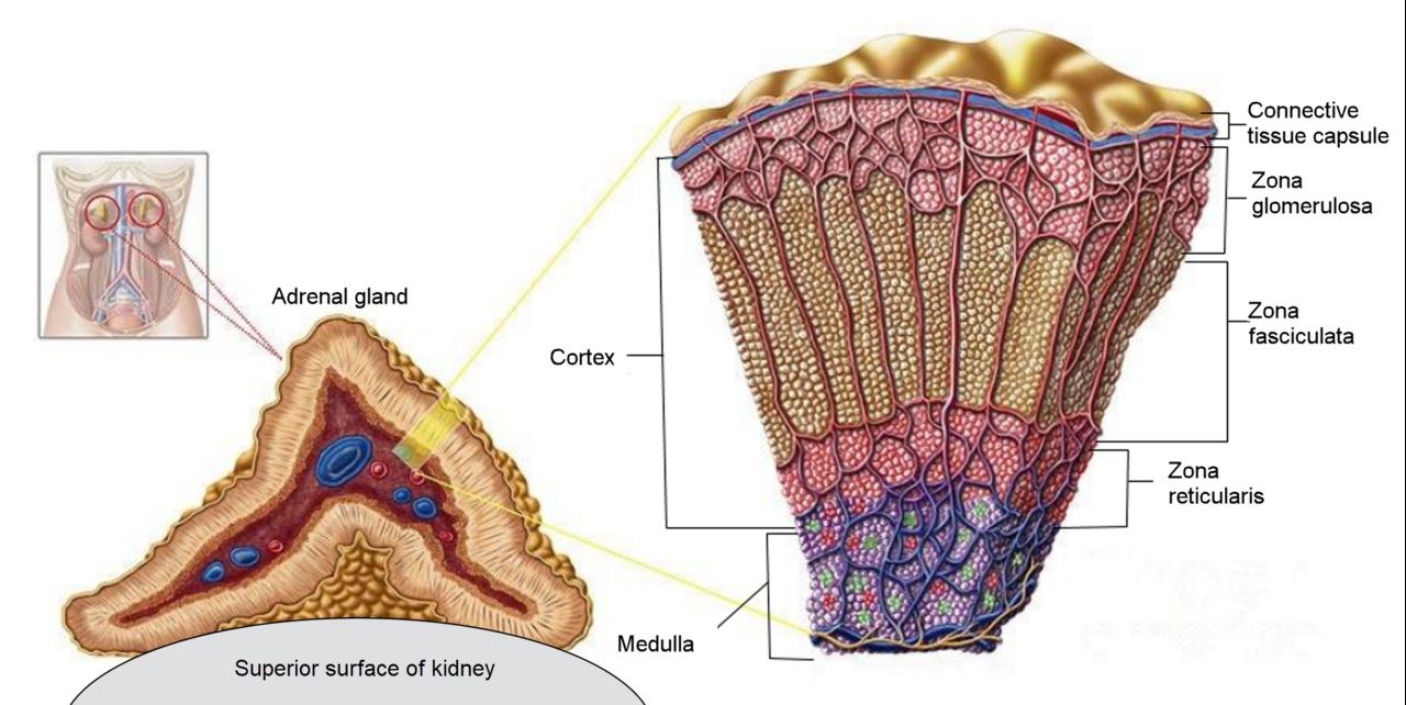
Cortisol excess: Cushing syndrome
Cortisol is a survival hormone: it raises glucose, breaks down protein and fat for fuel, suppresses inflammation and immunity, supports vascular tone, and retains sodium and water while wasting potassium. Give too much for too long and every one of those actions becomes a disease. Central obesity with a buffalo hump and moon face while the arms and legs waste; thin, fragile skin with purple striae wider than a centimetre; easy bruising and terrible wound healing; hyperglycaemia and frank diabetes; osteoporosis with vertebral compression fractures; hypertension; hypokalaemia and metabolic alkalosis; suppressed immunity with masked infection; mood lability, insomnia and psychosis; hirsutism and menstrual irregularity in women. The single most common cause is exogenous — the patient on prednisone for rheumatoid arthritis, transplant or inflammatory bowel disease. Endogenous causes split into ACTH-dependent (a pituitary corticotroph adenoma, which is properly called Cushing disease, and ectopic ACTH from small-cell lung cancer) and ACTH-independent (an adrenal adenoma or carcinoma).
Screening uses any two of three: 24-hour urinary free cortisol, late-night salivary cortisol (which exploits the fact that cortisol should be at its nadir at midnight), and the 1 mg overnight dexamethasone suppression test, where a morning cortisol above 1.8 mcg/dL fails to suppress. Once excess is confirmed, a plasma ACTH localises it: suppressed ACTH means the adrenal is the source; normal or high ACTH means pituitary or ectopic, and high-dose dexamethasone testing plus imaging or inferior petrosal sinus sampling sorts those. Treatment is surgical when there is a tumour — transsphenoidal resection for Cushing disease, adrenalectomy for an adrenal source — with ketoconazole, metyrapone, osilodrostat or mifepristone used medically. For exogenous Cushing, the answer is the lowest effective steroid dose and a slow taper, never an abrupt stop.
Nursing priorities read straight off the pathophysiology. Protect the skin: paper tape, gentle turning, no adhesive removal without an adhesive remover, meticulous pressure-injury prevention. Protect the bones: fall precautions, assistive devices, no independent ambulation until you have assessed proximal muscle strength, and remember a vertebral fracture can present as new back pain with no trauma. Protect against infection: hand hygiene, screening visitors, and a very low threshold for suspecting infection because the usual fever and leucocytosis may be blunted. Monitor glucose, potassium, weight and blood pressure daily. Teach a diet high in protein, calcium and vitamin D, low in sodium and refined carbohydrate. And after adrenalectomy or successful pituitary surgery, the patient swings the other way — they now need glucocorticoid replacement and are at risk of adrenal crisis for months.
Cortisol deficiency: Addison disease and adrenal crisis
Primary adrenal insufficiency, Addison disease, destroys the whole cortex, so the patient loses cortisol and aldosterone and adrenal androgens. Autoimmune adrenalitis is the leading cause in developed countries; tuberculosis, adrenal haemorrhage (including Waterhouse–Friderichsen syndrome in meningococcaemia), metastasis, HIV and drugs such as ketoconazole and etomidate account for the rest. Secondary insufficiency comes from a pituitary that cannot make ACTH — and by far the most common version is iatrogenic, from a patient who has taken more than about 5 mg of prednisone daily for more than three weeks and then stopped or got sick.
The distinction is clinically useful. In primary disease, ACTH is sky-high and its precursor peptide stimulates melanocytes, so the patient has hyperpigmentation of skin creases, scars, gums and the buccal mucosa; aldosterone is absent, so sodium is low, potassium is high, and the patient is salt-craving, orthostatic and volume depleted with a mild metabolic acidosis. In secondary disease there is no hyperpigmentation, and because the renin–angiotensin–aldosterone axis is intact, potassium is usually normal. Both share fatigue that is out of proportion, anorexia, weight loss, nausea, vague abdominal pain, myalgia, hypoglycaemia and hypotension.
Diagnosis: an 8 am serum cortisol below 3 mcg/dL is essentially diagnostic and above 15 to 18 mcg/dL essentially excludes it; anything between goes to a cosyntropin stimulation test, where a peak cortisol below roughly 18 mcg/dL confirms insufficiency. A simultaneous ACTH separates primary from secondary. Maintenance therapy is hydrocortisone 15 to 25 mg daily in divided doses that mimic the diurnal rhythm — the larger portion on waking — plus fludrocortisone 0.05 to 0.2 mg daily for primary disease only, since secondary patients still make aldosterone.
Adrenal crisis is the emergency. Triggers are infection, surgery, trauma, vomiting that prevents oral dosing, and abrupt steroid withdrawal. Presentation is profound hypotension and shock refractory to fluids and pressors, severe weakness, vomiting and abdominal pain that can mimic an acute abdomen, fever, confusion, hyponatraemia, hyperkalaemia and hypoglycaemia. Management: large-bore IV access, hydrocortisone 100 mg IV push immediately followed by 50 mg every 6 hours or a 200 mg per 24-hour infusion, and aggressive volume with isotonic saline — often 1 litre in the first hour — using D5 normal saline if hypoglycaemic. If the diagnosis is not yet made and you must preserve the cosyntropin test, dexamethasone 4 mg IV is given instead because it does not cross-react with the cortisol assay. Treat the hyperkalaemia, monitor the rhythm, find and treat the trigger. Do not delay steroids for a confirmatory test in a shocked patient.
Teaching is what keeps these patients out of your ICU. Never stop the steroid. Wear medical-alert identification. Carry an emergency injection kit of hydrocortisone 100 mg intramuscular and know how to use it — and teach a family member too. Follow sick-day rules: for fever or moderate illness, double or triple the usual oral hydrocortisone dose for two to three days; for vomiting or diarrhoea that prevents keeping tablets down, use the injection and call immediately; for major surgery, expect stress-dose steroids. Weigh daily, report weight loss, dizziness or salt craving, and increase salt in hot weather or with heavy exercise.
The medulla and the mineralocorticoid: pheochromocytoma and hyperaldosteronism
A pheochromocytoma is a catecholamine-secreting tumour of the adrenal medulla. It presents in paroxysms with the classic triad of pounding headache, palpitations and profuse diaphoresis, on a background of hypertension that may be sustained or episodic and is often severe and drug-resistant. Attacks can be provoked by exertion, anaesthesia, certain drugs, and by palpating the abdomen — which is why you do not deep-palpate the abdomen of a patient being worked up for one. Diagnosis is by plasma free metanephrines or 24-hour urinary fractionated metanephrines, then CT or MRI; teach the patient to avoid caffeine, acetaminophen, tricyclics and strenuous activity before the collection, and keep 24-hour urine on ice with the correct preservative.
The surgical rule here is the one students most often reverse: alpha blockade must come before beta blockade. Phenoxybenzamine or doxazosin is started 10 to 14 days preoperatively with liberal salt and fluid intake to re-expand the contracted vascular volume; only once alpha blockade is established is a beta blocker added for reflex tachycardia. Give a beta blocker first and you remove beta-2 mediated vasodilation while leaving alpha-1 vasoconstriction unopposed — a hypertensive crisis. Postoperatively, expect hypotension as the catecholamine surge disappears, and monitor glucose, which often falls.
Primary hyperaldosteronism (Conn syndrome) is an aldosterone-producing adenoma or bilateral hyperplasia and is a common cause of secondary hypertension. Look for hypertension that resists three drugs, hypokalaemia with muscle weakness, cramps and polyuria, and a metabolic alkalosis, with sodium high-normal and no oedema (aldosterone escape). Screen with a plasma aldosterone-to-renin ratio — a high aldosterone with a suppressed renin is the signature. Treatment is adrenalectomy for a unilateral adenoma, or a mineralocorticoid receptor antagonist — spironolactone or eplerenone — for bilateral disease. Monitor potassium closely once the antagonist starts, because it flips the patient toward hyperkalaemia, and warn men that spironolactone causes gynaecomastia.
Delegation across all adrenal disease: assistive personnel may obtain orthostatic vital signs when the RN has judged the patient stable enough, record weights and intake and output, and assist with hygiene. The RN retains the assessment of mentation, the interpretation of orthostatic changes, all steroid teaching including sick-day rules, and every decision about a hypotensive or hypoglycaemic patient.
Cortisol excess is a slow disease of skin, bone, sugar and infection risk; cortisol deficiency is a fast disease of shock, hyponatraemia, hyperkalaemia and hypoglycaemia — and in a shocked patient with suspected adrenal crisis, hydrocortisone and saline come before any confirmatory test.
Section 5The Pituitary: Water, Growth and the Transsphenoidal Patient
SIADH: too much antidiuretic hormone, too much water
Antidiuretic hormone (ADH, vasopressin) is made in the hypothalamus and released from the posterior pituitary; it inserts aquaporin channels in the renal collecting duct so water is reabsorbed. In the syndrome of inappropriate antidiuretic hormone, ADH keeps being released despite a low serum osmolality. Water is retained, the serum is diluted, and the patient becomes hyponatraemic while remaining euvolemic — there is no oedema, because the retained water distributes across all compartments and the mild volume expansion triggers a compensatory natriuresis — suppressed aldosterone plus released natriuretic peptides — that dumps the extra sodium, which is also why the urine sodium is high. Causes cluster: small-cell lung carcinoma (ectopic ADH), central nervous system insult — stroke, meningitis, subarachnoid haemorrhage, head injury, pituitary surgery — pulmonary disease such as pneumonia, and drugs including SSRIs, carbamazepine, oxcarbazepine, cyclophosphamide and thiazides — thiazides strictly produce a clinically identical hyponatraemia rather than true SIADH, and in older adults the combination of a thiazide and an antidepressant is one of the commonest causes you will meet.
The laboratory signature is worth memorising as a set: serum sodium below 135 mEq/L, serum osmolality below 275 mOsm/kg, urine osmolality inappropriately concentrated above 100 and usually above 300 mOsm/kg, urine sodium above 30 to 40 mEq/L, with normal thyroid, adrenal and renal function and no diuretic on board. Uric acid is characteristically low. Clinically, symptoms track the rate of fall more than the absolute number: headache, nausea, anorexia and muscle cramps early; then confusion, lethargy, weight gain without oedema, and hyporeflexia; then seizures, coma and respiratory arrest from cerebral oedema, usually below 120 mEq/L or with a rapid drop.
Management starts with fluid restriction, typically 800 to 1000 mL per 24 hours, counting everything including ice chips, the water with medications and the gravy on the tray. Add salt tablets or a high-sodium diet, sometimes a loop diuretic, and consider a vasopressin receptor antagonist (tolvaptan, conivaptan) or demeclocycline for chronic cases; tolvaptan requires inpatient initiation because it can correct sodium too quickly. For a seizing or comatose patient, hypertonic 3 percent saline is given — commonly 100 mL over 10 minutes, repeatable — through a monitored line. The nursing non-negotiable is the correction rate: raise the serum sodium by no more than about 8 mEq/L in 24 hours in a patient with chronic hyponatraemia. Correcting faster causes osmotic demyelination syndrome, in which the patient improves for a day or two and then develops dysarthria, dysphagia, spastic quadriparesis and locked-in syndrome that is often permanent. Check sodium every 2 to 4 hours during active correction, maintain strict intake and output, weigh daily on the same scale, institute seizure precautions, perform neurological checks every 2 to 4 hours, and provide frequent oral care because thirst under fluid restriction is genuinely miserable.
Diabetes insipidus: too little antidiuretic hormone, too much urine
Diabetes insipidus is the mirror image. In central DI the posterior pituitary does not release ADH — after head trauma, pituitary surgery, tumour, infection or infiltrative disease. In nephrogenic DI the kidney cannot respond to ADH — from lithium (the classic), demeclocycline, chronic hypokalaemia, hypercalcaemia or intrinsic renal disease. Either way, water pours out. Expect polyuria of more than 3 litres per day, often 5 to 20, with urine that is pale and dilute: specific gravity below 1.005 and urine osmolality below 300 mOsm/kg. Serum goes the other way — sodium above 145 mEq/L and serum osmolality above 295 mOsm/kg. The patient has extreme thirst and craves cold water, and as long as they can drink and the thirst mechanism works, they stay compensated. The danger is the patient who cannot drink: the postoperative patient, the intubated patient, the confused older adult. That patient becomes hypernatraemic and hypovolaemic fast, with tachycardia, hypotension, dry mucous membranes, weight loss, and neurological deterioration from cerebral cell shrinkage.
A water-deprivation test with desmopressin distinguishes the types: after deprivation, central DI concentrates the urine when desmopressin is given, nephrogenic DI does not. Copeptin assays are replacing the older test in many centres. Treatment of central DI is desmopressin — intranasal, oral or parenteral — titrated to urine output and serum sodium, with the caution that over-treatment produces iatrogenic SIADH. Nephrogenic DI is treated by removing the offending drug and using a low-sodium, low-protein diet with a thiazide diuretic (paradoxically antidiuretic here) or amiloride for lithium-induced disease.
Nursing care is measurement discipline. Hourly urine output in the acute phase, with a standing rule to report output greater than 200 to 250 mL per hour for two consecutive hours. Specific gravity every void or every shift. Daily weights at the same time on the same scale — a kilogram is a litre. Strict intake and output, serum sodium and osmolality on schedule, free access to water at the bedside for the patient who can drink, and safety measures for the patient with polyuria at night. Assistive personnel can measure and record output, weights and specific gravity; the RN interprets the trend and makes the call.
| SIADH | Diabetes insipidus | |
|---|---|---|
| ADH | Too much | Too little, or kidney cannot respond |
| Urine output | Low, concentrated | Massive, dilute |
| Urine specific gravity | Above 1.030 | Below 1.005 |
| Serum sodium | Low, below 135 mEq/L | High, above 145 mEq/L |
| Serum osmolality | Below 275 mOsm/kg | Above 295 mOsm/kg |
| Weight | Gains, without oedema | Loses |
| Main risk | Cerebral oedema and seizure | Hypovolaemic shock and hypernatraemia |
| Treatment | Fluid restriction; hypertonic saline if seizing; vaptans | Replace water; desmopressin for central; thiazide for nephrogenic |
Tumours, transsphenoidal surgery and hypopituitarism
Anterior pituitary tumours are almost always benign adenomas that cause trouble two ways: by secreting a hormone, or by taking up space. Mass effect gives headache and, when the tumour presses upward on the optic chiasm, bitemporal hemianopsia — ask the patient about clipping doorframes or missing cars merging beside them, and perform confrontation visual-field testing on every one of these patients every shift. Prolactinoma is the most common secreting type, causing galactorrhoea, amenorrhoea and infertility in women and erectile dysfunction, low libido and gynaecomastia in men; it is one of the few tumours treated medically first, with the dopamine agonists cabergoline or bromocriptine, which shrink it. Teach patients to take dopamine agonists with food at bedtime and to rise slowly, because orthostatic hypotension and nausea are common.
Growth-hormone excess causes gigantism before the epiphyses fuse and acromegaly after: enlarging hands, feet, jaw and tongue, coarsening features, rings and shoes that no longer fit, deepened voice, arthralgia, carpal tunnel syndrome, sleep apnoea, insulin resistance, hypertension, cardiomyopathy and colon polyps. Diagnosis is an elevated IGF-1 with failure of GH to suppress during an oral glucose tolerance test. Transsphenoidal resection is first-line, with octreotide or lanreotide, pegvisomant or cabergoline as adjuncts. Anticipate a difficult airway from soft-tissue overgrowth and flag it to anaesthesia. Hypopituitarism — from a large tumour, surgery, radiation, Sheehan syndrome after postpartum haemorrhage or pituitary apoplexy — means replacing each deficient axis, and the order matters: always give glucocorticoid before levothyroxine, because thyroid hormone accelerates cortisol clearance and can precipitate adrenal crisis in someone whose ACTH is also gone.
Transsphenoidal hypophysectomy, done through the nose and sphenoid sinus, has a distinctive postoperative plan. Elevate the head of bed 30 degrees. Absolutely no nose blowing, no sneezing with the mouth closed, no straws, no coughing without support, and no straining — give stool softeners and antiemetics preemptively, because a Valsalva can blow open the sellar repair. Expect a nasal drip pad and change it, documenting the amount and character of drainage. Watch for cerebrospinal fluid leak: clear drainage that tests positive for glucose or shows a halo sign on gauze, persistent postnasal drip, a salty or sweet taste, or a severe positional headache. Report it immediately; meningitis is the consequence. Perform oral care with rinses only and no toothbrushing near the incision line for about ten days, and warn the patient that upper-lip and gum numbness may last weeks. Monitor for the triphasic response: diabetes insipidus in the first one to three days as injured cells stop releasing ADH, then SIADH around days five to ten as damaged cells dump their stores, then possibly permanent DI. That means hourly urine output, specific gravity and serial sodium levels are not optional. Finally, monitor vision and mental status, and know that these patients go home on glucocorticoid replacement with full sick-day teaching.
The posterior pituitary is a water switch: SIADH holds water and drowns the brain in dilute sodium, diabetes insipidus dumps water and dries the brain out — and urine specific gravity with a serum sodium tells you in two minutes which one you are looking at.
The chapter in one breath
Endocrine disease is a conversation between the pituitary and a target gland, and reading the pair of hormones tells you which speaker is broken. A high TSH with a low free T4 is a failing thyroid, treated with levothyroxine taken on an empty stomach, started low in older adults and anyone with coronary disease, and rechecked in six to eight weeks. Decompensation runs in two directions: myxedema coma, where hypothermia and hypoventilation kill and hydrocortisone must precede IV thyroid hormone, and thyroid storm, where fever and tachyarrhythmia kill and the sequence is beta blocker, thionamide, then iodine at least an hour later, then steroid, and never aspirin. After thyroidectomy you watch three things — the airway for haematoma, the voice for recurrent laryngeal nerve injury, and the fingertips for hypocalcaemia. Calcium runs opposite to phosphate and opposite to the QT interval; low calcium means tetany, laryngospasm and IV calcium gluconate with a magnesium level. Adrenal disease is cortisol excess that erodes skin, bone and immunity, or cortisol deficiency that presents as refractory shock with low sodium, high potassium and low glucose and is treated with hydrocortisone and saline before any test. In the pituitary, SIADH retains water and dilutes sodium, diabetes insipidus dumps it — and specific gravity settles the question fast.
Words worth knowing
| Term | What it means |
|---|---|
| Negative feedback | The target hormone circulating back to shut off the hypothalamic and pituitary signals that produced it; the basis of every paired endocrine lab. |
| Primary vs secondary disorder | Primary means the target gland is broken (trophic hormone moves opposite to target hormone); secondary means the pituitary or hypothalamus is broken (both move in the same direction). |
| Trophic hormone | A pituitary hormone whose job is to stimulate another gland — TSH, ACTH, FSH, LH. |
| Myxedema | Non-pitting, boggy oedema from glycosaminoglycan deposition in hypothyroidism, classically periorbital and facial with generalised swelling of the hands and feet. The separate entity pretibial myxedema is Graves dermopathy over the shins and occurs in hyperthyroidism. |
| Myxedema coma | Decompensated hypothyroidism with hypothermia, hypoventilation, hyponatraemia, hypoglycaemia and depressed consciousness; a respiratory emergency. |
| Thyroid storm | Life-threatening thyrotoxicosis with fever above 38.5 degrees Celsius, heart rate above 140, delirium and high-output cardiac failure. |
| Thionamide | Methimazole or propylthiouracil; blocks thyroid peroxidase and therefore hormone synthesis. Agranulocytosis is the warning toxicity. |
| Wolff–Chaikoff effect | The transient block of iodide organification and thyroid hormone synthesis caused by a large iodine load; combined with an acute block of hormone release, it is why SSKI works within hours in storm and why the effect escapes after 10 to 14 days. |
| Exophthalmos | Forward protrusion of the globes from retro-orbital tissue expansion in Graves ophthalmopathy; risks corneal drying and ulceration. |
| Recurrent laryngeal nerve | The nerve running beside the thyroid whose injury causes hoarseness unilaterally and airway obstruction bilaterally; assessed by asking the patient to speak. |
| Chvostek sign | Facial twitch on tapping the facial nerve anterior to the earlobe; a sign of hypocalcaemic neuromuscular irritability. |
| Trousseau sign | Carpal spasm after a blood-pressure cuff is held above systolic for three minutes; more specific than Chvostek for hypocalcaemia. |
| Corrected calcium | Measured calcium plus 0.8 mg/dL for every 1 g/dL that albumin falls below 4.0 g/dL; prevents mislabelling a low-albumin patient as hypocalcaemic. |
| Calcimimetic | A drug such as cinacalcet that makes the parathyroid calcium-sensing receptor read calcium as high, turning PTH secretion off. |
| Cosyntropin stimulation test | Synthetic ACTH given IV or IM with cortisol drawn at 30 and 60 minutes; a peak below roughly 18 mcg/dL confirms adrenal insufficiency. |
| Dexamethasone suppression test | 1 mg at 11 pm with an 8 am cortisol; failure to suppress below 1.8 mcg/dL screens positive for Cushing syndrome. |
| Adrenal crisis | Acute cortisol deficiency with refractory hypotension, hyponatraemia, hyperkalaemia and hypoglycaemia; treated with IV hydrocortisone and isotonic saline immediately. |
| Sick-day rules | The patient teaching that doubles or triples oral glucocorticoid during illness and switches to the intramuscular kit if vomiting. |
| Osmotic demyelination syndrome | Irreversible pontine injury from correcting chronic hyponatraemia faster than about 8 mEq/L in 24 hours. |
| Triphasic response | The post-pituitary-surgery sequence of diabetes insipidus, then SIADH, then possible permanent diabetes insipidus. |
Check yourself
Neurologic Disorders: Stroke and Increased Intracranial Pressure
The brain is two percent of body weight and takes twenty percent of the cardiac output, it stores no fuel, and it lives inside a box that cannot expand. Every neurologic emergency in this chapter comes from one of two failures of that arrangement: flow stops, or the box fills. A stroke is flow — a clot that starves tissue or a vessel that ruptures into it — and the clock starts at the moment the patient was last known well, not the moment somebody noticed. Raised intracranial pressure is the box — blood, oedema or cerebrospinal fluid crowding out perfusion until the brainstem is pushed through the tentorium. What makes this content hard for students is that the two conditions share symptoms and share your patient, but the treatments oppose one another: the blood pressure you permit in an ischaemic stroke is the blood pressure that kills in a bleed. Learn the mechanism and the numbers stop being arbitrary.
By the end of this chapter you can
- Differentiate ischaemic from haemorrhagic stroke by mechanism, presentation, imaging and immediate treatment, and explain why only non-contrast CT can tell them apart at the bedside.
- Perform a focused neurologic assessment using level of consciousness, the Glasgow Coma Scale, pupillary response and motor exam, and describe what the NIHSS measures and how its score is used.
- Calculate time from last known well and apply the inclusion and exclusion criteria for intravenous thrombolysis within the 4.5-hour window, including the additional restrictions in the 3–4.5 hour period.
- Explain why blood pressure targets differ between ischaemic stroke (permissive hypertension), post-thrombolysis care, intracerebral haemorrhage and subarachnoid haemorrhage, and name the agents used to reach each target.
- Monitor a patient during and after alteplase or tenecteplase, recognising orolingual angioedema and symptomatic intracranial haemorrhage, and state the nursing actions for each.
- Relate the Monro–Kellie doctrine and cerebral perfusion pressure to every intervention used to lower intracranial pressure, including positioning, hyperosmolar therapy and cerebrospinal fluid drainage.
- Distinguish the early signs of rising intracranial pressure from the late Cushing triad, describe uncal herniation, and explain why prophylactic hyperventilation has been abandoned.
- Prioritise prevention of aspiration pneumonia, venous thromboembolism, pressure injury and shoulder subluxation, and delegate stroke care correctly between the RN, LPN and assistive personnel.
- Teach secondary prevention, escalate red flags using SBAR, and describe how stroke and raised intracranial pressure present differently in the older adult.
Section 1How the Brain Is Perfused and How You Assess It
The arterial map, and what each territory takes away
Two internal carotid arteries carry roughly eighty percent of cerebral blood flow into the anterior circulation; two vertebral arteries join to form the basilar artery and supply the posterior circulation — brainstem, cerebellum, occipital lobes and thalamus. They meet in the circle of Willis, a ring of communicating vessels at the base of the brain. That ring is why some patients occlude a carotid and walk into your unit talking: collateral flow rescued the territory. It is also why the same occlusion in the next patient causes a devastating infarct, because in many people the circle is incomplete. When you hear that a stroke is “bigger than the imaging predicted,” collaterals are usually the reason.
Territory dictates the deficit, and this is the fastest way to make sense of a chaotic first assessment. The middle cerebral artery is the most commonly occluded vessel and supplies the lateral hemisphere, where the face and arm are represented; an MCA stroke produces contralateral face and arm weakness greater than leg weakness, a gaze preference toward the side of the lesion, and, if the dominant hemisphere is involved, aphasia. The anterior cerebral artery supplies the medial surface, where the leg lives; leg weakness out of proportion to the arm, with incontinence and behavioural flatness, suggests ACA. The posterior cerebral artery supplies the occipital lobe and produces a contralateral visual field cut, often with the patient unaware of it.
Posterior circulation strokes are the ones students miss, because the symptoms sound like an inner-ear problem. Dizziness or vertigo, diplopia, dysarthria, dysphagia and dystaxia — the five Ds — plus crossed findings, meaning cranial nerve signs on one side of the face with weakness on the opposite side of the body, are brainstem until proven otherwise. Sudden-onset vertigo with a severe headache and inability to sit unsupported is a cerebellar stroke, and cerebellar strokes swell into the posterior fossa, where there is no room; they obstruct the fourth ventricle and cause hydrocephalus within twenty-four to seventy-two hours. Basilar occlusion can present as fluctuating consciousness and nothing else until the patient is locked in.
Perfusion arithmetic and autoregulation
Two equations carry this entire chapter. Mean arterial pressure is roughly the diastolic pressure plus one third of the pulse pressure, and cerebral perfusion pressure equals mean arterial pressure minus intracranial pressure. Normal intracranial pressure in an adult is 5–15 mmHg; sustained values above 20–22 mmHg are treated. The perfusion target in a patient with an intracranial pressure monitor is a CPP of 60–70 mmHg. Read the equation as a sentence and every intervention becomes obvious: you can raise perfusion by raising the mean arterial pressure or by lowering the intracranial pressure, and every drug, position and drain in this chapter does one of those two things.
Cerebral autoregulation normally holds flow constant across a mean arterial pressure of about 60–150 mmHg by constricting and dilating arterioles. Injured brain loses that ability. In an infarcted or contused region, flow becomes passively dependent on systemic pressure — drop the blood pressure and the tissue is simply less perfused, with no compensatory vasodilation to protect it. This single fact explains permissive hypertension in acute ischaemic stroke, why a hypotensive episode in a stroke patient is corrected urgently rather than tolerated, and why oversedating a head-injured patient can be as harmful as undersedating one.
Carbon dioxide is the most powerful physiologic dilator of cerebral vessels. A rising arterial carbon dioxide level dilates cerebral arterioles, increases cerebral blood volume and raises intracranial pressure; a falling level constricts them and lowers it. That relationship is the mechanism behind hyperventilation therapy, and also behind its abandonment as a routine measure — discussed in Section 4. Oxygen matters too but less linearly: hypoxaemia below a partial pressure of roughly 60 mmHg triggers vasodilation, and hypoxia is directly injurious, so oxygen is titrated to keep saturation above 94 percent and is not given routinely to patients who are already normoxic.
The neurologic assessment that actually changes decisions
Level of consciousness is the earliest and most sensitive indicator of neurologic deterioration — earlier than pupils, earlier than motor changes, far earlier than vital signs. The patient who was oriented at 0800 and is now restless, or who answers correctly but needs you to repeat the question, has changed. Describe what you see rather than labelling it: “opens eyes to name, follows one-step commands, oriented to person only” is documentation another nurse can compare against. Restlessness and agitation in a head-injured patient are neurologic findings, not behavioural problems, and sedating them without assessing is one of the classic errors on this content.
The Glasgow Coma Scale scores eye opening (1–4), verbal response (1–5) and best motor response (1–6) for a total of 3–15. A score of 13–15 is mild injury, 9–12 moderate, and 8 or less is severe — and a GCS of 8 or below is the conventional threshold for intubation, because the patient can no longer protect the airway. Score the best response in the best limb; a hemiplegic patient is scored on the unaffected side. Pupils are the next tier: assess size in millimetres, equality and reaction. Anisocoria greater than 1 mm that is new, a sluggish pupil, or a unilaterally fixed and dilated pupil are all escalation findings, and a fixed dilated pupil in a deteriorating patient is third cranial nerve compression from herniation until proven otherwise. Pinpoint non-reactive pupils suggest a pontine lesion or opioid effect.
Abnormal posturing tells you the level of the injury. Decorticate posturing is flexion of the arms toward the core with extension of the legs and indicates a lesion above the midbrain. Decerebrate posturing is rigid extension and internal rotation of all four limbs and indicates brainstem involvement; it is the worse of the two, and a patient who converts from decorticate to decerebrate is deteriorating. Flaccidity with no response is worse still. Test motor strength by drift — arms out, palms up, eyes closed, ten seconds — because pronator drift catches subtle hemiparesis that a hand squeeze will miss. Never assess “equal hand grips” alone and call it a neuro check.
| Glasgow Coma Scale component | Responses, best score first |
|---|---|
| Eye opening (max 4) | 4 spontaneous · 3 to voice · 2 to pressure · 1 none |
| Verbal response (max 5) | 5 oriented · 4 confused conversation · 3 words only · 2 sounds only · 1 none (record as T if intubated) |
| Motor response (max 6) | 6 obeys commands · 5 localises to pain · 4 withdraws from pain · 3 abnormal flexion (decorticate) · 2 extension (decerebrate) · 1 none |
| Interpretation | 13–15 mild · 9–12 moderate · 8 or less severe; GCS 8 or below generally means intubate |
| Feature | Left (usually dominant) hemisphere stroke | Right hemisphere stroke |
|---|---|---|
| Motor | Right-sided hemiparesis or hemiplegia | Left-sided hemiparesis or hemiplegia |
| Language | Aphasia — expressive (Broca, effortful, non-fluent, aware and frustrated) or receptive (Wernicke, fluent but meaningless, unaware) | Language usually intact; speech may be flat and monotone with poor emotional prosody |
| Perception | Right visual field cut; usually aware of the deficit | Left homonymous hemianopsia and left-sided unilateral neglect; may deny the deficit entirely (anosognosia) |
| Behaviour | Slow, cautious, anxious; high rate of post-stroke depression | Impulsive, distractible, overestimates ability, poor safety judgement |
| Nursing implication | Give time to respond, use yes/no questions and picture boards, never finish sentences for the patient | Highest fall risk on the unit; approach from the unaffected side at first, then teach deliberate visual scanning; never leave unattended in a chair without an alarm and a plan |
Perfusion equals mean arterial pressure minus intracranial pressure, injured brain cannot autoregulate, and a falling level of consciousness is the earliest warning that either side of that equation is going wrong.
Section 2Ischaemic Stroke: The Clock, the Score and the Clot
Mechanism, the penumbra and the transient event
About 87 percent of strokes are ischaemic. The mechanism is either thrombotic — a plaque in a large artery ruptures and clots locally, often producing a stuttering onset that worsens over hours — or embolic, in which a clot travels from elsewhere, most often the left atrium in atrial fibrillation or the carotid bifurcation, and produces a deficit that is maximal at onset. Small-vessel or lacunar strokes occur in the deep penetrating arteries damaged by chronic hypertension and diabetes and produce pure motor or pure sensory syndromes without cortical signs such as aphasia or neglect.
When flow stops, the core of the territory infarcts within minutes. Surrounding it is the ischaemic penumbra — tissue that is electrically silent but structurally alive, kept marginally perfused by collateral vessels. The penumbra is the reason for everything that follows. It is salvageable if flow is restored, and it dies progressively at an estimated rate of roughly 1.9 million neurons per minute. That is the origin of “time is brain,” and it is also why lowering an elevated blood pressure in the first hours can convert penumbra into infarct: those collaterals are pressure-dependent.
A transient ischaemic attack is a focal deficit from ischaemia that resolves — classically within an hour — with no infarction on imaging. It is not a benign event. It is unstable angina for the brain: the risk of completed stroke is highest in the first forty-eight hours and roughly three to five percent in the first two days. A TIA gets the same urgent workup as a stroke: imaging, carotid imaging, echocardiography, prolonged cardiac rhythm monitoring for occult atrial fibrillation, lipids and glucose. Do not let a patient whose symptoms resolved in triage go home without that workup being arranged.
The hyperacute pathway: what happens in the first sixty minutes
The single most important piece of history is last known well — the last moment anyone observed the patient at neurologic baseline, not the time symptoms were discovered. A patient who went to bed at 2200 fine and woke at 0600 with weakness was last known well at 2200, which is eight hours, not one. Get a phone number for the person who saw them last and get it before the patient leaves your area. Ask about anticoagulants, recent surgery, recent stroke or head trauma, and known bleeding disorders, because those are the exclusions you will need in ten minutes.
Concurrent priorities, not sequential ones: airway and breathing, oxygen only if saturation is below about 94 percent, cardiac monitoring, two large-bore intravenous lines, and a point-of-care glucose. Hypoglycaemia is the great stroke mimic and will resolve with dextrose; hyperglycaemia worsens outcome, and glucose is targeted to 140–180 mg/dL. Draw a complete blood count, coagulation studies, basic metabolic panel and troponin, get a twelve-lead ECG, and obtain an emergent non-contrast head CT. The CT is not there to prove infarction — early ischaemia is often invisible — it is there to exclude haemorrhage, because thrombolysis into a bleed is lethal. Do not delay the CT for laboratory results; in a patient with no anticoagulant history and no bleeding history, only the glucose must be back before thrombolysis. CT angiography follows to identify a large-vessel occlusion, and CT perfusion is used to define core and penumbra when the patient is outside the standard window.
The National Institutes of Health Stroke Scale is an eleven-item, 42-point standardised examination that scores consciousness, gaze, visual fields, facial palsy, arm and leg motor strength, limb ataxia, sensation, language, dysarthria and extinction or inattention. Higher is worse. It is not a diagnostic test and it does not decide treatment by itself; it quantifies severity, it standardises handoff between clinicians, it flags large-vessel occlusion when the score is high, and it is repeated so that any change is measurable rather than impressionistic. Note that a posterior circulation stroke can be devastating with a low NIHSS, because the scale is weighted toward hemispheric function. Institutional door-to-needle goals are 60 minutes or less, and many stroke centres now target 30 to 45.
| NIHSS total | Severity band | Practical meaning |
|---|---|---|
| 0 | No stroke symptoms | Deficit resolved or below detection; still work up as TIA |
| 1–4 | Minor | Candidate for short-course dual antiplatelet therapy when the NIHSS is 3 or less and no thrombolytic was given; still disabling if it is the dominant hand or speech |
| 5–15 | Moderate | Typical thrombolysis population |
| 16–20 | Moderate to severe | Strong suspicion of large-vessel occlusion; alert the thrombectomy team |
| 21–42 | Severe | Large territory at risk; anticipate airway compromise and malignant cerebral oedema at 48–72 hours |
Thrombolysis: who gets it, how it is given, and the twenty-four hours after
Alteplase is given at 0.9 mg/kg to a maximum total dose of 90 mg, with ten percent of the dose as an intravenous bolus over one minute and the remainder infused over sixty minutes. Many centres now use tenecteplase at 0.25 mg/kg to a maximum of 25 mg as a single bolus, which is faster to give and simplifies transfer for thrombectomy. The treatment window is within 4.5 hours of last known well; the 0–3 hour period uses the standard criteria, and the 3–4.5 hour period adds further exclusions. Weight matters: get an actual or best-estimate weight, because these are milligram-per-kilogram drugs and an overdose is an intracranial haemorrhage.
Blood pressure must be below 185/110 mmHg before thrombolysis begins and maintained below 180/105 mmHg for the following twenty-four hours. Labetalol 10–20 mg intravenously over one to two minutes, repeated once if needed, or a nicardipine infusion starting at 5 mg/hour and titrated by 2.5 mg/hour every five to fifteen minutes to a maximum of 15 mg/hour, are the standard agents. Nitroprusside and nitroglycerin are avoided as first-line agents because they dilate cerebral vessels and can raise intracranial pressure. If you cannot get the pressure safely under 185/110 and keep it there, the patient is not a thrombolysis candidate.
Nursing care during and after the infusion is where students earn their keep. Monitor blood pressure and perform neurologic checks every 15 minutes during the infusion and for two hours after it starts, then every 30 minutes for six hours, then hourly until twenty-four hours. Give no antiplatelet or anticoagulant for twenty-four hours, and repeat imaging at twenty-four hours before starting any antithrombotic. Avoid arterial punctures at non-compressible sites, avoid intramuscular injections, and defer indwelling urinary catheters and nasogastric tubes where clinically possible. Assess the tongue and lips about twenty minutes into the infusion for orolingual angioedema, which occurs in roughly one to five percent of patients, is more common in those taking an ACE inhibitor, and is often worse on the side opposite the infarct. This is the one thrombolysis complication where imaging is irrelevant and the airway is everything, because the swelling can close it within minutes. Assess the airway immediately and keep assessing it. Swelling confined to the anterior tongue or lip may be observed closely; swelling that is progressing, that involves the posterior tongue, oropharynx or larynx, or that produces stridor or any change in voice means stop the infusion and call for airway support early — anaesthesia or the difficult-airway team at the bedside, awake fibreoptic or nasotracheal intubation preferred, cricothyrotomy equipment ready. Give intravenous methylprednisolone, intravenous diphenhydramine and an H2 blocker such as famotidine, and add intramuscular epinephrine if there is no response and no contraindication. Hold the patient’s ACE inhibitor. A sudden severe headache, acute hypertension, nausea and vomiting, or any new neurologic deterioration means stop the infusion, obtain an emergent non-contrast head CT, and notify the provider — in that order, and you can do the first two while someone else pages. Anticipate a stat fibrinogen level, type and crossmatch, reversal with cryoprecipitate or fibrinogen concentrate, and neurosurgical consultation.
| Category | Criteria for intravenous thrombolysis in acute ischaemic stroke |
|---|---|
| Inclusion | Disabling neurologic deficit consistent with ischaemic stroke; treatment can begin within 4.5 hours of last known well; age 18 or older; non-contrast CT excludes haemorrhage |
| Absolute exclusions | Any intracranial haemorrhage on CT or by history; symptoms suggesting subarachnoid haemorrhage; ischaemic stroke, serious head trauma, or intracranial or intraspinal surgery within 3 months; intracranial neoplasm, arteriovenous malformation or aneurysm; active internal bleeding; suspected aortic arch dissection; infective endocarditis; blood pressure that cannot be lowered below 185/110 mmHg; CT hypodensity involving more than one third of the hemisphere |
| Haematologic exclusions | Platelet count below 100,000/mm³; INR above 1.7 or prothrombin time above 15 seconds; aPTT above 40 seconds or heparin within 48 hours with elevated aPTT; treatment-dose low-molecular-weight heparin within 24 hours; a direct oral anticoagulant within 48 hours unless coagulation testing is normal |
| Correct and reassess | Blood glucose below 50 mg/dL — treat and re-examine; if the deficit resolves it was hypoglycaemia, if it persists the patient may still be eligible |
| Additional exclusions, 3–4.5 hour window | Age over 80 years; NIHSS above 25; use of any oral anticoagulant regardless of INR; history of both diabetes mellitus and prior ischaemic stroke |
| Non-compressible arterial puncture | Within the previous 7 days — relative exclusion, weigh risk against benefit |
Thrombectomy, and the blood pressure question that decides everything
Mechanical thrombectomy retrieves the clot through a femoral or radial catheter and is indicated for large-vessel occlusion of the internal carotid or proximal middle cerebral artery. The standard window is within six hours of last known well; selected patients with favourable perfusion imaging — a small infarct core and a large salvageable penumbra — are treated out to twenty-four hours. Thrombolysis and thrombectomy are not alternatives: an eligible patient gets the drug and goes to the angiography suite. Post-procedure nursing includes groin site and distal pulse checks, bed rest with the affected leg straight per protocol, and neurologic checks at the same frequency as post-thrombolysis care, because reperfusion haemorrhage is the feared complication.
Now the point students most often get wrong. In an acute ischaemic stroke patient who is not receiving thrombolysis, hypertension is largely protective and is permitted — permissive hypertension. Treatment is generally withheld unless the pressure exceeds 220/120 mmHg, and even then the goal is a gentle reduction of about fifteen percent over the first twenty-four hours. The collateral vessels keeping the penumbra alive are pressure-dependent; an aggressive antihypertensive extends the infarct. The exceptions are the patient receiving a thrombolytic, in whom bleeding risk dominates, and the patient with a concurrent problem that demands lower pressure such as aortic dissection, acute coronary syndrome, acute heart failure or hypertensive encephalopathy.
Contrast this with haemorrhage. In an intracerebral bleed the vessel is already open; the pressure that maintains collateral flow in ischaemia instead expands the haematoma. So the target reverses. This is the single most testable comparison in neurologic nursing, and it is worth writing down until it is reflexive.
| Situation | Blood pressure target | Why |
|---|---|---|
| Ischaemic stroke, no thrombolytic | Treat only if above 220/120 mmHg; then lower about 15 percent in 24 hours | Penumbral collateral flow is pressure-dependent; lowering pressure grows the infarct |
| Before thrombolysis | Must be below 185/110 mmHg | Higher pressure plus a fibrinolytic multiplies intracranial haemorrhage risk |
| For 24 hours after thrombolysis | Keep below 180/105 mmHg | Same reason; the drug is still active and the tissue is freshly reperfused |
| Intracerebral haemorrhage, presenting systolic 150–220 mmHg | Lower systolic to approximately 140 mmHg (a 130–150 range); avoid dropping below 130 | Reduces haematoma expansion; excessive lowering risks hypoperfusion |
| Subarachnoid haemorrhage, aneurysm not yet secured | Systolic below 160 mmHg | Rebleeding risk is highest in the first 24 hours and rebleeding is frequently fatal |
| After vasospasm develops in SAH | Induced hypertension with euvolaemia, aneurysm already secured | The problem is now too little flow, not too much pressure |
Establish last known well, exclude haemorrhage with non-contrast CT, treat within 4.5 hours if criteria are met, and remember that blood pressure is permitted high in untreated ischaemic stroke but must be below 185/110 before a thrombolytic and below 180/105 for the next twenty-four hours.
Section 3Haemorrhagic Stroke: Intracerebral and Subarachnoid
Intracerebral haemorrhage: stop the expansion, reverse the anticoagulant
Intracerebral haemorrhage accounts for roughly ten percent of strokes and carries the highest thirty-day mortality of any stroke subtype. The commonest cause is chronic hypertension damaging the small penetrating arteries of the basal ganglia, thalamus, pons and cerebellum; in older adults, cerebral amyloid angiopathy produces lobar bleeds; anticoagulants, arteriovenous malformations, tumours and sympathomimetic drug use account for most of the rest. The presentation differs from ischaemia in tempo and company: a deficit that worsens progressively over minutes to hours, accompanied by severe headache, vomiting, markedly elevated blood pressure and early depression of consciousness, is a bleed until CT says otherwise. But no clinical rule is reliable enough to act on — the CT decides.
The first hours are about preventing haematoma expansion, which occurs in up to a third of patients and independently predicts death and disability. Three actions matter. First, blood pressure: for a patient presenting with systolic pressure between 150 and 220 mmHg, lowering to approximately 140 mmHg is reasonable and safe, using a titratable agent such as nicardipine or clevidipine rather than intermittent boluses that overshoot. Avoid dropping the systolic below 130. Second, reverse anticoagulation immediately and check the medication list yourself rather than trusting the triage note. Warfarin is reversed with four-factor prothrombin complex concentrate plus vitamin K 10 mg intravenously — the vitamin K is essential because the concentrate wears off long before the warfarin does. Dabigatran is reversed with idarucizumab. Apixaban and rivaroxaban are reversed with andexanet alfa where available or four-factor prothrombin complex concentrate. Third, correct any coagulopathy and treat platelet counts below 100,000/mm³; routine platelet transfusion for a patient on aspirin or clopidogrel is not recommended and may worsen outcomes.
Beyond that, care is neurocritical: head of bed at 30 degrees with the head midline, normothermia, glucose 140–180 mg/dL, isotonic fluids only, seizure treatment if seizures occur (prophylactic antiseizure medication is not routine), and serial neurologic examinations with repeat imaging for any change. Surgery is selective. Cerebellar haemorrhage larger than about 3 cm, or any cerebellar bleed causing brainstem compression or hydrocephalus, is a neurosurgical emergency because the posterior fossa has no reserve space. Supratentorial clot evacuation is considered case by case, and an external ventricular drain is placed for intraventricular extension with hydrocephalus.
Subarachnoid haemorrhage: the worst headache, the rebleed and the spasm
Subarachnoid haemorrhage is most often aneurysmal, and the classic history is a thunderclap headache — sudden, maximal within seconds, described as the worst headache of the patient’s life — often with brief loss of consciousness, vomiting, photophobia and nuchal rigidity from meningeal irritation. Up to half of patients report a lesser sentinel headache in the preceding days or weeks, a warning leak that was dismissed as a migraine. Focal deficits may be absent entirely. Non-contrast CT within six hours of onset detects nearly all cases; if the CT is negative and suspicion remains, lumbar puncture looks for xanthochromia and for red cells that do not clear between the first and fourth collection tubes. CT angiography or catheter angiography then defines the aneurysm.
The three killers, in order of appearance, are rebleeding, hydrocephalus and vasospasm. Rebleeding risk is highest in the first twenty-four hours and is frequently fatal, so the aneurysm is secured early by endovascular coiling or surgical clipping, and until it is secured the systolic pressure is kept below 160 mmHg with the patient on strict bed rest, in a quiet dim room, with stool softeners, analgesia and antiemetics — anything that provokes a Valsalva manoeuvre or a pressure surge is removed from the environment. Acute hydrocephalus from blood obstructing cerebrospinal fluid pathways presents as declining consciousness and is treated with an external ventricular drain.
Cerebral vasospasm is the delayed catastrophe, typically beginning on day 3 and peaking between days 4 and 14. Breakdown products of subarachnoid blood irritate the arteries into narrowing, producing delayed cerebral ischaemia. It is detected by a new focal deficit, a new confusion, or rising velocities on transcranial Doppler — and the nurse who notices that a previously alert patient is now inattentive on day 7 is the one who catches it. Every aneurysmal SAH patient receives nimodipine 60 mg orally or by nasogastric tube every 4 hours for 21 days. Three points about nimodipine are non-negotiable. It is a calcium channel blocker but it improves neurologic outcome by mechanisms beyond simple vasodilation, so it is given even if angiography looks fine. It must never be given intravenously — inadvertent intravenous administration of the oral capsule contents has caused cardiac arrest and death. And it lowers blood pressure, so check the pressure before every dose and hold or split the dose per protocol rather than pushing through hypotension. Established vasospasm is treated with euvolaemia and induced hypertension once the aneurysm is secured, and with endovascular angioplasty or intra-arterial vasodilators when medical therapy fails; the old triple-H protocol of hypertension, hypervolaemia and haemodilution has been abandoned because the volume loading caused pulmonary oedema without benefit.
Sodium, the heart and the rest of the body
Hyponatraemia occurs in up to a third of subarachnoid haemorrhage patients and has two very different causes that require opposite treatments. The syndrome of inappropriate antidiuretic hormone retains water; the patient is euvolaemic or slightly hypervolaemic with concentrated urine, and outside the brain-injured patient the treatment is fluid restriction. Cerebral salt wasting dumps sodium and water renally; the patient is hypovolaemic with a negative fluid balance, high urine output and high urine sodium, and treatment is sodium and volume replacement with isotonic or hypertonic saline. In aneurysmal subarachnoid haemorrhage the two cannot be reliably separated at the bedside, and the rule is the same whichever it is: correct the sodium while keeping the patient euvolaemic — isotonic or hypertonic saline, sodium supplementation, and fludrocortisone or hydrocortisone — and do not fluid-restrict. Hypovolaemia and a negative fluid balance are associated with delayed cerebral ischaemia, and the days the sodium falls are exactly the days vasospasm appears, so fluid-restricting a salt-waster in that window drops cerebral perfusion at the worst possible moment. Reserve the fluid-restriction rule for SIADH outside the subarachnoid haemorrhage and vasospasm setting. Assess volume status — intake and output, daily weights, orthostatic changes, central venous pressure if monitored — before assuming SIADH. Correct chronic hyponatraemia no faster than 8 mEq/L in 24 hours to avoid osmotic demyelination.
Neurogenic pulmonary oedema and stress cardiomyopathy occur after severe subarachnoid haemorrhage from a catecholamine surge; expect ST changes, T-wave inversions, prolonged QT and a rising troponin in a patient with normal coronary arteries. Report them, but do not let a positive troponin distract the team from the brain. Fever is common and is often central rather than infectious; treat it anyway, because every degree of fever raises cerebral metabolic demand. Screen for and treat hyperglycaemia. Deep vein thrombosis prophylaxis begins with intermittent pneumatic compression on admission; pharmacologic prophylaxis is added after the bleed is stable and the aneurysm secured, generally after 24 to 48 hours and always by provider order.
Families need honest, repeated, plain-language information here more than in almost any other diagnosis, because a young patient can be neurologically catastrophic within hours of a normal morning. Prepare them for the intensive care environment, for the drains and the monitors, and for the fact that the patient may look stable on day two and deteriorate on day seven from vasospasm. Chapter 14 revisits goals-of-care conversations; the groundwork for them starts on day one.
In haemorrhagic stroke the vessel is already broken, so the systolic pressure is driven down toward 140 mmHg, anticoagulation is reversed immediately, and in aneurysmal subarachnoid haemorrhage nimodipine is given enterally every four hours for twenty-one days to blunt the vasospasm that peaks on days 4 to 14.
Section 4Increased Intracranial Pressure, Herniation and Neurocritical Care
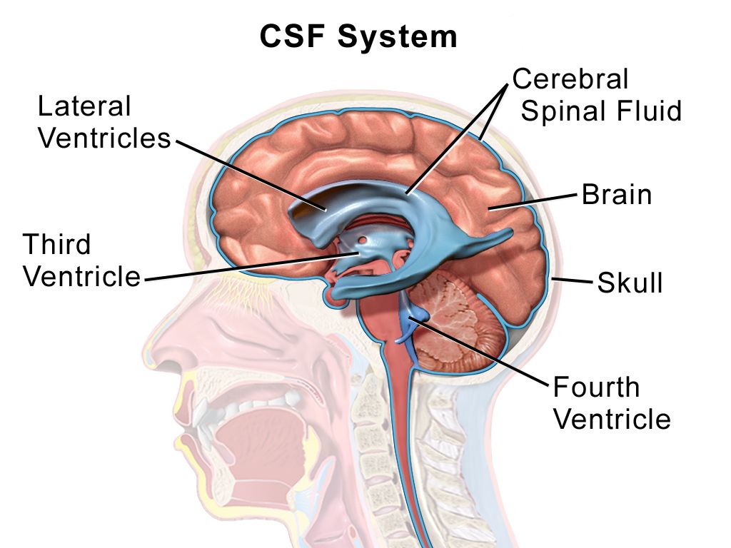
Monro–Kellie, compensation and the cliff
The Monro–Kellie doctrine states that the cranial vault is a fixed volume containing three components: brain tissue at roughly 80 percent, blood at about 10 percent and cerebrospinal fluid at about 10 percent. Because the container cannot expand, an increase in any one component must be matched by a decrease in another or the pressure rises. The brain compensates first by displacing cerebrospinal fluid into the spinal subarachnoid space and increasing its absorption, then by shunting venous blood out of the cranium. These mechanisms buy real time — which is exactly why a patient can look deceptively well while a haematoma grows.
Compensation is finite, and the pressure–volume relationship is not a straight line. Early on, a large added volume raises pressure very little. Once compensation is exhausted, a tiny additional volume — a coughing spell, a poorly timed suction pass, a few more millilitres of clot — produces a steep, sometimes catastrophic rise. That curve explains the clinical experience of a patient who is stable, stable, stable, then suddenly not. It also explains why the interventions in this section look trivial individually: each removes a few millilitres of volume, and near the cliff a few millilitres is the difference.
Causes fall into predictable buckets: mass lesions (haematoma, tumour, abscess), cerebral oedema (cytotoxic oedema after ischaemia and hypoxia, vasogenic oedema around tumours and abscesses), increased cerebrospinal fluid from hydrocephalus, and increased cerebral blood volume from hypercapnia, hypoxia, seizure, fever or venous outflow obstruction. Note that the last bucket is the one nursing care directly controls, which is the point of everything that follows.
Early signs, and why the Cushing triad is too late to be useful as a warning
The earliest sign of rising intracranial pressure is a change in level of consciousness: restlessness, irritability, confusion, needing louder or more repeated stimulation to rouse. Headache, often worse in the morning and worse with coughing or bending, and vomiting that may be projectile and unaccompanied by nausea follow. Pupillary changes come next — a sluggish pupil, then a unilaterally dilated one. Then motor changes: worsening hemiparesis, then abnormal posturing. Papilloedema on funduscopy is a real sign but a late and unreliable one at the bedside.
The Cushing triad — rising systolic pressure with a widening pulse pressure, bradycardia, and irregular or abnormal respirations — is a brainstem reflex to preserve cerebral perfusion when the pressure has already risen dangerously. It is a late sign of impending or ongoing herniation, not an early warning. If you are waiting for the Cushing triad to call the provider, you have waited too long. Be careful how you carry that into an examination question. When the Cushing triad is one of the options it is never the distractor: irregular respirations with bradycardia and a widening pulse pressure mean herniation is happening now, and under airway-breathing-circulation that is the patient you go to first. The teaching point runs the other way. When every option is still subtle, the newly drowsy or newly difficult-to-arouse patient is the one to escalate, because that is the finding that still leaves you time to act before the triad ever appears. Both require action; only one gives you time.
Herniation is displacement of brain across a dural boundary. Uncal (transtentorial) herniation compresses the third cranial nerve, producing an ipsilateral fixed, dilated pupil with contralateral hemiparesis and declining consciousness — the classic “blown pupil.” Central herniation produces progressive rostral-to-caudal deterioration. Tonsillar herniation forces the cerebellar tonsils through the foramen magnum onto the medulla and causes respiratory arrest and death. This is why a lumbar puncture is contraindicated when a mass lesion with raised pressure is suspected: removing fluid from below creates a pressure gradient that pulls the brain downward.
| Timing | Findings | What you do |
|---|---|---|
| Earliest | Restlessness, irritability, confusion, decreased arousal, subtle disorientation | Full neurologic assessment, vital signs, glucose, oxygenation; notify the provider now |
| Early | Headache worse with cough or bending, vomiting without nausea, blurred or double vision | Head of bed 30 degrees, head midline, antiemetic, minimise stimulation, escalate |
| Progressing | Sluggish then unilaterally dilated pupil, worsening hemiparesis, decorticate posturing | Emergent CT, prepare hyperosmolar therapy, alert neurosurgery, prepare for airway management |
| Late | Cushing triad: widening pulse pressure with rising systolic, bradycardia, irregular respirations; decerebrate posturing; fixed dilated pupil | Herniation is occurring — rapid response, airway, hypertonic saline or mannitol, immediate neurosurgical involvement |
| Terminal | Flaccidity, apnoea, bilateral fixed pupils | Full resuscitation per goals of care; outcome is usually determined |
Lowering the pressure: nursing measures, osmotherapy, ventilation and drains
Nursing measures are not filler; they are the first tier of treatment because they reduce cerebral blood volume by improving venous drainage. Keep the head of the bed at 30 degrees, the head in midline neutral alignment, and the neck free of tight tracheostomy ties or cervical collars — head rotation or neck flexion kinks the jugular veins and can raise intracranial pressure measurably. Avoid extreme hip flexion, which raises intra-abdominal and thus intrathoracic pressure. Prevent Valsalva: stool softeners, no straining, no vigorous coughing, and instruct patients not to hold their breath while repositioning. Preoxygenate before suctioning and limit each pass to ten seconds or less; suctioning is a potent pressure spike. Space nursing activities rather than clustering them — unlike most of your care, several stimulating tasks in a row are harmful here. Keep the room quiet and dim, treat pain and agitation (untreated pain raises pressure; oversedation removes your ability to assess), and treat fever aggressively toward normothermia because pyrexia raises cerebral metabolic demand. Use isotonic fluids only. Hypotonic fluids — dextrose 5 percent in water, half-normal saline — move free water into brain tissue and worsen oedema.
Hyperosmolar therapy draws water out of brain tissue across an intact blood–brain barrier. Mannitol 0.25–1 g/kg is infused over 15 to 30 minutes through a filter, because the solution crystallises; inspect every bag and never give one containing visible crystals. Mannitol is an osmotic diuretic, so it causes brisk urine output — an indwelling catheter is generally required — and it can produce hypovolaemia, hypotension, electrolyte disturbance and acute kidney injury. Monitor serum osmolality and hold the dose if it exceeds about 320 mOsm/kg, and watch the sodium, potassium and renal function. Hypertonic saline at 3 percent is infused to a target serum sodium of roughly 145–155 mEq/L with sodium checked every six hours; 23.4 percent hypertonic saline is given as a small bolus for acute herniation and must go through a central line. Hypertonic saline is often preferred in the hypotensive or hypovolaemic patient because it expands rather than depletes intravascular volume. Corticosteroids such as dexamethasone reduce the vasogenic oedema of tumours and abscesses; they are ineffective in ischaemic stroke and harmful in traumatic brain injury, and this distinction is heavily tested.
Hyperventilation is no longer routine. The physiology is real — lowering arterial carbon dioxide constricts cerebral arterioles, reduces cerebral blood volume and drops the pressure within minutes — but the effect is the problem. That vasoconstriction reduces cerebral blood flow to tissue that is already ischaemic, the benefit wanes within hours as bicarbonate buffering equilibrates, and rebound vasodilation follows when the carbon dioxide is allowed to normalise. Prolonged prophylactic hyperventilation worsens outcomes. Current practice keeps the arterial carbon dioxide in the normal 35–45 mmHg range and reserves brief, targeted hyperventilation to 30–35 mmHg as a rescue bridge for acute herniation while definitive therapy — osmotherapy, cerebrospinal fluid drainage, the operating room — is being arranged. Do not hyperventilate a head-injured patient with a bag-valve mask because it feels like doing something.
An external ventricular drain both monitors pressure and treats it by removing cerebrospinal fluid. The transducer and drip chamber are levelled to the foramen of Monro, approximated externally at the tragus of the ear or the outer canthus of the eye, and re-levelled every time the bed height or patient position changes — an unlevelled drain gives you a false number and can over-drain, which collapses ventricles and can cause a subdural haemorrhage. Close the drain before repositioning, before transport, and before any activity that raises pressure, then reopen it and document. Report cloudy drainage, a new fever or a sudden change in output volume. Maintain sterile technique religiously; ventriculitis is a devastating complication. Refractory pressure may require decompressive craniectomy, barbiturate coma with continuous electroencephalographic monitoring, or targeted temperature management.
The skull cannot expand, so a falling level of consciousness is the early warning and the Cushing triad is the late one; treat with midline positioning, venous drainage, hyperosmolar therapy and cerebrospinal fluid drainage, and reserve hyperventilation as a brief rescue rather than a routine.
Section 5From the First Seventy-Two Hours to Discharge
The swallow screen and the complication that kills most stroke patients
Dysphagia affects roughly half of acute stroke patients, and aspiration pneumonia is the leading preventable cause of death in the first weeks. The rule is absolute: keep the patient nil by mouth — no food, no fluids, not even the sip of water used to take a tablet — until a swallow screen has been passed. A validated bedside screen typically checks alertness, ability to sit upright, voice quality, volitional cough and a graded water trial, watching for coughing, throat clearing, a wet or gurgly voice, or a drop in oxygen saturation. Failing the screen means a formal speech-language pathology evaluation and possibly a videofluoroscopic swallow study. Silent aspiration — no cough at all — is common in stroke, so absence of coughing is not reassurance.
Once cleared, position the patient fully upright at 90 degrees for meals and for at least 30 minutes afterward, use the recommended texture and liquid consistency exactly as prescribed, feed slowly with small bites, place food on the unaffected side of the mouth, check for pocketing after every few bites, and perform oral care at least twice a day — oral hygiene measurably reduces pneumonia rates. Avoid straws unless specifically approved, and never rush a meal. If a patient will be unable to swallow safely for more than a few days, enteral feeding is started rather than allowing a week of malnutrition.
Other early complications cluster in a predictable order. Venous thromboembolism prevention begins with intermittent pneumatic compression on admission and early mobilisation. In ischaemic stroke that was not treated with a thrombolytic, prophylactic-dose subcutaneous heparin or low-molecular-weight heparin is started promptly — generally within 24 to 48 hours of admission — once the initial non-contrast CT has excluded haemorrhage, and no repeat scan is required first. It is only after intravenous thrombolysis that every antithrombotic, prophylactic dosing included, is deferred for 24 hours and until follow-up imaging is clear; in haemorrhagic stroke it waits until the haematoma is stable. Malignant cerebral oedema after a large middle cerebral artery infarct peaks between 48 and 72 hours — the patient who deteriorates on hospital day two or three is swelling, not re-stroking. Haemorrhagic transformation of an infarct, urinary retention and infection, pressure injury over the affected heel and sacrum, constipation, seizures and post-stroke depression all belong on the care plan from day one.
Mobility, the affected side and the mind
Protect the hemiplegic shoulder. The affected arm has no muscular support for the glenohumeral joint, and shoulder subluxation with a painful frozen shoulder is common and preventable. Never pull on the affected arm to reposition or transfer, support it on a pillow or lap tray when the patient is sitting, use a sling only as prescribed and not continuously, and perform passive range of motion within the prescribed limits. Position to prevent contracture and foot drop, turn at least every two hours, and inspect skin at every turn — the affected side has impaired sensation and the patient will not report the pain that would normally prompt a shift in position.
Unilateral neglect after a right hemisphere stroke and homonymous hemianopsia are safety issues, not curiosities. A patient who eats half the plate, shaves half the face and walks into the doorframe on the left is at high risk of falls and injury. Initially set up the environment on the unaffected side so the patient can function, then progressively teach deliberate head-turning and visual scanning toward the neglected side; a rehabilitation programme moves toward re-integration rather than permanent accommodation. Approach an impulsive right-hemisphere patient with structure, short instructions and constant supervision; they will attempt to stand and walk without warning and without insight into the deficit.
Post-stroke depression affects roughly a third of survivors and is under-recognised because clinicians attribute flat affect to the stroke itself. Screen for it, treat it, and tell families that emotional lability — sudden crying or laughing disproportionate to the situation — can be a neurologic symptom rather than grief. Communication support matters just as much: with expressive aphasia allow time and use yes/no questions, communication boards and gestures; with receptive aphasia use short simple sentences, one idea at a time, demonstration and consistent word choices. Speak to the patient, not about the patient.
| Task | RN | LPN/LVN | Assistive personnel |
|---|---|---|---|
| Initial and ongoing neurologic assessment, NIHSS, GCS | Yes | No | No |
| Bedside swallow screen before first oral intake | Yes (per policy, RN or SLP) | No | No |
| Administering and monitoring a thrombolytic | Yes | No | No |
| Care of the unstable or deteriorating patient | Yes | No | No |
| Patient and family teaching, evaluation of response | Yes | Reinforce only | No |
| Routine oral and enteral medications on a stable patient | Yes | Yes | No |
| Reinforcing a prescribed swallowing technique on a screened, stable patient | Yes | Yes | Feeding assistance only after the RN has assessed and set up |
| Vital signs on a stable patient, hygiene, turning, transfers, applying compression devices, intake and output, prescribed passive range of motion | Yes | Yes | Yes — with clear reporting parameters |
Secondary prevention, teaching and the older adult
Every ischaemic stroke or TIA patient leaves on antithrombotic therapy matched to the mechanism. For a non-cardioembolic stroke that is aspirin 81–325 mg daily or clopidogrel; after a minor stroke with an NIHSS of 3 or less or a high-risk TIA, dual antiplatelet therapy with aspirin plus clopidogrel is given for about 21 days and then stepped down to a single agent, because continuing both long-term increases bleeding without further benefit. For stroke with atrial fibrillation the answer is anticoagulation, and a direct oral anticoagulant — apixaban, rivaroxaban, edoxaban or dabigatran — is preferred over warfarin except in mechanical heart valves and moderate-to-severe mitral stenosis, where warfarin with an INR of 2.0–3.0 remains standard. High-intensity statin therapy, typically atorvastatin 80 mg, is standard regardless of the baseline cholesterol. Blood pressure control, glycaemic control, smoking cessation, weight and activity, and carotid endarterectomy or stenting for symptomatic stenosis of 70–99 percent complete the package.
Teaching should end with something the family can act on at 0300. Teach BE-FAST — Balance loss, Eyes or vision change, Face drooping, Arm weakness, Speech difficulty, Time to call emergency services — and be explicit that they call an ambulance rather than driving, because prehospital notification starts the stroke pathway before arrival. Teach that symptoms which resolve still require a 911 call. Review each medication by purpose and by the specific thing that would make them call: bleeding on an anticoagulant, muscle pain on a statin, dizziness on an antihypertensive. Confirm follow-up with neurology, primary care and outpatient therapy, arrange home safety modification, and address driving restrictions honestly — visual field cuts and neglect are legal and practical barriers, and the conversation is easier before discharge than after a crash.
Older adults present differently and you must adjust the index of suspicion. Acute confusion or delirium may be the only manifestation of a stroke, and a fall may be the presenting event rather than a consequence. Atrial fibrillation prevalence rises steeply with age, making cardioembolic stroke and anticoagulant-associated haemorrhage both more likely, and polypharmacy makes the medication reconciliation genuinely diagnostic. Age-related cerebral atrophy creates extra space inside the vault, so an older patient tolerates a growing subdural haematoma for far longer before signs appear — the presentation is often insidious confusion weeks after a minor head bump, and the classic pressure signs come late. Reduced renal reserve raises the risk from mannitol, contrast and osmotic shifts, so monitor creatinine and electrolytes closely. Baseline sensory impairment and cognitive decline make assessment harder, which is exactly why obtaining the family’s account of the patient’s true baseline is not optional.
Nothing by mouth until the swallow screen is passed, protect the affected shoulder and skin, mobilise early with the rehabilitation team, and discharge every patient on mechanism-matched antithrombotic therapy, a high-intensity statin, controlled blood pressure and a family who can recognise BE-FAST and call 911.
The chapter in one breath
Cerebral perfusion pressure is mean arterial pressure minus intracranial pressure, and every disorder in this chapter attacks one of those two terms. Ischaemic stroke, about 87 percent of cases, starves a territory and leaves a salvageable penumbra whose collateral flow is pressure-dependent — so blood pressure is permitted up to 220/120 mmHg unless a thrombolytic is being given, in which case it must be below 185/110 before and below 180/105 for twenty-four hours after. Establish last known well, exclude blood with non-contrast CT, quantify with the NIHSS, treat within 4.5 hours, and send large-vessel occlusions for thrombectomy. Haemorrhagic stroke reverses the logic: the vessel is open, so the systolic pressure comes down toward 140 mmHg and anticoagulation is reversed immediately, while aneurysmal subarachnoid haemorrhage adds early aneurysm securing, rigid pressure control until it is secured, and enteral nimodipine every four hours for twenty-one days against vasospasm that peaks on days 4 to 14. Raised intracranial pressure announces itself first as a falling level of consciousness; the Cushing triad is herniation already happening. Treat with midline positioning, venous drainage, osmotherapy and cerebrospinal fluid drainage — not routine hyperventilation. Then keep the patient nil by mouth until the swallow screen is passed, because aspiration pneumonia is what actually kills stroke survivors.
Words worth knowing
| Term | What it means |
|---|---|
| Last known well | The last moment the patient was observed at neurologic baseline; the clock for thrombolysis starts here, not at symptom discovery |
| Ischaemic penumbra | Hypoperfused but still living tissue surrounding an infarct core, salvageable if flow is restored and dependent on collateral perfusion pressure |
| Permissive hypertension | Deliberately tolerating an elevated blood pressure in acute ischaemic stroke, treating only above 220/120 mmHg, to preserve penumbral flow |
| NIHSS | National Institutes of Health Stroke Scale, an eleven-item 0–42 point standardised neurologic examination used to grade stroke severity and track change |
| Alteplase | Recombinant tissue plasminogen activator; 0.9 mg/kg to a maximum of 90 mg, ten percent as a bolus and the rest over 60 minutes, within 4.5 hours of last known well |
| Tenecteplase | A single-bolus fibrinolytic, 0.25 mg/kg to a maximum of 25 mg, increasingly used in place of alteplase for acute ischaemic stroke |
| Orolingual angioedema | Swelling of the tongue and lips occurring in a small percentage of patients receiving alteplase, more common with concurrent ACE inhibitor therapy |
| Mechanical thrombectomy | Catheter-based clot retrieval for large-vessel occlusion, standard within 6 hours and selectively out to 24 hours with favourable perfusion imaging |
| Haematoma expansion | Growth of an intracerebral bleed in the first hours, the main preventable driver of death and disability; limited by blood pressure control and anticoagulant reversal |
| Thunderclap headache | Sudden headache maximal within seconds, classic for subarachnoid haemorrhage and never to be dismissed as migraine |
| Xanthochromia | Yellow discolouration of cerebrospinal fluid from broken-down blood, supporting subarachnoid haemorrhage when the CT is negative |
| Nimodipine | Calcium channel blocker given 60 mg orally or enterally every 4 hours for 21 days after aneurysmal subarachnoid haemorrhage; never given intravenously |
| Cerebral vasospasm | Delayed arterial narrowing after subarachnoid haemorrhage beginning around day 3 and peaking days 4 to 14, causing delayed cerebral ischaemia |
| Cerebral salt wasting | Renal sodium and volume loss after brain injury producing hypovolaemic hyponatraemia; treated with salt and volume, unlike euvolaemic SIADH, which is fluid-restricted — but in aneurysmal subarachnoid haemorrhage neither is fluid-restricted, and hyponatraemia is corrected while maintaining euvolaemia |
| Monro–Kellie doctrine | The principle that brain, blood and cerebrospinal fluid occupy a fixed cranial volume, so an increase in one must be offset or the pressure rises |
| Cerebral perfusion pressure | Mean arterial pressure minus intracranial pressure; target 60–70 mmHg in a monitored patient |
| Cushing triad | Widening pulse pressure with rising systolic pressure, bradycardia and irregular respirations — a late sign of herniation, not an early warning |
| Uncal herniation | Displacement of the temporal lobe compressing the third cranial nerve, producing an ipsilateral fixed dilated pupil with contralateral hemiparesis |
| External ventricular drain | Catheter in a lateral ventricle that measures intracranial pressure and drains cerebrospinal fluid; levelled at the foramen of Monro and clamped before repositioning |
| Unilateral neglect | Failure to attend to one side of the body or environment, typical of right hemisphere stroke and a major fall and injury risk |
Check yourself
Neurologic Disorders: Seizures, Headache and CNS Infection
Three complaints in this chapter arrive looking almost identical — a patient who is confused, a patient whose head hurts, a patient who has just shaken on the floor — and the whole of your nursing judgement lies in telling the benign version from the one that kills within hours. A seizure is a burst of synchronous cortical firing; whether it is a nuisance or an emergency depends entirely on whether it stops. A headache is the commonest human symptom and the presentation of subarachnoid haemorrhage, giant cell arteritis and bacterial meningitis. An infection of the meninges is treatable with a drug you already have on the unit, but the outcome is measured in minutes to first antibiotic dose. So learn the mechanism briefly, then learn the timed algorithms: five minutes for status epilepticus, one hour for meningitis, and “before biopsy” for temporal arteritis. Your assessment, not the scan, is what starts those clocks.
By the end of this chapter you can
- Classify seizures as focal or generalized onset, describe the ictal and postictal phases, and recognise Todd paralysis as a mimic of stroke.
- Perform correct seizure precautions and bedside management, and document the ten observations that determine the diagnosis.
- Define status epilepticus by the five-minute rule and execute the timed algorithm — benzodiazepine first, then fosphenytoin, levetiracetam or valproate.
- Match antiseizure medications to their mechanism, monitoring parameter and signature toxicity, including phenytoin levels, lamotrigine rash and valproate teratogenicity.
- Distinguish migraine, tension-type and cluster headache, and apply the SNOOP red flags that mandate imaging before treatment.
- Interpret cerebrospinal fluid results against real reference values to separate bacterial from viral, fungal and tuberculous meningitis.
- Prioritise the first hour of suspected bacterial meningitis: cultures, dexamethasone, empiric antibiotics, droplet precautions and contact chemoprophylaxis.
- Recognise herpes simplex encephalitis and brain abscess, and state why acyclovir is started before the polymerase chain reaction result returns.
- Describe older-adult variation in all three presentations and construct an SBAR escalation for each red flag.
Section 1Seizures: What the Cortex Is Doing and What You Must See
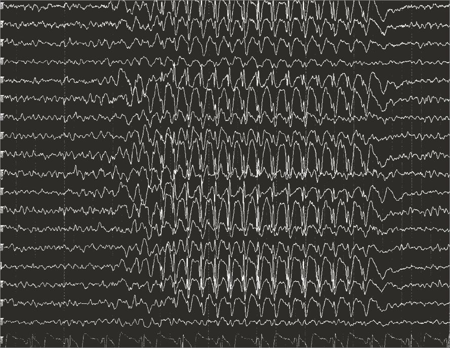
The Mechanism in One Paragraph
A normal cortex balances excitation carried by glutamate against inhibition carried by gamma-aminobutyric acid, and it keeps neighbouring neurons out of step with each other. A seizure is what happens when that balance tips and a population of neurons fires together, repetitively and without the usual braking. If the discharge stays in one region, the patient has a focal seizure and the symptoms are a caricature of whatever that region normally does — the motor strip produces jerking of a limb, the temporal lobe produces a rising epigastric sensation, an odd smell, lip-smacking or an intense sense of familiarity. If the discharge recruits networks in both hemispheres from the outset, the patient has a generalized seizure and consciousness is lost immediately.
That single mechanism explains almost every finding you will chart. Tonic stiffening is sustained motor cortex discharge; the clonic phase is that discharge being interrupted by returning inhibition. Cyanosis occurs because the diaphragm and intercostals are tonically contracted and the patient is not moving air — it is expected, it is brief, and it is not a reason to force an airway between clenched teeth. Incontinence follows loss of sphincter control. Tongue biting is lateral, not on the tip, because the jaw closes on a tongue caught between molars. The postictal state — sleepiness, confusion, headache, muscle soreness — is the cortex exhausted of neurotransmitter and running an oxygen debt, and it may last minutes to hours.
One postictal finding gets mistaken for catastrophe every week in every hospital. Todd paralysis is focal weakness of the limb or side that was seizing, present after the seizure ends and resolving within minutes to as long as forty-eight hours. It looks exactly like a stroke. It is not your job to decide which it is; it is your job to know that a new focal deficit after a witnessed seizure still gets a stroke evaluation, because a stroke can also cause the seizure.
Naming What You Saw
Modern classification asks three questions in order. Where did it start — focal, generalized or unknown onset? Was awareness preserved? Were the first prominent features motor or non-motor? A focal aware seizure is what students were taught to call an aura; the patient is awake and remembers it, and it is the most useful piece of localising information anyone will ever collect. A focal impaired-awareness seizure (previously complex partial) is the staring, unresponsive patient with automatisms — picking at bedding, chewing, fumbling — who afterwards cannot account for the last two minutes. Any focal seizure can spread; when it does, you chart a focal to bilateral tonic-clonic seizure, and the fact that it began focally is diagnostically precious.
Generalized types include tonic-clonic, myoclonic (brief shock-like jerks, often on waking), atonic (sudden loss of tone, the “drop attack” that produces facial and dental injury and often means a helmet at home), tonic, clonic and absence. Absence seizures are seconds of blank staring with abrupt onset and abrupt return, no postictal confusion, and often dozens per day — the school-age child labelled inattentive. Note the contrast that examinations love: the absence patient resumes mid-sentence, the focal impaired-awareness patient is groggy and disoriented for several minutes.
Epilepsy is a diagnosis, not an event. It means two unprovoked seizures more than twenty-four hours apart, or one unprovoked seizure with a recurrence risk of at least sixty per cent (for example a seizure arising from an old infarct visible on MRI), or a defined epilepsy syndrome. A single provoked seizure — from hypoglycaemia, alcohol withdrawal or hyponatraemia — is not epilepsy, and treating it with a lifelong drug instead of correcting the provocation is a classic error.
What You Do During and After — and What You Never Do
The first move is airway and safety, in that order and simultaneously if you can. Stay with the patient and call for help. Note the time on the clock — not “it felt like ages”, the actual time, because the entire treatment algorithm is timed. Ease the patient to the floor if standing, or lower the bed and raise the padded rails if in bed. Turn the patient onto the side so secretions drain, loosen anything tight at the neck, place something soft under the head and clear hard objects out of the arc of the limbs. Give oxygen and have suction at hand. Stay until the airway is patent and the patient is oriented.
Now the prohibitions, because they appear on every examination and, more importantly, because they injure people. Do not restrain the limbs — you will cause fractures and dislocations and you will not shorten the seizure. Do not put anything in the mouth, including an oral airway, a padded tongue blade or your fingers; the tongue cannot be swallowed, and forcing objects between clenched teeth breaks teeth and bites fingers. Do not attempt to insert an oral temperature probe on any patient with seizure precautions. Do not leave to fetch equipment; use the call bell.
Documentation is your clinical contribution, and it is more diagnostic than any scan. Chart what the patient was doing beforehand and any warning; the exact time of onset and of cessation; where the movement started and how it spread; head and eye deviation and to which side; whether awareness was preserved; automatisms; colour change; incontinence; tongue or other injury; the postictal state including orientation, speech and focal weakness; vital signs and capillary glucose. Ten observations, one minute of charting, and the neurologist can classify the seizure from your note.
| Seizure precautions | What it means at the bedside |
|---|---|
| Bed and rails | Bed in lowest position, side rails up and padded per facility policy, call bell within reach. |
| Airway equipment | Suction set up and tested, oxygen and a bag-valve-mask at the bedside — not down the hall. |
| Access | Patent IV for patients at risk, so a benzodiazepine can be given without a search for a vein. |
| Temperature route | Tympanic, temporal or axillary — never oral. |
| Ambulation and hygiene | Supervised showers, no tub baths, no locked bathroom door, assistance for transfers. |
| Medication timing | Antiseizure doses given on time, never held for “NPO” without contacting the prescriber for an IV equivalent. |
Provoked Seizures, the Workup, and the Older Adult
Every first seizure earns a search for a cause, and the reversible causes are ones you can identify from the chart before the neurologist arrives. Capillary glucose is the first test, always, because hypoglycaemia is instantly correctable and instantly fatal if missed. Serum sodium below roughly 120 mEq/L (normal 135–145) seizes; so does severe hypocalcaemia and severe hypomagnesaemia. Alcohol withdrawal seizures cluster six to forty-eight hours after the last drink. Drugs lower the threshold — bupropion, tramadol, meperidine through its metabolite normeperidine, high-dose beta-lactams in renal failure, and stimulant or cocaine intoxication. In pregnancy after twenty weeks and up to six weeks postpartum, a seizure is eclampsia until disproved, and the drug is magnesium sulfate, not lorazepam.
The structural workup is neuroimaging — MRI with epilepsy protocol where available, CT acutely to exclude haemorrhage or mass — plus electroencephalography. Remember what EEG can and cannot do: a normal interictal EEG never excludes epilepsy, because the study samples thirty minutes of a lifetime. Sleep deprivation, photic stimulation and hyperventilation are used to provoke discharges; prolonged video EEG is the gold standard when the diagnosis is uncertain. Add lumbar puncture when fever or immunosuppression suggests infection, and a toxicology screen when the history is thin.
Older adults are different and the difference is dangerous. New-onset seizures after age sixty are most often caused by cerebrovascular disease — a remote infarct is the single commonest substrate — followed by tumour, trauma, dementia-related change and drug effects. Presentation is frequently non-convulsive: hours to days of fluctuating confusion, staring, or unexplained aphasia rather than a dramatic convulsion. This is non-convulsive status epilepticus, it is missed because it is charted as “sundowning”, and the only way to find it is to think of it and ask for an EEG. Postictal states last far longer in older patients, and interactions with warfarin, antihypertensives and hepatically cleared drugs make antiseizure prescribing a careful business.
Delegation. Assistive personnel can pad rails, keep the bed low, place suction and oxygen at the bedside, take vital signs on a stable patient, assist with hygiene and stay with the patient while calling out for help. What may not be delegated: the neurologic assessment, interpreting what the seizure was, teaching about triggers and medications, evaluating whether care worked, and any care of an actively seizing or postictal unstable patient. Those are RN work, every time.
During a seizure your only jobs are time it, protect the airway and the head, and never restrain or place anything in the mouth — and afterwards your observations, not the scan, are what classify it.
Section 2Status Epilepticus and the Drugs That Stop Seizures
The Five-Minute Rule
Status epilepticus is five minutes or more of continuous seizure activity, or two or more seizures between which the patient does not return to baseline awareness. The old thirty-minute definition was abandoned for a practical reason: almost no untreated tonic-clonic seizure that has lasted five minutes will stop on its own, and the longer it runs the harder it becomes to stop. Receptors that benzodiazepines target are internalised within minutes, so a drug that works beautifully at four minutes may fail at forty. Mortality for convulsive status is roughly twenty per cent and climbs with duration, age and cause.
The damage is done three ways. Neurons die from excitotoxic calcium entry driven by relentless glutamate. Systemically, the patient develops hyperthermia from sustained muscle activity, lactic acidosis, hypoxaemia, hyperkalaemia, hypoglycaemia and, in prolonged cases, rhabdomyolysis with acute kidney injury. Autonomic surge produces tachycardia, hypertension then hypotension, and neurogenic pulmonary oedema. This is why the resuscitation is not just anticonvulsant — it is airway, oxygen, glucose, temperature and circulation at the same time.
The commonest precipitant on a medical floor is not new epilepsy. It is a known epileptic whose drug was held for a procedure, whose absorption failed with vomiting, or who ran out of a prescription. Second is acute brain injury — stroke, haemorrhage, trauma, CNS infection. Third is metabolic derangement and alcohol withdrawal. Ask, in this order: what medication was missed, what is the glucose, what is the sodium, and when did the drinking stop.
The Timed Algorithm
Zero to five minutes is stabilisation. Position, open the airway, high-flow oxygen by non-rebreather, suction, pulse oximetry, cardiac monitor, two large-bore IVs. Capillary glucose immediately; if below 60 mg/dL give dextrose, and in a patient with alcohol use disorder or malnutrition give thiamine 100 mg IV before or with the glucose to avoid precipitating Wernicke encephalopathy. Draw sodium, calcium, magnesium, renal and hepatic panel, complete blood count, toxicology and levels of any antiseizure drug the patient takes. Check temperature and start cooling if hyperthermic.
Five to twenty minutes is first-line therapy, and first line is always a benzodiazepine, which enhances GABA-mediated chloride influx and hyperpolarises the neuron. Give IV lorazepam 0.1 mg/kg, maximum 4 mg per dose, repeated once after three to five minutes if seizing continues. If there is no IV, give midazolam 10 mg intramuscularly for an adult over 40 kg — the intramuscular route is as effective as intravenous and faster to deliver in the field. Diazepam 0.15–0.2 mg/kg IV to a maximum of 10 mg is the alternative. The nursing watch is respiratory depression, hypotension and over-sedation; bag-valve-mask at the bedside, not in the supply room. Under-dosing is the commonest error — two milligrams of lorazepam given once is not a treated patient.
Twenty to forty minutes is second-line therapy, given even if the seizure has stopped, to prevent recurrence. Three agents are considered equivalent: fosphenytoin 20 mg phenytoin-equivalents/kg IV (maximum 1500 mg PE, infused no faster than 150 mg PE/min) with continuous cardiac monitoring and blood pressure checks for bradycardia, hypotension and arrhythmia; levetiracetam 60 mg/kg IV (maximum 4500 mg), the friendliest option because it has essentially no cardiac toxicity and no drug interactions; or valproate 40 mg/kg IV (maximum 3000 mg), which is avoided in hepatic disease, pancreatitis, mitochondrial disease and pregnancy.
Beyond forty minutes the patient is in refractory status: repeat a second-line agent or move to anaesthetic infusions of midazolam, propofol or pentobarbital, which means intubation, an intensive care bed and continuous EEG. That last point matters to floor nurses too. Once a patient is paralysed or deeply sedated, convulsions stop but electrical seizures may not, so “the shaking stopped” is no longer evidence of anything. Only the EEG tells you.
Maintenance Antiseizure Medications by Mechanism
Nearly every antiseizure drug does one of three things: block voltage-gated sodium channels so a neuron cannot fire repetitively, enhance GABA-mediated inhibition, or reduce glutamate release and calcium entry. Group them that way and the adverse effects become predictable rather than memorisable. Sodium-channel blockers (phenytoin, carbamazepine, oxcarbazepine, lamotrigine, lacosamide) produce dose-related dizziness, diplopia and ataxia because the cerebellum and brainstem use those same channels. GABA enhancers (benzodiazepines, phenobarbital) sedate. All of them can cause rash, and a few of those rashes are fatal.
| Drug | Mechanism | Therapeutic level | Signature toxicity and nursing monitoring |
|---|---|---|---|
| Phenytoin | Sodium-channel blocker | Total 10–20 mcg/mL; free 1–2 mcg/mL | Nystagmus near 20, ataxia above 30, lethargy and coma above 40 mcg/mL. Gingival hyperplasia — teach meticulous oral care and dental visits. Hirsutism, coarse facies, folate and vitamin D deficiency, osteoporosis. Rash may herald Stevens–Johnson syndrome. Highly protein-bound — check a free level in hypoalbuminaemia or renal failure. Potent enzyme inducer: reduces efficacy of oral contraceptives and warfarin control. Teratogenic. |
| Carbamazepine | Sodium-channel blocker | 4–12 mcg/mL | Hyponatraemia from SIADH — check sodium. Agranulocytosis and aplastic anaemia — baseline and serial CBC, report sore throat, fever, bruising. Stevens–Johnson syndrome and toxic epidermal necrolysis strongly linked to HLA-B*15:02; screen patients of Asian ancestry before starting. Autoinduces its own metabolism, so levels fall after a few weeks. Avoid grapefruit juice. |
| Valproate | Multiple: sodium channels, GABA | 50–100 mcg/mL | Hepatotoxicity — baseline and serial LFTs, report anorexia, vomiting, jaundice. Pancreatitis — report severe abdominal pain. Hyperammonaemia with encephalopathy even when LFTs are normal (ammonia 15–45 mcg/dL). Thrombocytopenia. Weight gain, tremor, hair loss. Neural tube defects — not for patients who can become pregnant without effective contraception and full counselling. |
| Levetiracetam | SV2A synaptic vesicle protein | Not routinely monitored | Behavioural adverse effects are the whole story: irritability, agitation, aggression, depression, occasionally suicidal ideation. Ask the family, not just the patient. Renally cleared — dose reduced in kidney disease. Minimal interactions, which is why it is so widely used. |
| Lamotrigine | Sodium-channel blocker | Not routinely monitored | Serious rash including Stevens–Johnson syndrome; risk rises with rapid titration. Titrate slowly over weeks; when combined with valproate the level roughly doubles, so the starting dose is halved and titration slowed further. Any rash stops the drug and calls the prescriber. Good mood profile, favoured in pregnancy. |
| Phenobarbital | GABA-A enhancer | 15–40 mcg/mL | Sedation, cognitive slowing, respiratory depression, dependence. Strong enzyme inducer. Still first-line for neonatal seizures. |
| Topiramate | Mixed: sodium channels, GABA, carbonic anhydrase | Not routinely monitored | Word-finding difficulty and cognitive slowing, weight loss, paraesthesia, metabolic acidosis, kidney stones (push fluids), reduced sweating with heat intolerance, and acute angle-closure glaucoma — sudden eye pain and blurred vision is an emergency. |
| Oxcarbazepine | Sodium-channel blocker | Monitor sodium, not level | Hyponatraemia more common than with carbamazepine, especially in older adults on diuretics. Fewer haematologic effects; cross-reactive rash with carbamazepine in about one quarter of patients. |
Two rules cut across the whole table. First, never stop an antiseizure medication abruptly — abrupt withdrawal is itself a leading cause of status epilepticus, and that includes holding doses for a procedure without arranging a parenteral equivalent. Second, the enzyme inducers (phenytoin, carbamazepine, phenobarbital) accelerate the metabolism of oral contraceptives, warfarin, direct oral anticoagulants, statins, some antiretrovirals and many chemotherapy agents; every new prescription for these patients needs an interaction check.
Living With Epilepsy: Teaching and Escalation
Adherence is the whole game, and the reasons people miss doses are boring and fixable: cost, forgetting, sedation, and stopping because they “feel fine”. Teach a pill organiser and phone alarms, a written seizure diary with dates, duration and suspected triggers, and never running the last week of a prescription without a refill. Triggers to control are sleep deprivation, missed doses, alcohol, febrile illness, physiologic stress and, in a minority, flashing lights. Teach the family how to time a seizure, when to give a rescue benzodiazepine if one is prescribed (rectal diazepam gel, intranasal midazolam), and precisely when to call emergency services: a seizure lasting more than five minutes, a second seizure without recovery, injury, difficulty breathing, or a first-ever seizure.
Discuss safety honestly. Shower rather than bathe, do not swim alone, use the back burners of the stove, avoid ladders and heights, and consider a helmet for atonic seizures. Driving is regulated by the state and typically requires a seizure-free interval of three to twelve months with prescriber notification. Discuss pregnancy planning early: folic acid supplementation, avoidance of valproate, and never stopping medication on discovering pregnancy without speaking to the neurologist, because convulsive seizures harm a fetus more than most drugs do. And discuss SUDEP — sudden unexpected death in epilepsy — because the biggest modifiable risk factor is uncontrolled generalized tonic-clonic seizures, and patients who understand that take their tablets.
For drug-resistant epilepsy, defined as failure of two appropriately chosen and tolerated drugs, options include epilepsy surgery (resection of a defined focus after video EEG localisation), vagus nerve stimulation, responsive neurostimulation and the ketogenic diet. Referral to a comprehensive epilepsy centre after two failed drugs is the standard, and it is often the nurse who notices that the patient is on their fourth agent and still seizing weekly.
A seizure lasting five minutes is status epilepticus and needs an adequately dosed benzodiazepine immediately, followed by a second-line agent even if the convulsing has stopped.
Section 3Headache: Sorting the Miserable From the Catastrophic

The Three Primary Headaches and How They Differ
Primary headache means the headache is the disease; there is nothing structurally wrong. Migraine is a disorder of a hyperexcitable brain in which a slow wave of neuronal depolarisation followed by suppression — cortical spreading depression — produces the aura, and activation of the trigeminovascular system with release of calcitonin gene-related peptide (CGRP) produces the pain, the meningeal vasodilation and the sensitisation that makes light, sound and even hair-brushing intolerable. Clinically: four to seventy-two hours, unilateral more often than not, pulsating, moderate to severe, worse with routine activity, with nausea or vomiting and photophobia and phonophobia. Roughly a quarter have aura — visual most commonly, sensory or speech occasionally — developing over five or more minutes and lasting under an hour.
Tension-type headache is the opposite phenotype and the commonest headache overall: bilateral, pressing or tightening like a band, mild to moderate, not worsened by walking upstairs, without vomiting and with at most one of photophobia or phonophobia. Patients function through it. It responds to simple analgesia, sleep, hydration, posture correction and stress management, and it is the headache most often over-treated into a bigger problem.
Cluster headache is the most severe pain in clinical medicine and the easiest to recognise if you have seen one. Strictly unilateral, orbital or temporal, boring or stabbing, lasting fifteen to a hundred and eighty minutes untreated, occurring one to eight times daily in bouts of weeks to months, often waking the patient at the same hour each night. The give-away is the pairing of ipsilateral autonomic features — tearing, conjunctival redness, nasal congestion or rhinorrhoea, eyelid oedema, ptosis and miosis — with restlessness. The migraine patient lies still in a dark room; the cluster patient paces, rocks and cannot keep still. It affects men more than women, and alcohol reliably triggers attacks during a bout.
Treating Migraine and Cluster
Abortive migraine therapy works best taken early, at the first sign of pain, and works poorly once central sensitisation has set in — teach that explicitly, because patients are trained to “wait and see”. Mild attacks: NSAIDs or aspirin or acetaminophen, with an antiemetic such as metoclopramide or prochlorperazine, which treats the nausea and also has independent antimigraine effect (watch for akathisia and acute dystonia; diphenhydramine is the antidote at hand). Moderate to severe attacks: a triptan, a serotonin 1B/1D agonist that constricts dilated cranial vessels and inhibits CGRP release. Sumatriptan comes oral, nasal and subcutaneous; the subcutaneous route works in ten minutes when vomiting defeats tablets.
The triptan contraindications are the ones you must know cold, because they are vascular: known coronary artery disease, prior myocardial infarction, uncontrolled hypertension, peripheral vascular disease, ischaemic stroke, hemiplegic or brainstem-aura migraine, and use of an ergot within twenty-four hours. Teach the patient that chest tightness, jaw or throat tightness and flushing can occur and must be reported. Where triptans are unsafe, the newer classes are a genuine advance: gepants (ubrogepant, rimegepant) are small-molecule CGRP receptor antagonists that do not vasoconstrict, and lasmiditan is a 5-HT1F agonist without vascular effect — but it causes dizziness and sedation and the patient must not drive for eight hours after a dose.
Prevention is offered when attacks occur four or more days a month, or are disabling. Classes: beta-blockers (propranolol, metoprolol — avoid in asthma and check heart rate), topiramate, valproate, amitriptyline or venlafaxine, candesartan, and the monoclonal antibodies against CGRP or its receptor (erenumab, fremanezumab, galcanezumab, eptinezumab) given monthly or quarterly. OnabotulinumtoxinA is licensed for chronic migraine, defined as fifteen or more headache days a month. Every preventive needs a two-to-three-month trial at an adequate dose before it is called a failure — tell the patient that at the first visit or they will abandon it in week three.
Cluster headache is treated differently and the difference is examinable. The acute treatment is high-flow oxygen, 12–15 L/min via a non-rebreather mask for fifteen to twenty minutes, which aborts most attacks, plus subcutaneous sumatriptan 6 mg or intranasal zolmitriptan. Oral drugs are useless — the attack is over before they absorb. Preventive treatment during a bout is verapamil at high dose, which requires a baseline and serial ECG because of heart block, with a steroid taper or occipital nerve block as a bridge. Galcanezumab is approved for episodic cluster. Lithium remains an option.
Secondary Headache: The Red Flags You Cannot Miss
Secondary headache means something else is causing it, and the screening mnemonic is SNOOP. Systemic features — fever, weight loss, immunosuppression, cancer, pregnancy. Neurologic signs — any focal deficit, papilloedema, seizure or altered mentation. Onset that is sudden — the thunderclap headache reaching maximum intensity within one minute. Older — a new headache after age fifty. Pattern change, Positional worsening, Precipitation by Valsalva, cough or exertion, Papilloedema, Pregnancy or postpartum state, and Painkiller overuse. Any of these means imaging and a full evaluation before symptomatic treatment.
A thunderclap headache — “the worst headache of my life”, maximal in under a minute, often with vomiting, neck stiffness or brief loss of consciousness — is subarachnoid haemorrhage until proven otherwise. Non-contrast CT within six hours of onset detects over ninety-five per cent; beyond that a negative CT is followed by lumbar puncture looking for xanthochromia, or by CT angiography. Other thunderclap causes include cervical artery dissection, cerebral venous sinus thrombosis (think postpartum, oral contraceptives, thrombophilia, dehydration), reversible cerebral vasoconstriction syndrome and pituitary apoplexy. Your action at the bedside is the same for all of them: stop, do not give a triptan, take vital signs and a neurologic check, and escalate immediately.
A new headache in a patient over fifty with scalp tenderness, jaw claudication when chewing, shoulder-girdle aching or any visual change is giant cell arteritis until proved otherwise. The erythrocyte sedimentation rate is usually well above 50 mm/hr and C-reactive protein is elevated; temporal artery biopsy or vascular ultrasound confirms it. The critical teaching point is sequencing: high-dose corticosteroids are started immediately, before the biopsy, because the untreated disease causes permanent, irreversible blindness and biopsy remains informative for one to two weeks after steroids begin. If vision is already affected, treatment is intravenous methylprednisolone.
Two more patterns worth holding. A headache that is worse lying flat and on waking, worse with cough or straining, with vomiting and papilloedema, suggests raised intracranial pressure from a mass or from idiopathic intracranial hypertension — classically a young woman with obesity, papilloedema, pulsatile tinnitus and a CSF opening pressure above 25 cm H2O, treated with weight loss and acetazolamide. And in an older adult, especially one on an anticoagulant, a headache that has crept up over days to weeks after a minor knock — sometimes a bump they have forgotten — is a chronic subdural haematoma; look for gait change, personality change or fluctuating confusion.
| Feature | Migraine | Tension-type | Cluster |
|---|---|---|---|
| Site | Unilateral, may alternate | Bilateral, band-like | Strictly unilateral, orbital or temporal |
| Quality and severity | Pulsating, moderate to severe | Pressing, mild to moderate | Boring or stabbing, excruciating |
| Duration | 4–72 hours | 30 minutes to 7 days | 15–180 minutes, 1–8 per day |
| Associated features | Nausea, vomiting, photophobia, phonophobia, aura in about 25 per cent | Few or none | Ipsilateral tearing, nasal congestion, ptosis, miosis |
| Behaviour | Lies still in a dark, quiet room | Continues activity | Restless, pacing, cannot sit still |
| Acute treatment | NSAID, triptan, gepant, antiemetic | Simple analgesia, non-drug measures | Oxygen 12–15 L/min plus subcutaneous sumatriptan |
Treat headache as primary only after the SNOOP red flags are clear — thunderclap onset, new headache over fifty, fever with neck stiffness, focal deficit or papilloedema all buy imaging and escalation, not analgesia.
Section 4Meningitis: The Hour That Decides the Outcome
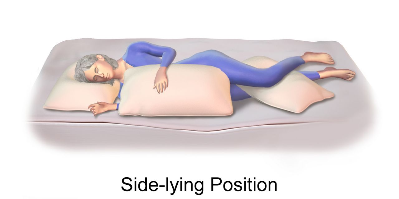
Mechanism and Presentation
Bacteria reach the subarachnoid space by colonising the nasopharynx and invading the bloodstream, by contiguous spread from sinusitis, otitis media or mastoiditis, or directly through a skull fracture, neurosurgical wound or shunt. Once inside, they replicate in a space with almost no immune surveillance. The inflammatory response — not the organism — does most of the damage: cytokines increase blood–brain barrier permeability, producing vasogenic cerebral oedema; purulent exudate obstructs cerebrospinal fluid outflow, producing hydrocephalus and rising intracranial pressure; and inflamed vessels thrombose, producing infarcts and cranial nerve palsies. That is why steroids given before the antibiotic matter, and it is why the patient can deteriorate in the first hours of apparently correct treatment.
The organisms follow age and host. Streptococcus pneumoniae is the commonest cause in adults and the most likely to leave deafness or focal deficit behind. Neisseria meningitidis favours adolescents and young adults in congregate settings and produces the petechial or purpuric rash that must never be dismissed as a viral exanthem. Listeria monocytogenes matters in adults over fifty, pregnancy, alcohol use disorder, malignancy and immunosuppression — and it is the reason ampicillin is added to the empiric regimen in those patients. Gram-negative bacilli and staphylococci dominate after neurosurgery or head trauma. Viral (aseptic) meningitis, most often enteroviral, is far commoner overall and far milder.
The classic triad of fever, nuchal rigidity and altered mental status is present in fewer than half of adults, so do not wait for it. Almost all patients have at least two of headache, fever, neck stiffness and altered mentation. Add photophobia, vomiting, seizures in roughly one in five, and focal deficits. Kernig sign (resistance and pain on extending the knee with the hip flexed) and Brudzinski sign (involuntary hip and knee flexion on passive neck flexion) are famous but insensitive — their absence proves nothing. In older adults the presentation is often confusion alone, with a low-grade or absent fever and a supple neck, which is precisely why the diagnosis is missed in the population with the highest mortality.
Diagnostics: Reading the Cerebrospinal Fluid
Lumbar puncture is the definitive test. Normal CSF is crystal clear with an opening pressure of 6–20 cm H2O, 0–5 white cells/mm3 (lymphocytes), protein 15–45 mg/dL and glucose 45–80 mg/dL, which should be about two-thirds of a simultaneous serum glucose. Nursing role: have the patient empty the bladder, position curled in lateral decubitus or sitting leaning forward to open the interspaces, provide a consent check and support during the procedure, label tubes in order, send them promptly, and afterwards position the patient for comfort — routine flat bed rest after the tap does not prevent post-dural-puncture headache and is no longer required — encourage fluids, and monitor the site for leakage and the patient for post-dural-puncture headache (positional, worse upright, treated with fluids, caffeine and if persistent an epidural blood patch).
| CSF parameter | Normal | Bacterial | Viral | Fungal or tuberculous |
|---|---|---|---|---|
| Appearance | Clear, colourless | Cloudy, turbid, purulent | Clear | Clear or slightly cloudy |
| Opening pressure (cm H2O) | 6–20 | Elevated, often above 30 | Normal or mildly raised | Elevated, sometimes markedly |
| White cells per mm3 | 0–5 | 1000–5000, neutrophils above 80 per cent | 50–1000, lymphocyte predominant | 100–500, lymphocyte predominant |
| Protein (mg/dL) | 15–45 | 100–500 | Normal to about 100 | Very high, often above 200 |
| Glucose (mg/dL) | 45–80; CSF-to-serum ratio at or above 0.6 | Below 40; ratio below 0.4 | Normal | Low |
| Other | Sterile | Gram stain positive in 60–90 per cent; culture | Enterovirus or HSV PCR | India ink, cryptococcal antigen, acid-fast stain, PCR |
Head CT is obtained before lumbar puncture only when herniation risk is real: immunocompromise, known CNS disease, new-onset seizure, papilloedema, reduced or fluctuating consciousness, focal neurologic deficit, or age sixty and above. Blood cultures — two sets — are drawn in every case, and they are positive in roughly half to three-quarters of bacterial cases when they are drawn before antibiotics. Prior antibiotic exposure sharply reduces the yield of both blood and CSF cultures, which is exactly why cultures precede the first dose in the first-hour sequence. Expect elevated white count with left shift, elevated CRP and procalcitonin, and check sodium at baseline because SIADH is common.
The First Hour
Here is the sequence that saves lives, and the order is the point. Recognise the patient. Draw blood cultures. Give dexamethasone 0.15 mg/kg IV ten to twenty minutes before or together with the first antibiotic dose, continued every six hours for four days — it reduces hearing loss and mortality in pneumococcal disease, and it works only if it precedes the bacterial lysis the antibiotic causes. Give empiric antibiotics. Then arrange CT and lumbar puncture if they are indicated. Antibiotics are never delayed for a scan or a tap. A student who answers “get the LP first” has just cost a patient their hearing.
Empiric therapy for community-acquired bacterial meningitis in an adult is vancomycin plus ceftriaxone 2 g IV every 12 hours (or cefotaxime), with ampicillin 2 g IV every 4 hours added for patients over fifty, pregnant patients and the immunocompromised to cover Listeria, which cephalosporins do not touch. Add acyclovir if encephalitis is in the differential. Vancomycin is dosed to a trough of 15–20 mcg/mL or by AUC, with renal function and infusion-rate monitoring — infuse over at least an hour to avoid the histamine flushing reaction. Therapy is narrowed once cultures return and typically runs seven days for meningococcus, ten to fourteen for pneumococcus and twenty-one for Listeria or gram-negatives.
Nursing management runs in parallel. Neurologic checks with Glasgow Coma Scale and pupils at least hourly while unstable. Head of bed at 30 degrees with the neck midline to aid venous drainage. A quiet, dimly lit room, with nursing activities spaced rather than clustered so intracranial pressure can settle back to baseline between them, because photophobia and hyperacusis are genuine and stacked stimulation drives the pressure up. Seizure precautions in place from admission. Antipyretics and cooling for fever, which increases cerebral metabolic demand. Strict intake and output with daily weights and serum sodium, because SIADH presents as falling sodium with concentrated urine and may require fluid restriction. Pain management, oral care, deep-vein thrombosis prophylaxis and pressure-injury prevention for the patient who cannot move.
Isolation, Prophylaxis and What Is Left Behind
Place any patient with suspected bacterial meningitis on droplet precautions immediately — private room, surgical mask donned on entry to the room rather than only when you come within a metre, mask on the patient during transport — and maintain them until twenty-four hours of effective antibiotic therapy have been completed. Do not wait for confirmation of the organism; you are protecting staff and other patients against meningococcus, which is transmitted by respiratory droplets.
Close contacts of a meningococcal case need chemoprophylaxis within twenty-four hours: household members, childcare contacts, anyone directly exposed to oral secretions, and healthcare workers who performed mouth-to-mouth resuscitation, intubation or unprotected airway suctioning. Simply being in the room, or handing a chart across the desk, does not qualify. Regimens are ciprofloxacin 500 mg orally as a single dose, rifampin 600 mg orally twice daily for two days (warn about orange discoloration of urine, sweat and tears, permanent staining of soft contact lenses, and reduced oral contraceptive efficacy), or ceftriaxone 250 mg intramuscularly once, which is preferred in pregnancy. Prevention long-term is vaccination: MenACWY and MenB for adolescents and at-risk adults, pneumococcal conjugate vaccine for older adults and the immunocompromised, and Hib in childhood.
Complications to anticipate and chart against: rising intracranial pressure and herniation, seizures, communicating hydrocephalus, cerebral infarction from vasculitis, cranial nerve palsies, septic shock, disseminated intravascular coagulation and — the commonest permanent sequela — sensorineural hearing loss. Every survivor of bacterial meningitis needs formal audiology testing before or shortly after discharge, and families need to know that a child who seems fine may have lost hearing.
In suspected bacterial meningitis the order is cultures, dexamethasone, antibiotics, then imaging and lumbar puncture — the tap never delays the drug, and droplet precautions start on suspicion.
Section 5Encephalitis, Brain Abscess and the Long Tail of CNS Infection
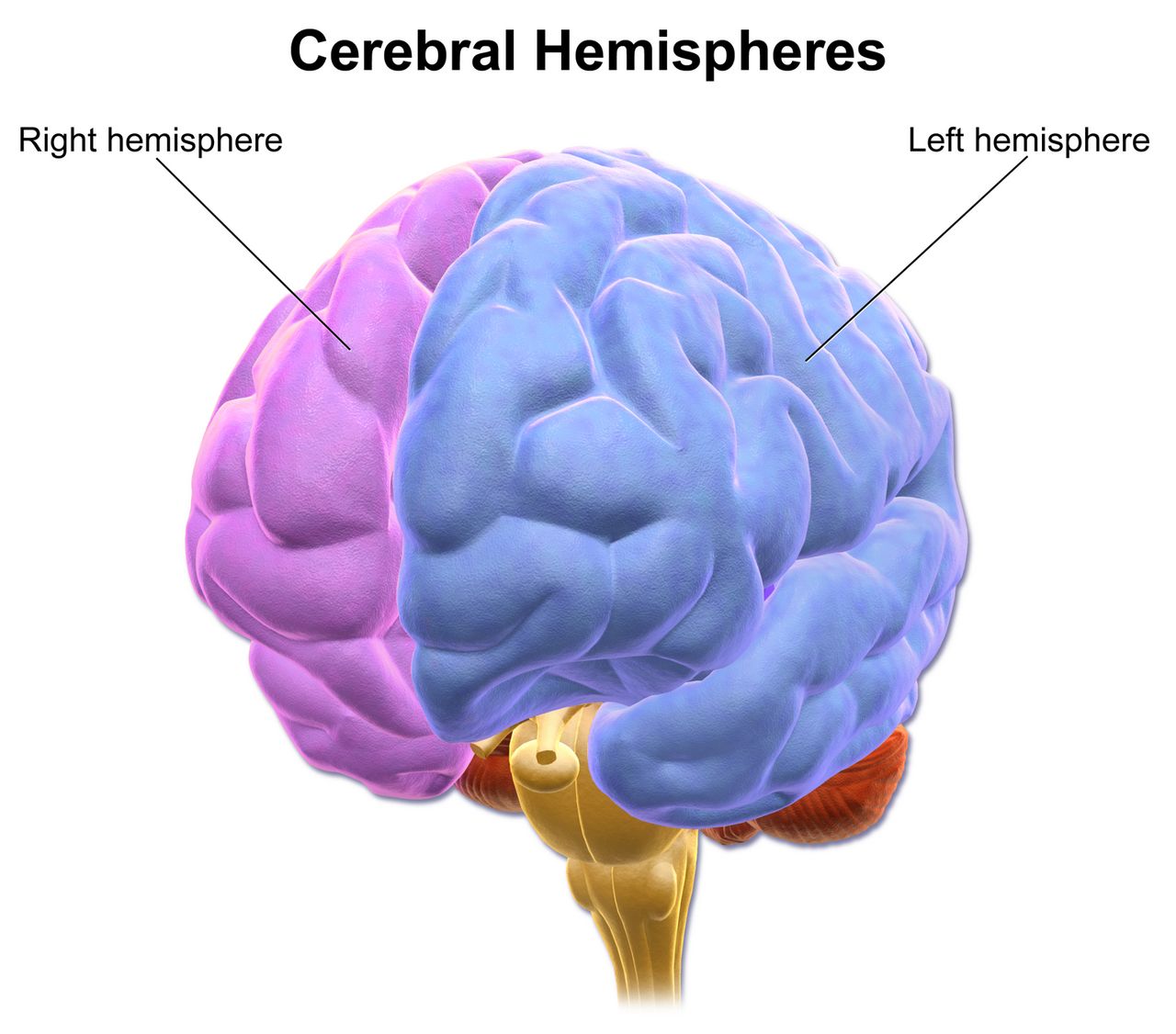
Encephalitis: When the Brain Itself Is Inflamed
Meningitis inflames the coverings; encephalitis inflames the brain substance. That single distinction predicts the clinical difference. Meningitis gives you headache, fever, neck stiffness and a patient who is irritable but whose cortex works. Encephalitis gives you altered mental status as the defining feature — personality change, psychosis, disorientation, aphasia, focal deficits, movement disorders and seizures — often with fever and often with meningeal signs too, since the two overlap as meningoencephalitis.
Herpes simplex virus type 1 is the commonest sporadic cause in adults and the one that must never be missed, because it is treatable and untreated mortality approaches seventy per cent. It has a predilection for the temporal and inferior frontal lobes, which is why it presents with bizarre behaviour, olfactory or gustatory hallucinations, memory failure, aphasia and focal seizures. MRI shows temporal lobe hyperintensity; EEG may show periodic lateralised discharges; CSF shows a lymphocytic pleocytosis, elevated protein, normal glucose, often red cells, and a positive HSV polymerase chain reaction. Intravenous acyclovir 10 mg/kg every 8 hours for fourteen to twenty-one days is started empirically on suspicion and never held for the PCR result — every hour of delay costs neurologic function. The nursing watch is renal: acyclovir crystallises in the tubules, so infuse over at least one hour, hydrate generously, and monitor creatinine and urine output. Phlebitis at the site is common.
Other causes worth recognising. Arboviral encephalitis follows mosquito season: West Nile virus classically adds an asymmetric flaccid paralysis resembling polio; eastern equine and La Crosse viruses are regional. Rabies, once symptomatic, is essentially always fatal — the intervention is post-exposure prophylaxis at the time of the bite. And a category students miss entirely: autoimmune encephalitis, of which anti-NMDA-receptor encephalitis is the archetype — typically a young woman with a viral prodrome, then psychiatric symptoms severe enough to reach a psychiatric ward, then seizures, orofacial dyskinesias and autonomic instability, sometimes with an underlying ovarian teratoma. Treatment is immunotherapy and tumour removal, not antivirals, and the diagnosis is made only when someone thinks of it.
Brain Abscess and Other Space-Occupying Infection
A brain abscess begins as a focal cerebritis that walls itself into a pus-filled cavity with a capsule. Bacteria arrive by contiguous spread from otitis media, mastoiditis, sinusitis or dental infection, by haematogenous seeding from endocarditis, lung abscess or a right-to-left cardiac shunt, or directly from penetrating trauma or surgery. The classic triad of headache, fever and focal deficit is complete in a minority; headache is nearly universal, fever occurs in about half, and the tempo is days to weeks rather than hours. New focal seizures, progressive hemiparesis or a papilloedematous fundus in a patient with a chronic ear or sinus infection should prompt imaging.
Contrast-enhanced MRI or CT shows the ring-enhancing lesion with surrounding vasogenic oedema, and diffusion-weighted imaging distinguishes abscess from tumour. Lumbar puncture is contraindicated when there is mass effect, because withdrawing fluid below a pressure gradient invites herniation; the diagnosis is made by imaging and by culture of aspirated material. Management is stereotactic aspiration or excision plus prolonged intravenous antibiotics, commonly vancomycin with a third or fourth-generation cephalosporin and metronidazole for anaerobes, for six to eight weeks, with dexamethasone if oedema threatens and antiseizure prophylaxis in many cases. Nursing care is neurologic surveillance, long-line management, and the practical work of a six-week outpatient antibiotic course.
Two other patterns deserve a line. Cryptococcal meningitis presents subacutely in advanced HIV with a CD4 count usually below 100 cells/mm3, with headache and fever but often minimal neck stiffness; diagnosis is by cryptococcal antigen or India ink; treatment is amphotericin B with flucytosine followed by fluconazole, and the defining nursing issue is serial therapeutic lumbar punctures to control the very high opening pressure that causes blindness and death. Tuberculous meningitis is basilar, subacute over weeks, with cranial nerve palsies, very high CSF protein and low glucose, and requires four-drug therapy with adjunctive steroids. And any patient with a ventriculoperitoneal shunt who develops fever, headache, vomiting or altered mentation has a shunt infection or malfunction until proved otherwise.
Complications, the Older Adult, and Priority Setting
Whatever the organism, CNS infection kills through a small number of final common pathways, and these are what your assessments are hunting for. Raised intracranial pressure from oedema, hydrocephalus or abscess: declining level of consciousness first, then headache and vomiting, then the late and ominous Cushing triad of widening pulse pressure, bradycardia and irregular respirations, with a fixed dilated pupil signalling herniation. Seizures, including non-convulsive status in the persistently obtunded. SIADH with dilutional hyponatraemia. Septic shock and DIC. Cerebral infarction from inflamed, thrombosed vessels. And after recovery, cognitive impairment, hearing loss, epilepsy and fatigue that outlast the infection by months.
The priority framework does not change. Airway and breathing before circulation, circulation before neurologic status, and any change in level of consciousness outranks every other neurologic finding because it is the earliest and most sensitive sign of rising pressure. Unstable before stable; acute before chronic; actual problems before risks. Applied concretely: the postictal patient with noisy respirations is seen before the febrile confused patient, who is seen before the migraine patient waiting for her second dose of analgesia, who is seen before the stable epilepsy patient wanting discharge teaching — but the teaching still happens before the shift ends, because it is what prevents the next admission.
Older adults change every one of these presentations. Meningitis appears as confusion without fever or neck stiffness, and Listeria coverage becomes mandatory. New seizures are usually cerebrovascular and often non-convulsive. New headache demands consideration of giant cell arteritis, subdural haematoma and mass lesion before anyone says migraine, since primary headache rarely begins after fifty. Antiseizure drugs interact with polypharmacy, and drugs like phenytoin and phenobarbital add sedation, ataxia and fall risk to a patient who already has both. Renal and hepatic clearance are reduced, so start low and go slow, and reassess gait and orthostatic vitals after every dose change.
| Red flag you observe | What it may mean | Your first action |
|---|---|---|
| Seizure lasting past five minutes, or a second seizure without waking | Status epilepticus | Airway, oxygen, glucose, IV access; call for a benzodiazepine order now |
| Falling GCS, widening pulse pressure, bradycardia, irregular breathing | Raised ICP with impending herniation | Airway and oxygen first, then head of bed 30 degrees with neck midline; call rapid response, prepare hyperosmolar therapy |
| Fever with headache and any spreading petechial rash | Meningococcaemia | Droplet precautions, cultures, escalate for immediate antibiotics and fluid resuscitation |
| Thunderclap headache, maximal within one minute | Subarachnoid haemorrhage | Vital signs and neurologic check, escalate immediately for stat non-contrast CT; keep nothing by mouth and give no triptan |
| New headache over age fifty with jaw claudication or visual change | Giant cell arteritis | Stat ESR and CRP, escalate for high-dose steroids before biopsy |
| Any rash in a patient recently started on lamotrigine or carbamazepine | Stevens–Johnson syndrome | Hold the next dose and notify the prescriber before giving it |
| Serum sodium falling with concentrated urine after CNS infection | SIADH | Strict intake and output, daily weight, seizure precautions, notify for fluid restriction |
Every CNS infection converges on the same killers — rising intracranial pressure, seizures, SIADH and shock — so a falling level of consciousness is the single finding that outranks all others and triggers escalation.
The chapter in one breath
A seizure is synchronous cortical firing, and what you see depends on where it fires: focal seizures caricature the region involved, generalized seizures take consciousness at once. At the bedside you time it, protect the airway and head, never restrain and never put anything in the mouth, then document the ten observations that classify it. Five minutes of continuous seizing is status epilepticus, and the algorithm is timed — stabilise and check glucose, then an adequately dosed benzodiazepine, then fosphenytoin, levetiracetam or valproate whether or not the shaking has stopped. Maintenance drugs are grouped by mechanism, and their monitoring follows: phenytoin levels 10–20 mcg/mL and gum care, carbamazepine sodium and blood counts, valproate liver and ammonia and pregnancy risk, lamotrigine rash, levetiracetam behaviour. Headache is sorted by phenotype and screened by SNOOP: thunderclap means subarachnoid haemorrhage, new headache over fifty means giant cell arteritis and steroids before biopsy, fever with stiff neck means meningitis. In suspected bacterial meningitis the order is cultures, dexamethasone, antibiotics, then the scan and the tap — with droplet precautions from the moment you suspect it. And in encephalitis, acyclovir starts before the PCR returns.
Words worth knowing
| Term | What it means |
|---|---|
| Aura | A focal aware seizure that precedes spread; the patient is conscious and remembers it, and it localises the seizure onset. |
| Postictal state | The period after a seizure of confusion, drowsiness, headache and muscle soreness as the exhausted cortex recovers. |
| Todd paralysis | Transient focal weakness after a seizure, lasting minutes up to 48 hours, that mimics stroke and still warrants stroke evaluation. |
| Status epilepticus | Five minutes or more of continuous seizure activity, or repeated seizures without return to baseline awareness between them. |
| Non-convulsive status | Ongoing electrical seizure activity presenting as prolonged confusion or unresponsiveness without convulsions; diagnosed only by EEG. |
| Automatism | Repetitive purposeless movement — lip-smacking, chewing, picking at clothing — typical of focal impaired-awareness seizures. |
| SUDEP | Sudden unexpected death in epilepsy; risk rises with uncontrolled generalized tonic-clonic seizures and poor medication adherence. |
| Phenytoin equivalents (PE) | The dosing unit for fosphenytoin, expressing the amount of phenytoin the prodrug will yield; misreading it causes overdose. |
| Cortical spreading depression | A slow wave of neuronal depolarisation followed by suppression that produces migraine aura. |
| CGRP | Calcitonin gene-related peptide, the neuropeptide central to migraine pain and the target of gepants and the monoclonal antibodies. |
| Thunderclap headache | Headache reaching maximum intensity within one minute; subarachnoid haemorrhage until proved otherwise. |
| Medication-overuse headache | Daily or near-daily headache caused by frequent acute analgesic use; requires withdrawal plus a preventive agent. |
| Giant cell arteritis | Large-vessel vasculitis of older adults causing new headache, scalp tenderness, jaw claudication and irreversible blindness if steroids are delayed. |
| Nuchal rigidity | Resistance and pain on passive neck flexion from meningeal irritation; absent in many older adults with meningitis. |
| Kernig and Brudzinski signs | Classic meningeal irritation manoeuvres; highly specific when present but far too insensitive to rule meningitis out. |
| Opening pressure | Cerebrospinal fluid pressure measured at lumbar puncture; normal 6–20 cm H2O, markedly elevated in bacterial and cryptococcal disease. |
| Pleocytosis | An increased white cell count in cerebrospinal fluid; neutrophilic suggests bacterial, lymphocytic suggests viral, fungal or tuberculous. |
| Waterhouse–Friderichsen syndrome | Bilateral adrenal haemorrhage with vascular collapse and DIC complicating fulminant meningococcaemia. |
| Ring-enhancing lesion | A rounded lesion whose capsule takes up contrast on CT or MRI; in the febrile patient it suggests brain abscess. |
| Aseptic meningitis | Meningeal inflammation with negative routine bacterial cultures, most often enteroviral and usually self-limiting. |
Check yourself
Neurologic Disorders: Degenerative and Neuromuscular Disease
Stroke announces itself; these diseases do not. Parkinson disease, multiple sclerosis, myasthenia gravis, Guillain–Barré syndrome, amyotrophic lateral sclerosis and Alzheimer disease arrive over months or slide in over a weekend, and the nurse is usually the person who notices that something has changed. What unites them is a single motor pathway — cortex to corticospinal tract to anterior horn cell to peripheral nerve to neuromuscular junction to muscle — and a single shared endpoint that kills people: the diaphragm gets weak, secretions pool, the swallow fails and the patient aspirates or stops ventilating. This chapter teaches you to localise the lesion from the bedside exam, to read the diagnostics that confirm it, to give the drugs safely (levodopa is time-critical; magnesium can put a myasthenic on a ventilator), and to recognise the moment when a chronic disease becomes an airway emergency. Everything else — teaching, mobility, dignity — is built on getting that moment right.
By the end of this chapter you can
- Differentiate upper motor neuron, lower motor neuron, neuromuscular junction and muscle lesions using reflexes, tone, atrophy, fasciculations and sensory findings.
- Monitor neuromuscular respiratory failure with forced vital capacity, negative inspiratory force and bedside screens, and state the values that trigger escalation.
- Explain the dopaminergic basis of Parkinson disease and administer carbidopa–levodopa and adjuncts safely, including the dopamine blockers that are contraindicated.
- Apply the McDonald criteria concept of dissemination in space and time to multiple sclerosis, and distinguish a true relapse from a pseudo-relapse.
- Compare myasthenic crisis and cholinergic crisis and prioritise airway assessment over diagnostic labelling.
- Describe albuminocytologic dissociation, autonomic instability and the immunotherapy of Guillain–Barré syndrome, including why corticosteroids are not used.
- Plan care for amyotrophic lateral sclerosis and Alzheimer disease, including noninvasive ventilation timing, enteral access decisions and behavioural management without restraints.
- Identify red flags in each disorder and escalate using SBAR, and delegate correctly — assessment, teaching, evaluation and unstable patients stay with the RN.
- Describe how older adults present differently, and how polypharmacy and anticholinergic burden change the risk calculation.
Section 1Reading the failing motor unit
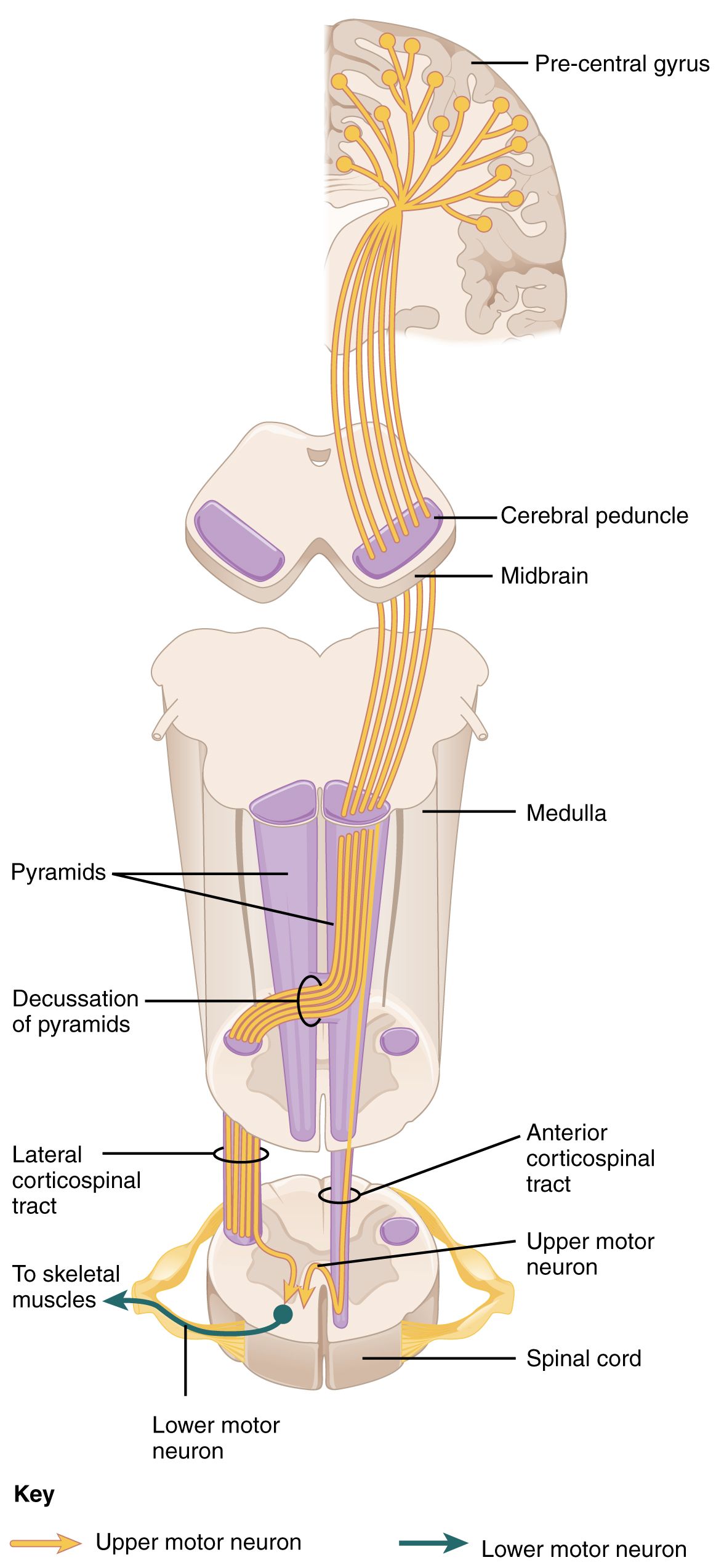
Upper motor neuron, lower motor neuron, and everything between
Voluntary movement travels a two-neuron relay. The upper motor neuron begins in the precentral gyrus, descends through the internal capsule, brainstem and corticospinal tract, and synapses in the anterior horn of the spinal cord. The lower motor neuron leaves the anterior horn, becomes the peripheral nerve, crosses the neuromuscular junction and contracts the muscle. Damage anywhere along that relay produces weakness — but weakness with a different accent depending on where the break is. Learning that accent is the single most useful skill in this chapter, because it lets you turn a vague complaint (“my legs feel heavy”) into a localisation, and a localisation into a plan.
Upper motor neuron lesions release the cord from cortical inhibition. The result is spasticity, hyperreflexia, clonus, a positive Babinski sign (upgoing great toe on plantar stimulation) and weakness in a pyramidal pattern — arm extensors and leg flexors give out first. Muscle bulk is preserved early because the muscle is still innervated; any atrophy is from disuse and appears late. Lower motor neuron lesions cut the muscle off from its nerve entirely: tone falls, reflexes diminish or vanish, the muscle wastes, and denervated fibres twitch spontaneously as fasciculations visible under the skin.
Two further levels have their own signatures. Neuromuscular junction disease produces fatigable weakness that worsens with repeated use and improves with rest, spares sensation entirely, leaves reflexes normal, and preferentially strikes the extraocular, eyelid and bulbar muscles — ptosis and diplopia at the end of the day. Primary muscle disease produces symmetric proximal weakness (trouble rising from a chair, trouble reaching a shelf) with normal sensation and a creatine kinase raised above the usual reference range, which in most laboratories runs about 30 to 200 U/L.
The presence or absence of sensory findings is a powerful discriminator that students consistently under-use. Myasthenia gravis and amyotrophic lateral sclerosis do not cause numbness. If a patient with suspected ALS reports glove-and-stocking sensory loss, something else is going on — vitamin B12 deficiency (level below roughly 200 pg/mL), diabetic polyneuropathy, cervical myelopathy or a paraneoplastic process. Guillain–Barré syndrome and multiple sclerosis, by contrast, involve sensory pathways and produce paraesthesia, numbness and pain alongside weakness.
| Level of lesion | Tone | Reflexes | Atrophy / fasciculations | Sensation | Classic example |
|---|---|---|---|---|---|
| Upper motor neuron | Increased (spastic, clasp-knife) | Brisk, clonus, Babinski present | Late disuse atrophy; no fasciculations | Variable, depends on tracts involved | Stroke, MS spinal cord plaque |
| Lower motor neuron | Decreased (flaccid) | Diminished or absent | Early marked atrophy; fasciculations | Intact if pure anterior horn | ALS anterior horn loss, polio |
| Peripheral nerve | Decreased | Absent, often distal first | Atrophy over weeks | Impaired — numbness, pain | Guillain–Barré, diabetic neuropathy |
| Neuromuscular junction | Normal | Normal | None | Normal | Myasthenia gravis |
| Muscle | Normal or decreased | Normal until severe | Proximal wasting; no fasciculations | Normal | Inflammatory myopathy, statin myopathy |
The vital sign these patients share: respiratory reserve
Whatever the diagnosis on the whiteboard, the neuromuscular patient dies of the same thing — ventilatory failure and aspiration. The diaphragm is driven by the phrenic nerve from C3, C4 and C5 (“C3, 4, 5 keeps the diaphragm alive”). When intercostal and diaphragmatic strength fall, tidal volume falls, the patient compensates with a fast shallow pattern, carbon dioxide climbs, and only at the very end does oxygen saturation drop. This is the trap: pulse oximetry is reassuring right up until it is catastrophic, particularly if the patient is on supplemental oxygen. Never chart “respiratory status stable, sat 96 percent” in a patient whose neck flexors will not lift their head off the pillow.
Bedside measures that actually detect the problem are forced vital capacity (FVC) and negative inspiratory force (NIF, also called maximal inspiratory pressure). Normal FVC is roughly 60 to 70 mL/kg of ideal body weight, so about 4 to 5 L in an average adult. Normal NIF is more negative than −60 cm H2O. In Guillain–Barré syndrome and myasthenic crisis, the widely taught “20/30/40 rule” flags impending failure: vital capacity below 20 mL/kg, NIF less negative than −30 cm H2O, or maximal expiratory pressure below 40 cm H2O. Elective intubation is usually undertaken by the time FVC falls below about 15 mL/kg or NIF is less negative than roughly −20 to −25 cm H2O — and elective is the word that matters, because emergent intubation in a patient with a full stomach, a weak cough and autonomic instability is a far more dangerous event.
You will not always have a spirometer in your hand at 0300. Two bedside screens buy you information immediately. Ask the patient to take a full breath and count aloud at a normal pace — counting comfortably into the twenties suggests reasonable reserve, while stopping in the low teens or below, or needing to breathe mid-count, is a warning. Then watch them speak in a full sentence, watch for accessory muscle use, paradoxical abdominal movement when supine, and a weak or wet cough. A patient who cannot lift their head off the bed for five seconds has neck flexor weakness that correlates with diaphragmatic weakness, and that patient does not go to a monitored floor bed — they go where an airway can be secured.
When a single nerve fails: Bell palsy and trigeminal neuralgia
Not every cranial nerve problem is a stroke, but the differentiation must be deliberate. Bell palsy is an acute idiopathic lower motor neuron palsy of cranial nerve VII, often preceded by a viral prodrome and thought to involve herpes simplex reactivation with oedema of the nerve in the facial canal. Because the lower facial nucleus receives only contralateral cortical input while the forehead receives bilateral input, a peripheral (Bell) lesion paralyses the entire half of the face — the patient cannot wrinkle the forehead or close the eye — whereas a central lesion such as a stroke spares the forehead. Patients may also report hyperacusis, loss of taste on the anterior two-thirds of the tongue and decreased tearing. Onset is over hours to about 72 hours; anything slower, bilateral, or accompanied by limb weakness or a rash deserves urgent reconsideration.
Management is prednisone, typically 60 to 80 mg daily for about a week with a short taper, started within 72 hours of onset because that window drives recovery; an antiviral such as valaciclovir may be added in severe cases. The nursing priority is the cornea. An eye that cannot close will dry, ulcerate and scar. Teach artificial tears every hour while awake, lubricating ointment and taping or patching the eye at night, and protective eyewear outdoors. Reassure the patient that roughly seven in ten recover completely, most within three to six months, and that facial exercises and warm compresses help comfort even if they do not change the timeline. Screen for the emotional impact — a paralysed face is disfiguring, and patients frequently withdraw socially.
Trigeminal neuralgia is the opposite complaint: not weakness but pain. Brief, electric, lancinating attacks in the V2 or V3 distribution, triggered by chewing, talking, a cold breeze, brushing teeth or even light touch to a specific point. Most cases stem from vascular compression of the nerve root; in a patient under 40 or with bilateral symptoms, suspect multiple sclerosis and image the brain. First-line therapy is carbamazepine, which requires real monitoring: baseline and periodic CBC (aplastic anaemia and agranulocytosis), sodium (hyponatraemia from a syndrome of inappropriate antidiuretic hormone effect — normal sodium is 135 to 145 mEq/L), liver enzymes, and HLA-B*15:02 screening in patients of Han Chinese, Thai and other Southeast Asian ancestry before starting because of severe cutaneous reactions. Oxcarbazepine, gabapentin and baclofen are alternatives; microvascular decompression and gamma-knife radiosurgery are options when medication fails. Nursing care means clustering care to avoid triggering an attack, offering room-temperature fluids and soft foods, avoiding drafts, and never casually touching the patient’s face.
Localise the lesion from tone, reflexes, atrophy, fasciculations and sensation — then monitor the one thing every neuromuscular patient can lose overnight, which is the strength to breathe and swallow.
Section 2Parkinson disease and disorders of movement
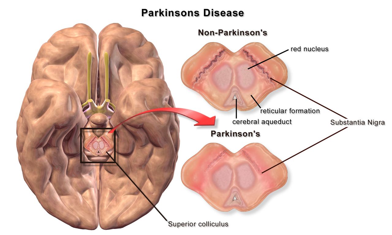
Dopamine, the basal ganglia and TRAP
Parkinson disease is the progressive loss of dopamine-producing neurons in the substantia nigra pars compacta, with intracellular aggregates of misfolded alpha-synuclein called Lewy bodies. The nigra normally feeds dopamine to the striatum, where it biases the basal ganglia circuits toward permitting movement. Strip that input away and the inhibitory output wins: movements become smaller, slower and harder to initiate. Because the system has enormous reserve, symptoms do not appear until roughly 60 to 80 percent of nigral neurons are gone — which is why the disease is already well advanced on the day of diagnosis, and why patients often say in hindsight that their handwriting shrank or their arm stopped swinging years earlier.
The cardinal motor features are remembered as TRAP. Tremor at rest, classically a 4 to 6 Hz pill-rolling tremor of the hand that damps down with purposeful movement and worsens with stress — the opposite of essential tremor, which appears with action and posture and often improves transiently with alcohol. Rigidity, felt as ratcheting cogwheel resistance when you passively flex the wrist or elbow, present in flexors and extensors equally. Akinesia and bradykinesia — slow, small movements, masked facies, decreased blink, hypophonic monotone speech, micrographia, difficulty turning in bed. Postural instability, which appears later and produces the retropulsion, festinating shuffle and freezing at doorways that generate falls.
The non-motor features are what actually degrade life and frequently precede the tremor by a decade: hyposmia, constipation, REM sleep behaviour disorder (acting out dreams), depression and anxiety, orthostatic hypotension, urinary urgency, drooling from reduced automatic swallowing, and eventually cognitive impairment or dementia in a substantial proportion of long-standing cases. Diagnosis remains clinical — bradykinesia plus rest tremor or rigidity, asymmetric onset, and a clear response to levodopa. A dopamine transporter SPECT scan (DaTscan) supports the diagnosis when the picture is muddy, and MRI is used to exclude vascular parkinsonism, normal-pressure hydrocephalus and structural mimics. There is no blood test.
Drug therapy: mechanism, monitoring and the doses you must not miss
Levodopa remains the most effective drug in neurology. Dopamine itself cannot cross the blood–brain barrier, but its precursor can; carbidopa is added to block peripheral decarboxylation so more levodopa reaches the brain and far less becomes circulating dopamine causing nausea and hypotension. Over years, as the storage capacity of remaining neurons fails, the smooth response fragments: patients develop wearing off before the next dose, unpredictable on–off fluctuations, and peak-dose dyskinesias — writhing involuntary movements that families often mistake for the disease worsening when they actually signal too much drug at the peak.
Two administration facts belong in your muscle memory. First, dietary protein competes with levodopa for the same large neutral amino acid transporter in the gut and at the blood–brain barrier, so doses work best about 30 minutes before meals; if nausea is limiting, a few crackers — not a protein shake — are the workaround. Second, carbidopa–levodopa is a time-critical medication, meaning it must be given within about 30 minutes either side of the scheduled time rather than within the usual one-hour administration window. A dose given “with the 0900 pass” when it was due at 0730 can leave a patient rigid, immobile, unable to swallow and at risk of aspiration for hours; abrupt discontinuation can precipitate a parkinsonism-hyperpyrexia syndrome resembling neuroleptic malignant syndrome, with fever, severe rigidity, autonomic instability and rhabdomyolysis. Home schedules are individualised and must be reconciled and honoured, not folded into standard hospital times.
| Class / example | Mechanism | What to monitor and teach |
|---|---|---|
| Carbidopa–levodopa | Dopamine precursor plus peripheral decarboxylase inhibitor | Give on time and before meals; nausea, orthostatic hypotension, dyskinesia, vivid dreams, darkened sweat and urine. Do not crush extended-release tablets. Never stop abruptly. |
| Dopamine agonists — pramipexole, ropinirole, rotigotine patch | Direct postsynaptic D2/D3 stimulation | Impulse-control disorders (gambling, hypersexuality, shopping — ask directly and ask the family), sudden sleep attacks, hallucinations, ankle oedema, orthostasis. Higher confusion risk in older adults. |
| MAO-B inhibitors — selegiline, rasagiline, safinamide | Block central dopamine breakdown | Insomnia; avoid meperidine and use caution with SSRIs, SNRIs and tramadol because of serotonin syndrome; teach tyramine awareness at higher doses. |
| COMT inhibitors — entacapone, opicapone | Extend levodopa half-life peripherally | Only useful with levodopa; brownish-orange urine discolouration is harmless; diarrhoea; may amplify dyskinesia. Tolcapone requires liver enzyme monitoring. |
| Amantadine | NMDA antagonist with dopaminergic effects | Best drug for levodopa-induced dyskinesia; livedo reticularis, ankle oedema, confusion and hallucinations especially in older adults; renal dose adjustment. |
| Anticholinergics — trihexyphenidyl, benztropine | Rebalance striatal acetylcholine | Tremor-predominant younger patients only; dry mouth, blurred vision, urinary retention, constipation, confusion. Listed on Beers criteria — avoid in older adults. |
| Apomorphine subcutaneous / sublingual | Rescue dopamine agonist for sudden off episodes | Profound hypotension and vomiting; contraindicated with 5-HT3 antagonists — ondansetron with apomorphine can cause severe hypotension and loss of consciousness. |
Deep brain stimulation of the subthalamic nucleus or globus pallidus internus is offered to patients with good levodopa response but disabling fluctuations or dyskinesia. It does not cure the disease and does not reliably help axial symptoms such as freezing or falls. Post-implant nursing points: the patient carries a controller and identification card, MRI is restricted per device labelling, and the device may need to be turned off for certain procedures — check before diathermy or cardioversion.
Nursing care: falls, swallowing, and the two conditions that share the ward
Aspiration pneumonia is the leading cause of death in Parkinson disease, and it grows out of quiet dysphagia that no one screened for. Any Parkinson patient who coughs during meals, holds food in the cheek, takes a long time to eat, has a wet voice after drinking, or has lost weight needs a bedside swallow screen and a speech-language pathology evaluation before you feed them. Practical measures: sit fully upright, remain upright 30 to 60 minutes after eating, small bites with a chin-tuck if recommended, no straws if that is the plan of care, no distractions or conversation during swallowing, and timing meals to the medication “on” period. Oral care twice daily meaningfully reduces aspiration pneumonia risk and is one of the highest-value tasks on the unit.
Falls occur at predictable moments — turning, standing up, starting to walk, passing through a doorway, and reaching for something while walking. Teach turning in a wide arc rather than pivoting, one task at a time, no carrying objects while ambulating, and cueing strategies for freezing: count a marching rhythm, step over an imaginary line or a laser projected from a cane, rock side to side to initiate. Check orthostatic vital signs — a fall of 20 mm Hg systolic or 10 mm Hg diastolic within three minutes of standing defines orthostatic hypotension and is extremely common here from both the disease and its drugs. Constipation is managed with fluid, fibre, activity and a scheduled regimen; it also delays levodopa absorption, so it is a motor issue, not just a comfort one.
Two other movement disorders appear on medical floors. Huntington disease is an autosomal dominant trinucleotide repeat disorder (40 or more CAG repeats in the HTT gene on chromosome 4) producing chorea, behavioural change and dementia in midlife. Nursing focus is safety from injurious involuntary movement, enormous caloric need (choreic patients may require several thousand extra calories daily), aspiration precautions, suicide risk screening, and genetic counselling for children who have a 50 percent risk. VMAT2 inhibitors such as tetrabenazine and deutetrabenazine reduce chorea but carry a boxed warning for depression and suicidality — screen mood at every encounter. Essential tremor, by contrast, is benign, symmetric, action-related, often familial, and treated with propranolol or primidone; the most important nursing act is telling the patient this is not Parkinson disease.
Older adults deserve a specific caution. Bradykinesia, stooped posture and a soft voice are frequently written off as “just getting old,” delaying diagnosis by years. At the same time older patients tolerate dopamine agonists and anticholinergics poorly, developing confusion, hallucinations and urinary retention where a 55-year-old would not. When an older Parkinson patient becomes acutely confused in hospital, work the delirium algorithm — infection, retention, constipation, pain, hypoxia, new drug — before assuming disease progression.
In Parkinson disease the two nursing acts that change outcomes are giving levodopa exactly on the home schedule and protecting the airway from a swallow that is failing silently.
Section 3Multiple sclerosis and demyelinating disease
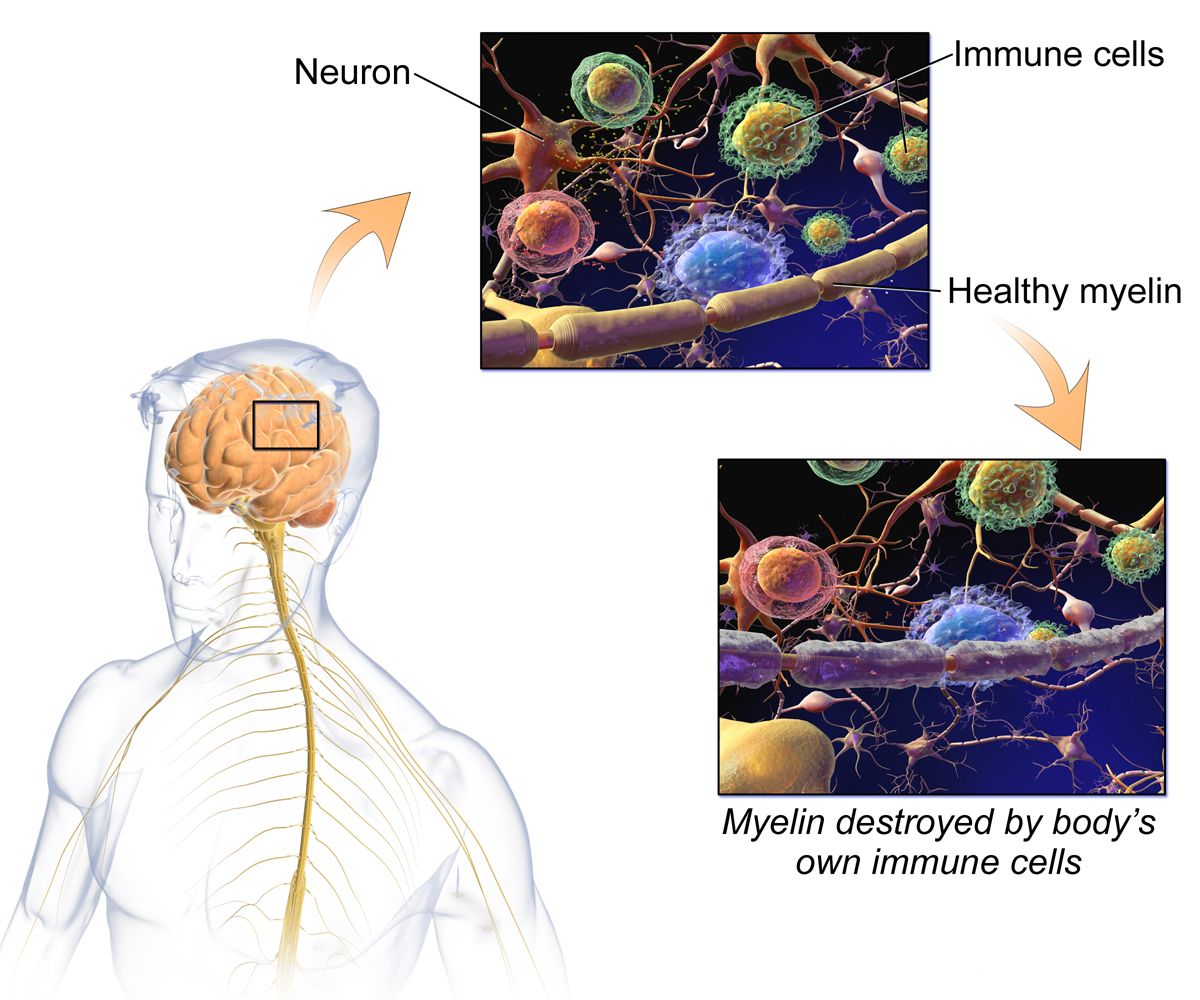
Demyelination, and why the symptoms wander
Multiple sclerosis is an immune-mediated attack on central nervous system myelin. Autoreactive T and B lymphocytes cross the blood–brain barrier and destroy oligodendrocytes and the myelin sheaths they maintain, leaving plaques of demyelination in periventricular white matter, the optic nerves, brainstem, cerebellum and spinal cord. Because myelin exists to make saltatory conduction fast, demyelinated axons conduct slowly, unreliably, or not at all — which is why symptoms appear, partially remit as inflammation subsides and remyelination occurs, then recur elsewhere. Over years, repeated inflammation causes irreversible axonal transection, and that is what converts relapsing disease into fixed disability.
Onset is typically between ages 20 and 40, with women affected two to three times as often as men. Risk rises with distance from the equator, low vitamin D, smoking, obesity in adolescence, and prior Epstein–Barr virus infection. Roughly 85 percent begin with a relapsing–remitting course; many eventually transition to secondary progressive disease with steady accumulation of disability, while about 10 to 15 percent have primary progressive MS from the outset with no distinct relapses.
The clinical picture is genuinely scattered, because a plaque can land anywhere. Optic neuritis — unilateral blurring or loss of central vision with pain on eye movement and a relative afferent pupillary defect — is a common first event. Internuclear ophthalmoplegia produces double vision with impaired adduction on lateral gaze. Sensory complaints, spasticity, ataxia, intention tremor, scanning dysarthria, bladder urgency or retention, bowel dysfunction, cognitive slowing and depression all occur. Two signs are worth memorising: Lhermitte sign, an electric shock down the spine on neck flexion, and Uhthoff phenomenon, the temporary worsening of symptoms with any increase in body temperature — a hot shower, a fever, exercise, a summer afternoon. Fatigue, which patients describe as a wall rather than tiredness, is consistently rated the most disabling symptom of all.
Diagnostics and telling a relapse from an imposter
Diagnosis rests on demonstrating lesions disseminated in space (multiple central nervous system sites) and in time (new lesions appearing, or the coexistence of enhancing and non-enhancing lesions), with no better explanation — the substance of the McDonald criteria. MRI of brain and spinal cord with gadolinium is the anchor: gadolinium enhancement indicates a lesion less than roughly four to six weeks old with an open blood–brain barrier, while T2/FLAIR hyperintensities map old and new burden. Lumbar puncture is no longer mandatory but is used when the picture is atypical; cerebrospinal fluid shows oligoclonal bands — two or more immunoglobulin bands present in CSF but not in serum — and an elevated IgG index. CSF protein is normal or only mildly raised (normal roughly 15 to 45 mg/dL) and cell counts are normal or show mild lymphocytosis. Evoked potentials can reveal clinically silent slowed conduction. Blood work exists to exclude mimics: vitamin B12, TSH, ANA, HIV, Lyme serology, and aquaporin-4 and MOG antibodies to rule out neuromyelitis optica spectrum disorder, which is treated very differently.
A true relapse is a new or clearly worsening neurologic symptom lasting more than 24 hours in the absence of fever or infection, developing over hours to days. A pseudo-relapse is the temporary re-emergence of old deficits caused by anything that raises body temperature or physiologic stress — most commonly a urinary tract infection, but also pneumonia, dehydration, heat, or poor sleep. The nursing implication is concrete and heavily tested: when an MS patient arrives with worsening weakness, check the temperature and obtain a urinalysis and culture before anyone reaches for high-dose steroids. Treating an infection with immunosuppression is the wrong direction.
Acute relapses that impair function are treated with methylprednisolone 1 g intravenously daily for three to five days, sometimes followed by an oral taper. Steroids shorten the relapse; they do not change long-term disability. Monitor capillary glucose (steroids will unmask or worsen hyperglycaemia, particularly in diabetes), blood pressure, mood and sleep — insomnia, irritability and frank steroid psychosis are common — provide gastric protection, and warn about metallic taste and facial flushing. Plasma exchange is used for severe steroid-refractory relapses.
Disease-modifying therapy and daily life
Disease-modifying therapies reduce relapse rate and new lesion formation in relapsing forms; they do not repair existing damage, which is why early initiation is emphasised. Adherence teaching is a nursing job, and so is anticipatory guidance about side effects, because patients who are surprised by side effects stop the drug.
| Therapy | Mechanism in one line | Key monitoring / teaching |
|---|---|---|
| Interferon beta-1a / beta-1b (injection) | Immunomodulation, reduced lymphocyte trafficking | Flu-like symptoms — dose at bedtime, pre-treat with acetaminophen or ibuprofen; rotate injection sites; monitor CBC and liver enzymes; screen for depression. |
| Glatiramer acetate (injection) | Decoy antigen shifting T-cell response | Injection-site lipoatrophy; a self-limited post-injection reaction of chest tightness, flushing, palpitations and anxiety lasting 15 to 30 minutes — frightening but not cardiac. Teach it in advance. |
| Dimethyl fumarate / diroximel (oral) | Nrf2 antioxidant pathway activation | Flushing (take with food, aspirin may help) and GI upset; monitor CBC for lymphopenia and liver enzymes. |
| Teriflunomide (oral) | Pyrimidine synthesis inhibition | Boxed warning — hepatotoxicity and teratogenicity; effective contraception essential for both sexes; hair thinning; accelerated elimination protocol with cholestyramine if pregnancy occurs. |
| Sphingosine-1-phosphate modulators — fingolimod, siponimod, ozanimod (oral) | Trap lymphocytes in lymph nodes | First-dose bradycardia monitoring is drug-specific: fingolimod requires about 6 hours of observation with an ECG before and after the first dose in every patient; siponimod uses a titration pack and needs first-dose observation only with pre-existing sinus bradycardia, first- or second-degree AV block, or prior myocardial infarction or heart failure; ozanimod uses dose escalation with no routine first-dose observation. Across the class — macular oedema screening at 3 to 4 months; varicella titres and vaccination before starting; infection risk. |
| Natalizumab (IV monthly) | Blocks alpha-4 integrin lymphocyte entry to CNS | Progressive multifocal leukoencephalopathy risk — JC virus antibody testing and index monitoring; report any new cognitive, visual or behavioural change immediately; restricted distribution programme. |
| Anti-CD20 antibodies — ocrelizumab, ofatumumab, rituximab | B-cell depletion | Infusion reactions — premedicate and monitor; hepatitis B screening before therapy; hypogammaglobulinaemia and infection over time; hold live vaccines. |
Symptom management often matters more to the patient than the DMT. Spasticity responds to baclofen or tizanidine — tizanidine causes hypotension, sedation and requires liver enzyme monitoring, and oral baclofen must be tapered, while abrupt withdrawal of intrathecal baclofen is a medical emergency producing rebound spasticity, hyperthermia, altered mental status and rhabdomyolysis. Fatigue is approached with energy conservation, scheduled rest, cooling, exercise, and sometimes amantadine or modafinil. Neuropathic pain responds to gabapentin, pregabalin or duloxetine. Dalfampridine, a potassium-channel blocker, may improve walking speed but is contraindicated with any seizure history or creatinine clearance of 50 mL/min or less. Bladder dysfunction must be characterised before it is treated: a spastic bladder causes urgency and frequency and responds to antimuscarinics or mirabegron, while a flaccid bladder retains urine and needs post-void residual measurement and intermittent catheterisation — giving an anticholinergic to a retaining bladder causes exactly the infection you are trying to prevent.
Teaching priorities: avoid heat (hot tubs, saunas, hot yoga), use cooling vests and cool showers, treat fevers promptly, keep vaccinations current but coordinate live vaccines with the DMT, stop smoking, maintain vitamin D, exercise in a cool pool, plan pregnancy with the neurologist because most DMTs are stopped beforehand, and report any new symptom lasting more than a day. Older adults with long-standing MS carry an added burden of osteoporosis from steroids and immobility, and their DMTs are often de-escalated because infection risk rises while relapse rate falls with age.
Multiple sclerosis is inflammation scattered in space and time; nurses change outcomes by separating true relapse from infection-driven pseudo-relapse, protecting patients from heat, and making disease-modifying therapy tolerable enough to continue.
Section 4Neuromuscular emergencies: myasthenia gravis and Guillain–Barré syndrome
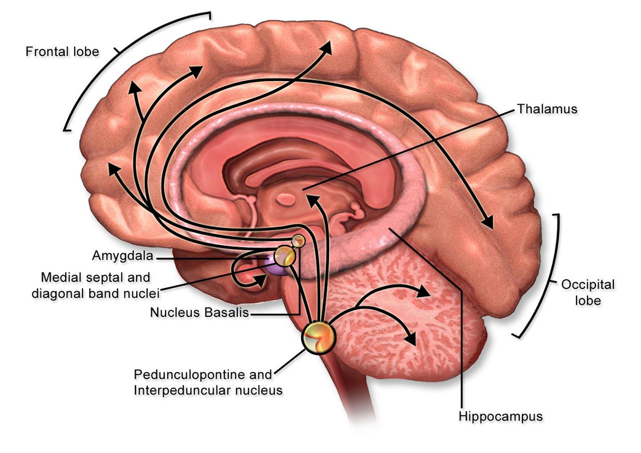
Myasthenia gravis: the junction that runs out
Myasthenia gravis is an autoimmune attack on the postsynaptic membrane of the neuromuscular junction. Antibodies against the nicotinic acetylcholine receptor — present in roughly 85 percent of generalised cases and about half of purely ocular cases — cross-link and internalise receptors, activate complement and flatten the postsynaptic folds. Fewer receptors means a smaller endplate potential; add the physiologic run-down of acetylcholine release with repeated stimulation and you get the defining clinical feature: weakness that worsens with sustained use and recovers with rest. A subset of patients instead carry MuSK antibodies (about 5 to 8 percent), which cause more bulbar and respiratory involvement and respond less well to cholinesterase inhibitors. Thymic abnormality is central — hyperplasia in about two-thirds and thymoma in 10 to 15 percent.
Assessment findings follow the fatigability rule. Ptosis and diplopia are the presenting complaint in roughly half and are worse in the evening, after reading or after driving. Bulbar involvement gives a nasal or slurred voice that deteriorates during a conversation, jaw fatigue partway through a meal, difficulty chewing and swallowing, and nasal regurgitation of liquids. Proximal limb weakness makes stairs and hair-brushing hard; neck extensor weakness produces a dropped head. Crucially, sensation is normal, reflexes are normal and pupils are normal — a distinction that separates myasthenia from botulism, which produces a descending paralysis with fixed dilated pupils and prominent dry mouth. On exam, sustained upgaze for 60 seconds reproduces ptosis; an ice pack applied to the closed lid for two minutes improves it, because cooling slows acetylcholinesterase.
Diagnostics: serum acetylcholine receptor binding antibodies first, then MuSK if negative. Repetitive nerve stimulation shows a decremental response greater than about 10 percent; single-fibre electromyography is the most sensitive test. CT of the chest is mandatory to look for thymoma. Check TSH, because autoimmune thyroid disease frequently travels with myasthenia, and screen for other autoimmune conditions. The edrophonium (Tensilon) test is largely historical and no longer generally available.
Treatment has two arms. Symptomatic therapy is pyridostigmine, a cholinesterase inhibitor typically dosed around 60 mg orally every 4 to 6 hours, with onset in 30 to 60 minutes and peak effect at about one to two hours — time the dose 30 to 45 minutes before meals in a patient with chewing or swallowing weakness, and time physical therapy to the peak. Its side effects are muscarinic: abdominal cramping, diarrhoea, salivation, lacrimation, bradycardia, increased bronchial secretions. Disease-modifying therapy is immunosuppression: corticosteroids (which can transiently worsen weakness during initiation, so severe bulbar patients are started in hospital), azathioprine or mycophenolate for maintenance with a slow onset over many months, rituximab particularly for MuSK disease, complement inhibitors such as eculizumab and ravulizumab (meningococcal vaccination is required before the first dose), and neonatal Fc receptor blockers such as efgartigimod that accelerate IgG clearance. Thymectomy is indicated for any thymoma and improves outcomes in many acetylcholine-receptor-positive generalised patients.
| Drug class | Examples | Why it matters |
|---|---|---|
| Aminoglycosides | Gentamicin, tobramycin, amikacin | Impair presynaptic acetylcholine release — can precipitate crisis. Flag the order. |
| Fluoroquinolones and macrolides | Ciprofloxacin, levofloxacin, azithromycin; telithromycin is contraindicated | Fluoroquinolones carry a boxed warning for exacerbation of myasthenia; macrolides can also worsen it. Choose an alternative when possible. |
| Magnesium (especially IV) | Magnesium sulfate for eclampsia or repletion | Blocks calcium-dependent acetylcholine release — a classic cause of iatrogenic crisis. |
| Cardiac agents | Beta blockers, procainamide, quinidine, some calcium channel blockers | Blunt neuromuscular transmission; monitor closely if unavoidable. |
| Non-depolarizing blockers | Rocuronium, vecuronium | Marked sensitivity — a standard dose can produce profound, prolonged paralysis, so the dose is reduced substantially, quantitative train-of-four monitoring is essential and sugammadex is the preferred reversal. Anaesthesia must know the diagnosis. |
| Depolarizing blocker | Succinylcholine | The opposite problem — with fewer acetylcholine receptors these patients are relatively resistant, so a standard dose may not relax them and a higher dose is needed. Duration may still be prolonged in a patient taking pyridostigmine, because cholinesterase inhibition slows the drug’s breakdown. Anaesthesia must know the diagnosis. |
| Other | Penicillamine, checkpoint inhibitors, botulinum toxin, high-dose corticosteroid initiation | Can induce or unmask myasthenia, or transiently worsen it. |
Crisis: what you do first, and only then what you call it
Myasthenic crisis is respiratory failure from disease severity, usually triggered by infection, surgery, aspiration, pregnancy or the postpartum period, tapering of immunosuppression, or one of the drugs above. Cholinergic crisis is respiratory failure from excess cholinesterase inhibitor, and it is now uncommon. Both present as a weak patient who cannot breathe. Students burn precious minutes trying to decide which one it is; the correct first action is neither — it is to assess and support ventilation. Raise the head of the bed, suction, stop oral intake, apply continuous pulse oximetry and cardiac monitoring, obtain a vital capacity and NIF, call the provider and rapid response, and prepare for intubation. Diagnosis follows stabilisation.
| Myasthenic crisis | Cholinergic crisis | |
|---|---|---|
| Cause | Too little drug, or a trigger (infection, surgery, drug) | Too much cholinesterase inhibitor |
| Timing clue | Weakness improves after a dose; often a missed or late dose | Weakness begins within about an hour of a dose |
| Pupils | Normal | Constricted (miosis) |
| Autonomic signs | Dry, often tachycardic, anxious | SLUDGE — salivation, lacrimation, urination, diarrhoea, GI cramping, emesis; bradycardia, sweating, fasciculations |
| Secretions | Usually manageable | Copious oral and bronchial secretions |
| Treatment | Airway support plus IVIG or plasma exchange; corticosteroids; treat the trigger | Hold the cholinesterase inhibitor; atropine for muscarinic symptoms; airway support |
In crisis, intravenous immune globulin (commonly 2 g/kg divided over 2 to 5 days) or plasma exchange (typically five exchanges) produces improvement within days. IVIG carries risks of aseptic meningitis, acute kidney injury, thrombosis, volume overload and anaphylaxis in IgA-deficient patients — hydrate, infuse slowly at the start, monitor renal function and watch for headache. Plasma exchange requires large-bore or central access and causes citrate-induced hypocalcaemia (watch for perioral tingling, Chvostek and Trousseau signs), hypotension and coagulopathy. Pyridostigmine is usually held while the patient is intubated to reduce secretions and restarted as weaning begins.
Guillain–Barré syndrome: paralysis that climbs
Guillain–Barré syndrome is an acute immune-mediated polyradiculoneuropathy in which antibodies raised against a preceding infection cross-react with peripheral nerve myelin or axolemma — molecular mimicry. About two-thirds of patients report a respiratory or gastrointestinal illness one to three weeks earlier; Campylobacter jejuni is the most frequently identified trigger, followed by cytomegalovirus, Epstein–Barr virus, Mycoplasma, influenza, hepatitis E and Zika virus. The acute inflammatory demyelinating polyradiculoneuropathy (AIDP) form predominates in North America and Europe; axonal variants and the Miller Fisher variant (ophthalmoplegia, ataxia and areflexia, associated with anti-GQ1b antibodies) also occur.
The classic presentation is symmetric ascending flaccid weakness beginning in the legs, with early loss of deep tendon reflexes that is often more striking than the weakness itself. Paraesthesias precede or accompany the weakness, and severe aching back and limb pain occurs in a majority and is frequently under-treated. Progression is rapid — hours to two weeks, reaching nadir within four weeks by definition — and ascends toward the trunk, arms, and then bulbar and respiratory muscles. Roughly one in four patients requires mechanical ventilation. Facial diplegia and difficulty handling secretions are ominous.
Autonomic instability is the feature that kills the ones who do not die of respiratory failure: labile blood pressure swinging from hypertension to hypotension, tachyarrhythmias and sudden bradyarrhythmias, ileus, urinary retention, diaphoresis and abnormal thermoregulation. Practical consequences — keep the patient on cardiac monitoring, change position slowly, be cautious with suctioning (vagal stimulation can precipitate profound bradycardia and asystole in these patients), avoid abrupt dosing of antihypertensives because the pressure may reverse in minutes, listen for bowel sounds, and monitor for retention.
Diagnosis is clinical and supported by lumbar puncture and nerve conduction studies. After about the first week, CSF shows the hallmark albuminocytologic dissociation: elevated protein (above the usual upper limit of about 45 mg/dL, often 100 to several hundred) with a normal white cell count, typically fewer than 10 cells/mm3. A high CSF cell count should redirect you toward HIV seroconversion, Lyme disease, sarcoidosis or malignant infiltration. Nerve conduction studies show slowed conduction, prolonged distal latencies and conduction block.
Treatment is intravenous immune globulin 0.4 g/kg daily for five days or plasma exchange — they are equally effective, they are not combined, and neither reverses damage already done; both shorten the course. Corticosteroids are ineffective in Guillain–Barré syndrome and are not recommended, which is a favourite examination point precisely because steroids are used in so many other neuroimmune conditions. Everything else is supportive and it is almost entirely nursing: serial vital capacity every 2 to 4 hours during the progressive phase, elevation of the head of bed, aspiration precautions and swallow assessment, venous thromboembolism prophylaxis (these patients are immobile and at high clot risk), pressure injury prevention with a turning schedule, passive range of motion and splinting to prevent contractures, eye care if the lids will not close, bowel and bladder programmes, and aggressive neuropathic pain management with gabapentin or pregabalin, using opioids cautiously because of ileus.
Do not overlook the psychological dimension. A patient with severe Guillain–Barré syndrome may be fully alert, oriented and terrified inside a body that will not respond, unable to speak around an endotracheal tube. Establish a communication method early — an eye-blink code, a letter board, a call system adapted to whatever movement remains — explain every procedure before touching them, orient them to time and progress, and keep family present. Most patients recover: roughly 80 percent walk independently by six months, though fatigue and residual weakness can persist for a year or more, and mortality remains around 3 to 7 percent, higher in older adults, who also recover more slowly and less completely.
Myasthenic crisis and Guillain–Barré syndrome are the same emergency wearing different masks — serial vital capacity and negative inspiratory force, not pulse oximetry, tell you when to secure the airway before the patient decompensates.
Section 5ALS, dementia and the long arc of progressive disease
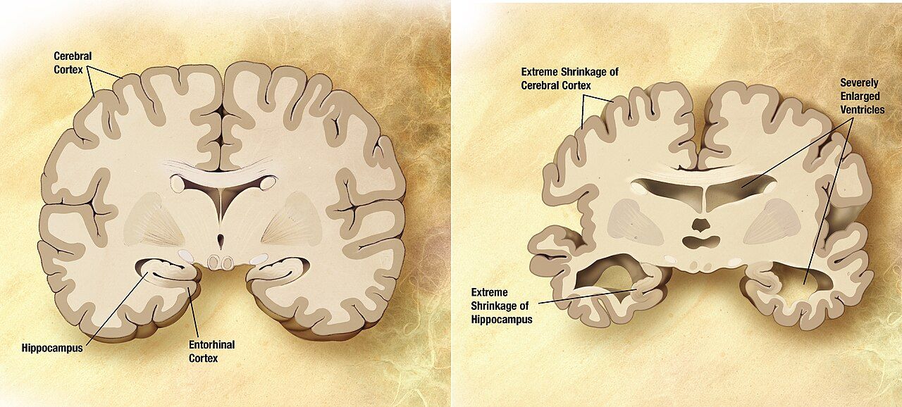
Amyotrophic lateral sclerosis: both motor neurons, intact senses
ALS is the progressive degeneration of upper and lower motor neurons simultaneously, which is why it produces a combination of findings that occurs in no other common disease: spasticity and hyperreflexia in a limb that is also wasting and fasciculating. Roughly 90 percent of cases are sporadic; about 10 percent are familial, with C9orf72 repeat expansions and SOD1 mutations among the most common. Sensation, extraocular movement, bowel and bladder control, and sexual function are characteristically spared, and most cognition is preserved — although about 15 percent develop frontotemporal dementia and up to half show some executive or behavioural change.
Presentation is limb-onset in about two-thirds — asymmetric foot drop with tripping, or a hand that drops objects and loses fine dexterity — and bulbar-onset in about a third, with slurred speech, difficulty swallowing, and a wasted fasciculating tongue. Bulbar onset carries a worse prognosis. Pseudobulbar affect, uncontrollable laughing or crying disproportionate to feeling, distresses families and is treatable. Median survival is around three to five years from symptom onset, with respiratory failure the usual cause of death.
Diagnosis is clinical, supported by electromyography showing active and chronic denervation across multiple body regions, and requires excluding treatable mimics: cervical spondylotic myelopathy, myasthenia gravis, vitamin B12 deficiency, thyroid disease, lead toxicity, Lyme disease and multifocal motor neuropathy. Creatine kinase may be modestly elevated. Disease-modifying options are limited — riluzole 50 mg orally every 12 hours, taken one hour before or two hours after meals, with liver enzymes checked at baseline, monthly for the first three months and periodically thereafter, extends survival by a few months; edaravone is an antioxidant option; tofersen targets SOD1-associated disease. The real work is multidisciplinary symptom management.
| Problem | Approach | Nursing focus |
|---|---|---|
| Respiratory decline | Noninvasive ventilation, usually initiated as FVC falls below about 50 percent predicted or with orthopnoea and nocturnal hypoventilation | Serial FVC at each visit; NIV improves survival and quality of life; teach mask fit and troubleshoot early intolerance so the patient stays with it. Cough assist devices clear secretions. |
| Dysphagia and weight loss | Diet texture modification, then gastrostomy tube — placed before FVC drops below roughly 50 percent, when the procedure is safer | Weigh at every visit; a falling weight predicts poorer survival. Raise the enteral access conversation early, not at the crisis. |
| Sialorrhoea and thick secretions | Glycopyrrolate, amitriptyline, atropine drops, botulinum toxin; nebulised saline and mucolytics for thick secretions | Portable suction at home; teach caregivers to distinguish thin drooling from thick plugging, which need opposite treatments. |
| Spasticity, cramps, pain | Baclofen, tizanidine, mexiletine for cramps; range of motion and positioning | ALS pain is largely musculoskeletal from immobility — repositioning and stretching are as valuable as drugs. |
| Pseudobulbar affect | Dextromethorphan–quinidine, or an SSRI | Explain to the family that the emotion displayed is not the emotion felt. |
| Communication loss | Voice banking early, speech-generating and eye-gaze devices | Start before speech fails. A patient who can still speak clearly is the one who should be recording their voice. |
Alzheimer disease and other dementias
Alzheimer disease is a progressive neurodegeneration characterised by extracellular beta-amyloid plaques and intraneuronal neurofibrillary tangles of hyperphosphorylated tau, with early hippocampal and entorhinal degeneration spreading to association cortex, and loss of cholinergic projections from the basal forebrain. Clinically it begins with episodic memory failure — the same question asked repeatedly, misplaced objects, missed appointments — and advances through the four A’s: amnesia, aphasia (word-finding failure, then empty speech), apraxia (inability to perform learned motor tasks such as dressing) and agnosia (failure to recognise objects and eventually people). Other dementias have their own signatures worth recognising: vascular dementia progresses in steps with focal findings, dementia with Lewy bodies brings visual hallucinations, fluctuating cognition, parkinsonism and severe neuroleptic sensitivity, and frontotemporal dementia presents in a younger patient with behavioural disinhibition or progressive aphasia rather than memory loss.
Workup exists to find what is reversible. Obtain a structured cognitive assessment — the Montreal Cognitive Assessment is commonly used, with a score of 26 or above out of 30 considered normal — along with CBC, comprehensive metabolic panel, TSH, vitamin B12, and neuroimaging; depression, hypothyroidism, B12 deficiency, normal-pressure hydrocephalus, subdural haematoma and medication effects all masquerade as dementia. Amyloid PET imaging and CSF or plasma biomarkers such as phosphorylated tau are increasingly used to confirm Alzheimer pathology, particularly when an anti-amyloid therapy is being considered.
Pharmacology is modest. Cholinesterase inhibitors — donepezil, rivastigmine (including a patch), galantamine — raise synaptic acetylcholine and may slow functional decline; they cause nausea, diarrhoea, anorexia, vivid dreams, bradycardia and syncope, so check the apical pulse before administration and be cautious in patients on beta blockers or with conduction disease. Memantine, an NMDA receptor antagonist, is added in moderate to severe disease and requires renal dose adjustment. Anti-amyloid monoclonal antibodies such as lecanemab and donanemab are available for early symptomatic Alzheimer disease with confirmed amyloid; their defining safety issue is amyloid-related imaging abnormalities (ARIA) — brain oedema or microhaemorrhage — requiring scheduled surveillance MRI, extra caution in APOE ε4 homozygotes and in patients on anticoagulation, and immediate reporting of new headache, confusion, visual change, dizziness or seizure.
Most of what helps is not a drug. For agitation, wandering and sundowning, work non-pharmacologically first: consistent routine and caregivers, orientation cues, adequate daytime light and activity, limited napping and caffeine, reduced noise and clutter, familiar objects, simple one-step instructions, validation and redirection rather than argument, and a systematic search for an unmet need — pain, hunger, thirst, a full bladder, constipation, or being too hot or cold. Antipsychotics carry a boxed warning for increased mortality in older adults with dementia-related psychosis and are a last resort requiring an explicit risk conversation; physical restraints increase injury, delirium and death and are not a solution. Home safety planning covers wandering and identification, stove and firearm access, driving cessation, medication supervision and fall-proofing, and financial and legal planning — including advance directives and health care proxy designation — should be completed while the patient still has capacity.
The long arc: prioritising, delegating and planning
These diseases overlap on a busy floor, and prioritisation follows the same hierarchy every time: airway and ventilation, then circulation and acute neurologic deterioration, then safety, then chronic disease management and teaching. If you hold assignments for a Parkinson patient whose 0730 levodopa is due, a myasthenic patient with a new nasal voice, a Guillain–Barré patient asking for pain medication, and a dementia patient calling out, you see the myasthenic first (progressing bulbar and respiratory weakness), then give the levodopa before it is late (a time-critical medication whose omission causes rigidity and dysphagia), then assess the dementia patient for a treatable cause of the calling out, then treat pain. Acute beats chronic, unstable beats stable, and physiologic need beats comfort — but note that the chronic drug given late becomes an acute problem, which is why levodopa outranks routine comfort measures.
Delegation in this population is unforgiving. Unlicensed assistive personnel may take vital signs, assist with hygiene and repositioning, perform passive range of motion under direction, apply splints already fitted, ambulate a stable patient with a documented plan, and feed a patient whose swallow has already been assessed and whose plan is established. The RN retains all assessment including neurologic and swallow screening, vital capacity and NIF measurement, interpretation of any change, all teaching, evaluation of response, the care of any unstable patient, IVIG and plasma exchange management, and the first dose of a new medication. Reporting expectations must be explicit: tell the assistive personnel exactly what to report immediately — coughing during meals, a new change in voice, difficulty being understood, a heart rate below a stated threshold, any fall or near-fall.
Older adults change every equation in this chapter. They present atypically (myasthenia mistaken for a stroke or for “ageing eyelids”, ALS mistaken for arthritis or deconditioning, Parkinson mistaken for depression); they carry an anticholinergic burden from bladder drugs, antihistamines and antidepressants that compounds every centrally acting agent you add; they have less physiologic reserve for aspiration, immobility and infection; and they are more likely to live alone with a caregiver who is themselves elderly. Assess the caregiver as deliberately as you assess the patient — burden, sleep, depression, finances and respite — because caregiver collapse is the most common reason a plan of care fails.
Finally, plan for the trajectory rather than the visit. Every one of these diagnoses warrants advance care planning early, while the patient can speak for themselves: health care proxy, advance directive, and specific decisions about noninvasive ventilation, tracheostomy and long-term ventilation in ALS, and about enteral feeding in advanced dementia — where the evidence is clear that feeding tubes do not prolong survival, do not prevent aspiration and do not improve pressure injuries, so careful hand feeding is preferred. Palliative care runs alongside disease-modifying therapy from the beginning, not after it fails; Chapter 14 develops that conversation in full.
Progressive neurologic disease is managed by anticipating the next loss — ventilation, swallowing, communication, safety — and having the equipment, the teaching and the goals-of-care conversation in place before the loss arrives.
The chapter in one breath
Every disorder here breaks the same motor relay at a different point, and the exam tells you where: spasticity with brisk reflexes points upstream to the upper motor neuron, flaccid areflexic wasting with fasciculations points to the lower motor neuron and peripheral nerve, fatigable painless weakness with normal reflexes and sensation points to the neuromuscular junction. Parkinson disease is nigral dopamine loss producing tremor, rigidity, bradykinesia and postural instability — give levodopa exactly on the home schedule, keep dopamine blockers such as metoclopramide and haloperidol away from the patient, and guard the swallow, because aspiration is what kills. Multiple sclerosis scatters demyelinating plaques in space and time, confirmed by MRI and oligoclonal bands, treated acutely with methylprednisolone and long term with disease-modifying therapy — but new weakness is a urinary tract infection until proven otherwise. Myasthenia gravis and Guillain–Barré syndrome are airway emergencies waiting to happen: trend vital capacity and negative inspiratory force rather than oxygen saturation, remember IVIG or plasma exchange (and no steroids in Guillain–Barré), and screen every order for magnesium, aminoglycosides and fluoroquinolones. ALS and Alzheimer disease demand anticipation — ventilation, nutrition, communication, safety, caregiver support and advance directives arranged before the crisis, never during it.
Words worth knowing
| Term | What it means |
|---|---|
| Albuminocytologic dissociation | Elevated cerebrospinal fluid protein (above about 45 mg/dL) with a normal cell count (under 10 cells/mm3) — the hallmark CSF finding of Guillain–Barré syndrome after the first week. |
| ARIA | Amyloid-related imaging abnormalities — brain oedema or microhaemorrhage caused by anti-amyloid monoclonal antibodies, monitored with scheduled MRI. |
| Babinski sign | Upgoing great toe with fanning on plantar stimulation; abnormal after infancy and indicates an upper motor neuron lesion. |
| Bradykinesia | Slowness and progressive reduction in the amplitude of voluntary movement; the required feature for a Parkinson disease diagnosis. |
| Bulbar weakness | Weakness of muscles supplied by the lower cranial nerves — producing dysarthria, dysphagia, nasal speech and impaired secretion handling. |
| Cogwheel rigidity | Ratcheting resistance felt during passive limb movement, caused by tremor superimposed on increased tone in Parkinson disease. |
| Fasciculation | Visible spontaneous twitching of a denervated motor unit; a lower motor neuron sign, prominent in ALS. |
| Festinating gait | Short, accelerating shuffling steps with a forward-leaning trunk, seen in Parkinson disease and associated with falls. |
| Fatigable weakness | Weakness that worsens with sustained or repeated use and improves after rest — the defining feature of neuromuscular junction disease. |
| Lhermitte sign | An electric-shock sensation running down the spine and into the limbs on neck flexion, indicating cervical cord demyelination. |
| Lower motor neuron | The neuron from anterior horn cell to muscle; damage causes flaccidity, hyporeflexia, atrophy and fasciculations. |
| Myasthenic crisis | Respiratory failure from myasthenia gravis itself, usually triggered by infection, surgery or a contraindicated drug, requiring ventilatory support plus IVIG or plasma exchange. |
| Negative inspiratory force (NIF) | Maximal negative pressure generated on inspiration against an occluded airway; normally more negative than −60 cm H2O, with values less negative than −30 cm H2O signalling impending failure. |
| Oligoclonal bands | Two or more immunoglobulin bands found in cerebrospinal fluid but not in serum, indicating intrathecal antibody production; supports multiple sclerosis. |
| On–off phenomenon | Abrupt, sometimes unpredictable swings between mobility and immobility in advanced Parkinson disease on long-term levodopa. |
| Plasma exchange (plasmapheresis) | Extracorporeal removal of circulating antibodies; used in myasthenic crisis, Guillain–Barré syndrome and steroid-refractory MS relapse. Watch for hypocalcaemia, hypotension and coagulopathy. |
| Pseudo-relapse | Temporary re-emergence of old MS deficits caused by fever, infection, heat or metabolic stress rather than new demyelination; treat the cause, not with steroids. |
| Uhthoff phenomenon | Transient worsening of multiple sclerosis symptoms with any rise in body temperature, including exercise, fever or a hot bath. |
| Upper motor neuron | The neuron from motor cortex to anterior horn cell; damage causes spasticity, hyperreflexia and a positive Babinski sign without early atrophy. |
| Vital capacity | The maximum volume exhaled after a maximal inspiration; normally about 60 to 70 mL/kg, and the most useful serial measure of neuromuscular respiratory reserve. |
Check yourself
Musculoskeletal Disorders and Orthopaedic Trauma
Bone is living tissue with an arterial supply, a nerve supply and a metabolic life, and almost everything that goes wrong with it goes wrong through one of those three channels. A fracture is not simply a broken stick: it is a haemorrhaging, swelling, inflamed compartment of tissue wrapped in fascia that does not stretch, threaded with nerves that die after four to six hours of ischaemia, sitting on a patient who is now immobile and therefore at risk of clot, pneumonia, pressure injury, delirium and deconditioning. The orthopaedic patient rarely dies of the broken bone. They die of the pulmonary embolus, the fat embolism, the sepsis from an infected implant, or the cascade that begins when an eighty-four-year-old breaks a hip and never walks again. This chapter teaches you the assessment that catches those complications early, the priorities that keep a limb attached, and the teaching that keeps the next fracture from happening.
By the end of this chapter you can
- Perform and document a structured neurovascular assessment using the six Ps, and state which finding is earliest and which is latest.
- Interpret common musculoskeletal diagnostics — radiograph, CT, MRI, bone scan, DEXA T-score, alkaline phosphatase, calcium, ESR and CRP — against real reference ranges.
- Classify fractures, describe the four stages of bone healing, and provide safe nursing care for casts, skin traction, skeletal traction, external fixation and ORIF.
- Recognise acute compartment syndrome, fat embolism syndrome, venous thromboembolism, rhabdomyolysis and osteomyelitis, and take the correct first action for each.
- Escalate limb-threatening and life-threatening orthopaedic findings using SBAR within a defined time frame.
- Deliver pre-operative and post-operative care for total hip arthroplasty, total knee arthroplasty and amputation, including approach-specific precautions and prevention of flexion contracture.
- Differentiate osteoarthritis, osteoporosis, osteomalacia and Paget disease by mechanism, laboratory pattern and treatment.
- Teach bisphosphonate administration, fall prevention and assistive-device use, and identify which orthopaedic tasks may and may not be delegated.
- Describe how fracture, infection and bone disease present differently in the older adult, including atypical pain and delirium.
Section 1Reading the Musculoskeletal System: Assessment and Diagnostics
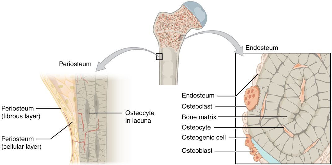
Why bone hurts, swells and stops working
Three structures generate almost every musculoskeletal symptom you will assess. The periosteum is the fibrous sleeve over the bone surface and it is densely innervated — which is why a fracture, a bone tumour or an expanding infection under the periosteum produces pain that is deep, boring, unrelieved by position and often worse at night. The cortex and marrow hold a rich blood supply, so a fractured femur can sequester one to two litres of blood into the thigh without a drop of it appearing on the sheets. And the fascial compartments that bundle muscle, nerve and vessel are effectively inelastic; once bleeding and oedema raise the pressure inside one of them, the small vessels collapse and the tissue inside starts to die while the pulse distal to it is still perfectly palpable.
Function loss follows mechanism. A torn ligament (a sprain) makes a joint unstable and painful with stress in one direction. A torn muscle or tendon (a strain) makes a movement weak and painful with resisted contraction. A fracture produces pain, deformity, crepitus, loss of function, shortening and ecchymosis that often blooms twelve to twenty-four hours later, far from the injury, as blood tracks along fascial planes. A compressed nerve root produces pain in a dermatome, weakness in a myotome and a diminished deep tendon reflex; the pain radiates in a stripe rather than spreading in a blob.
Your general survey therefore has a shape: look at the limb (deformity, swelling, colour, wounds, shortening, rotation), touch it (temperature, capillary refill, pulses, tenderness), move it or watch the patient move it (active before passive; never move an obviously deformed limb before imaging unless the limb is pulseless), and always compare with the other side. The uninjured extremity is your control, and no textbook range of motion beats it.
The neurovascular assessment: the skill that saves the limb
The neurovascular check — also charted as circulation, motion and sensation (CMS) — is the single most important repeated assessment in orthopaedic nursing. Perform it on admission, after any manipulation, cast, splint or surgery, then hourly for the first twenty-four hours (many units use every hour for four hours, then every two hours for eight, then every four), and any time the patient complains. Compare it to the other limb and to the previous entry. A trend is worth more than a snapshot.
| The six Ps | What you actually do | What it means |
|---|---|---|
| Pain | Rate at rest and on passive stretch of the muscles in the compartment (extend the toes or fingers) | Pain out of proportion to injury, escalating despite opioids, and severe pain on passive stretch is the earliest sign of compartment syndrome |
| Paraesthesia | Light touch and two-point discrimination in each nerve distribution | Numbness or tingling is the earliest reliable neurological sign; sensory fibres die before motor fibres |
| Pallor | Colour and capillary refill (normal under 3 seconds) | Late arterial finding; a dusky or mottled limb is worse than a pale one |
| Pulselessness | Palpate, then Doppler if not palpable; mark the spot | Latest and least reliable sign in compartment syndrome — never wait for it; in true arterial injury it is decisive |
| Paralysis | Active wiggle of fingers or toes, then strength against resistance | Late; motor loss suggests irreversible damage is starting |
| Poikilothermia | Back of your hand on the limb, compared to the other side | The limb takes on room temperature when perfusion fails |
Two nerve checks are worth memorising because they are commonly missed. After an upper extremity injury or cast, ask the patient to make the “OK” sign (anterior interosseous or median), spread the fingers against resistance (ulnar), and extend the wrist and thumb (radial); check sensation in the web space between thumb and index finger (radial), the tip of the index finger (median) and the tip of the little finger (ulnar). After a lower extremity injury, cast or knee surgery, check dorsiflexion of the ankle and extension of the great toe and sensation in the first web space of the foot — that is the peroneal nerve, the nerve most often injured by a tight cast, a poorly padded knee immobiliser, a fibular neck fracture or prolonged leg-crossed positioning.
Diagnostics and the numbers that matter
Plain radiograph is the first study for suspected fracture; two views at ninety degrees to each other, plus the joint above and below. Remember that a normal X-ray does not exclude a fracture. Occult hip fractures, scaphoid fractures and stress fractures are commonly invisible for seven to ten days; if the older adult cannot bear weight and the hip film is clean, the next step is MRI (most sensitive) or CT, not reassurance. CT defines complex intra-articular and pelvic fractures and is the study of choice for operative planning. MRI is the study for soft tissue — disc herniation, meniscus, rotator cuff, ligament, spinal cord compression and early osteomyelitis. A bone scan (technetium-99m) lights up anywhere osteoblasts are busy, which makes it sensitive but non-specific: it detects metastases, osteomyelitis and stress fractures but cannot tell them apart. DEXA measures bone mineral density. Arthrocentesis samples joint fluid; bone biopsy with culture is the gold standard for osteomyelitis and for tumour diagnosis.
| Test | Adult reference | Orthopaedic meaning |
|---|---|---|
| Total serum calcium | 8.6–10.2 mg/dL | Normal in osteoporosis and Paget disease; low in osteomalacia; high in hyperparathyroidism and malignant bone disease |
| Ionised calcium | 4.6–5.3 mg/dL | The physiologically active fraction; check before denosumab and IV bisphosphonate |
| Phosphate | 2.5–4.5 mg/dL | Low in osteomalacia and vitamin D deficiency |
| Alkaline phosphatase (ALP) | 30–120 U/L | Marker of osteoblast activity; markedly elevated in Paget disease and osteosarcoma; modestly elevated in healing fracture and osteomalacia |
| 25-hydroxy vitamin D | Sufficient 30–100 ng/mL; deficient under 20 ng/mL | Correct before starting any antiresorptive drug |
| ESR | Roughly under 20 mm/h (men), under 30 mm/h (women) | Non-specific inflammation; used to trend osteomyelitis and prosthetic joint infection |
| CRP | Under 1.0 mg/dL (10 mg/L) | Rises and falls faster than ESR; the better day-to-day infection trend |
| Creatine kinase (CK) | 30–170 U/L | Rhabdomyolysis after crush or prolonged compression is CK above roughly five times the upper limit of normal — about 1,000 U/L against this range; above 5,000 U/L marks severe muscle injury and a high risk of acute kidney injury |
| Synovial fluid WBC | Under 200/mm³ normal | 2,000–50,000/mm³ inflammatory; over 50,000/mm³ with high neutrophils suggests septic arthritis — a surgical emergency |
| DEXA T-score | Normal above −1.0 | −1.0 to −2.5 is low bone mass (osteopenia); −2.5 or lower is osteoporosis |
Two cautions. First, ESR and CRP are elevated for days to weeks after any surgery, so a single raised value on post-operative day two means nothing; the alarming pattern is a value that was falling and starts to climb again. Second, D-dimer is essentially useless after orthopaedic surgery for the same reason — it is elevated in nearly everyone, so a suspected post-operative pulmonary embolism goes to CT pulmonary angiography, not to a D-dimer.
The neurovascular check — performed on a schedule, compared to the other limb and to the last entry, and charted in findings rather than adjectives — is the assessment that separates a limb that is saved from a limb that is lost.
Section 2Fractures and Soft-Tissue Injury: From Field to Fixation
Patterns, healing and the timeline you will be asked about
A fracture is complete or incomplete, displaced or non-displaced, and — the distinction that changes everything — closed or open. An open (compound) fracture communicates with the skin surface and is contaminated by definition. It needs intravenous antibiotics within one hour of arrival (typically cefazolin, with gram-negative coverage added for larger wounds), tetanus prophylaxis, sterile moist covering of exposed bone, and urgent operative irrigation and debridement. Patterns tell you mechanism: transverse from direct blow, oblique and spiral from twisting (a spiral fracture in a non-ambulatory infant is a child-abuse red flag), comminuted (more than two fragments) from high energy or from weak bone, impacted from axial load, greenstick in children, compression in the osteoporotic vertebra, and pathologic through a bone already weakened by tumour, infection or osteoporosis.
Healing proceeds in four overlapping stages: haematoma formation in the first hours to days, when bleeding at the fracture site forms the scaffold; fibrocartilaginous (soft) callus over roughly days three to fourteen; bony callus from about week three to week twelve, when woven bone bridges the gap and the fracture becomes clinically stable; and remodelling, which continues for months to years as osteoclasts and osteoblasts convert woven bone to lamellar bone along lines of stress. Practical timelines: upper extremity fractures generally unite in six to eight weeks, lower extremity in eight to twelve or more, and femur or tibia in an older adult with diabetes considerably longer. Smoking, corticosteroids, poor protein intake, diabetes, infection, inadequate immobilisation and distraction at the fracture site all delay healing. Delayed union is healing that is slower than expected; non-union is healing that has stopped; malunion is healing in the wrong position.
Nutrition is nursing therapy here, not a footnote. Fracture healing needs adequate protein (roughly 1.2 to 1.5 g/kg/day in an older adult unless renal disease dictates otherwise), calcium, vitamin D, vitamin C for collagen cross-linking, and enough total calories that the patient is not catabolising the muscle they will need for rehabilitation. The frail hip-fracture patient who eats twenty per cent of their tray needs a dietitian referral and oral nutrition supplements, and that is a nursing judgement you make without an order.
Immobilisation: casts, traction and fixation
Casts. Plaster is heavy, moulds beautifully, and takes twenty-four to seventy-two hours to dry fully; fibreglass is lighter, water-resistant and sets in about thirty minutes. Handle a wet cast with the palms only — fingertips leave indentations that become pressure points. Leave the cast uncovered to air-dry, support it on a cloth-covered pillow, and elevate the limb above heart level for the first twenty-four to forty-eight hours with ice over (not under) the fracture site for twenty minutes at a time. Teach the patient never to insert anything under a cast to scratch: use cool air from a hairdryer instead. Report immediately any drainage staining through, foul odour, a warm spot on the cast surface, increasing pain, numbness, tingling, or an inability to move the fingers or toes. If circulation is compromised the cast is bivalved or a window is cut — and a nurse never cuts a cast without an order and the correct tool.
Traction. Skin traction (Buck extension, Russell traction) applies pull through a boot or adhesive strapping and is limited to about 5 to 10 pounds (2.3 to 4.5 kg) because more will shear the skin; it is used chiefly for comfort and muscle-spasm control before hip-fracture surgery. Skeletal traction pulls through a pin or wire in the bone and tolerates 15 to 30 pounds (7 to 13 kg); it is continuous and the weights are never removed. For any traction: weights hang freely and never rest on the floor or the bed frame, ropes stay in the pulley grooves and are not frayed, knots are clear of the pulleys, the patient stays aligned in the middle of the bed so that body weight provides counter-traction, and the pull remains in the correct line. Assess pin sites at least every shift for the expected serous crusting versus the abnormal purulent drainage, erythema spreading beyond the pin, tenderness, loosening or fever. Standard pin care is once-daily cleansing with chlorhexidine solution after the first forty-eight to seventy-two hours, following the facility protocol.
Surgical fixation. ORIF (open reduction with internal fixation) uses plates, screws, rods or intramedullary nails and permits earlier mobilisation — the reason it is now standard for the older adult with a hip fracture. External fixation uses pins connected to an outside frame, chosen for open fractures with soft-tissue loss, comminuted fractures or when the patient is too unstable for definitive surgery. Its advantage is access to the wound; its price is pin-tract infection and a device that looks alarming to the patient and family, so build reassurance and body-image support into your care plan. Never use the external fixator frame as a handle to lift or turn the limb.
Soft-tissue injury and repetitive strain
Sprains (ligament) and strains (muscle or tendon) are managed acutely with rest, ice for twenty minutes several times daily for the first twenty-four to forty-eight hours, compression and elevation, followed by early protected motion — prolonged immobilisation produces stiffness and weakness, not healing. A grade III sprain is a complete tear with instability and may require surgery. Teach patients that the inability to bear weight for four steps immediately and in the emergency department, or bony tenderness over the malleoli, is what prompts an ankle radiograph.
Rotator cuff tear usually involves the supraspinatus; the patient reports pain with overhead reach, night pain lying on the shoulder, and weakness in abduction, and a positive drop-arm test (the arm falls when lowered from ninety degrees of abduction) suggests a full-thickness tear. Meniscal injury presents with a twisting mechanism, joint-line tenderness, clicking and locking. Carpal tunnel syndrome is median nerve compression at the wrist producing numbness and burning in the thumb, index, middle and radial half of the ring finger, worse at night, with thenar wasting late; Tinel and Phalen signs support the diagnosis, and first-line treatment is a neutral-position wrist splint worn at night plus ergonomic modification. Epicondylitis, bursitis and plantar fasciitis follow the same pattern: overuse, localised tenderness, pain reproduced by loading the involved structure, treated with relative rest, ice, NSAIDs, stretching and correction of the offending activity.
The older adult and the fragility fracture
A fragility fracture is a fracture from a fall at standing height or less — the definition matters because it establishes osteoporosis clinically regardless of the DEXA result, and it obliges you to think about the next fracture before this one has healed. The classic presentation of an extracapsular hip fracture is a shortened, externally rotated, adducted leg with severe groin pain and inability to bear weight. But do not anchor on that picture: an impacted femoral neck fracture can leave the patient able to walk with a limp, and a vertebral compression fracture may present only as sudden mid-back pain when lifting a laundry basket, or as progressive height loss and kyphosis with no remembered injury at all.
Older adults also present atypically in ways that catch students out. Pain may be reported as vague or denied entirely in the patient with dementia, in whom the fracture announces itself as agitation, refusal to transfer, or new delirium. Untreated pain is itself a leading cause of post-operative delirium, so the answer is scheduled analgesia and a validated observational pain scale (PAINAD) rather than withholding opioids out of fear. Best practice for hip fracture is surgical repair within twenty-four to forty-eight hours, because delay increases mortality, pneumonia, pressure injury and delirium; one-year mortality after hip fracture remains roughly twenty to thirty per cent, and only about half of survivors regain their previous level of function. Regional nerve blocks (fascia iliaca) reduce opioid requirement and delirium. Screen and treat for the reason they fell: orthostatic hypotension, polypharmacy, sedative-hypnotics, vision, footwear, home hazards and vitamin D deficiency.
Immobilisation devices are only as safe as the assessment that surrounds them: elevate and ice a new cast, never remove skeletal traction weights, keep the patient in line with the pull, and re-check circulation, motion and sensation after every device change.
Section 3When a Fracture Turns Dangerous
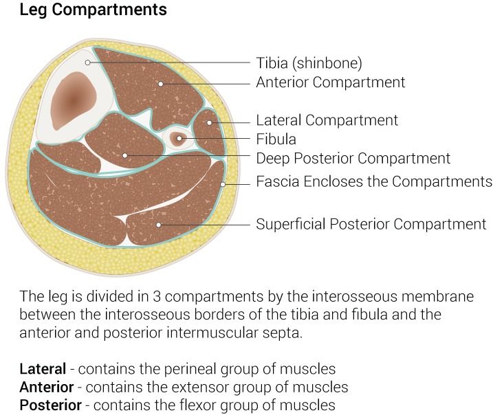
Acute compartment syndrome: the six-hour clock
Pressure inside a closed fascial compartment rises above the pressure in the capillaries that feed it, perfusion stops, muscle and nerve become ischaemic, and after roughly four to six hours the damage is permanent. Causes divide into things that increase the contents (bleeding, oedema after fracture, reperfusion after vascular repair, infiltrated intravenous fluid, burn oedema) and things that decrease the space (a tight cast, a circumferential dressing or burn eschar, a poorly applied splint). The tibia is the most common site, followed by the forearm; a supracondylar humerus fracture in a child is the classic precursor of Volkmann ischaemic contracture, the permanent claw deformity that follows untreated forearm compartment syndrome.
The presentation is deceptive because the limb often looks fine. The cardinal findings are pain out of proportion to the injury and escalating despite escalating opioid doses, pain on passive stretch of the compartment muscles, and a compartment that feels tense and woody. Paraesthesia follows. Pallor, pulselessness and paralysis are late; the artery is a large high-pressure vessel and it keeps flowing long after the capillaries have collapsed, so a palpable pulse never rules this out. Diagnosis is clinical, supported by compartment pressure measurement: normal is 0 to 8 mmHg, and either an absolute pressure above about 30 mmHg or a delta pressure (diastolic blood pressure minus compartment pressure) below 30 mmHg indicates fasciotomy.
Your first actions, in order: stop and do a focused neurovascular assessment; loosen or remove anything constricting — unwrap the elastic bandage, cut the cast padding after the cast is bivalved by the provider (bivalving a cast without removing the underlying padding relieves only about half the pressure); place the limb at heart level; remove ice; and notify the surgeon immediately. Do not wait for the next scheduled check, do not medicate and reassess in an hour, and do not elevate. Fasciotomy wounds are left open and later closed or grafted. Follow-up laboratory studies matter because devitalised muscle releases myoglobin and potassium: watch CK, potassium, creatinine and urine colour for rhabdomyolysis and give aggressive intravenous crystalloid to protect the kidneys.
Fat embolism, venous thromboembolism and haemorrhage
Fat embolism syndrome (FES) is a distinct entity from pulmonary embolism, and the timing is the giveaway. Marrow fat globules enter the venous circulation after long-bone or pelvic fracture and produce inflammation in the lung and brain, typically twenty-four to seventy-two hours after injury — often in a young adult with a femur fracture who was fine yesterday. The classic triad is hypoxaemia (dyspnoea, tachypnoea, PaO₂ below 60 mmHg on room air), neurological change (restlessness, confusion, agitation, sometimes the very first sign) and a petechial rash across the chest, axillae, neck and conjunctivae, which appears in only a minority of patients but is close to diagnostic when it does. Laboratory findings include falling platelets and haematocrit. There is no specific treatment: management is oxygen, sometimes mechanical ventilation, and supportive care. Prevention is what nursing controls — early immobilisation of the fracture, early operative fixation within twenty-four hours where possible, and careful handling of the fractured limb.
Venous thromboembolism is the leading preventable cause of death after major orthopaedic surgery. Hip fracture, hip arthroplasty and knee arthroplasty carry among the highest venous thromboembolism risk of any surgery, because the triad is complete: stasis from immobility, endothelial injury from the operation, and a hypercoagulable post-operative state. Prophylaxis combines mechanical measures (sequential compression devices worn whenever the patient is in bed, early ambulation, ankle pumps) with pharmacologic prophylaxis for ten to thirty-five days — commonly enoxaparin 40 mg subcutaneously daily (or 30 mg every twelve hours), a direct oral anticoagulant such as rivaroxaban 10 mg daily, or aspirin in selected low-risk arthroplasty protocols. Assess daily for unilateral calf swelling, warmth, tenderness and a calf circumference difference; never massage a suspicious calf. Sudden dyspnoea, pleuritic chest pain, tachycardia, anxiety or unexplained hypoxaemia is a pulmonary embolism until proven otherwise: oxygen, rapid response, ECG and CT pulmonary angiography.
Haemorrhage and hypovolaemia are quiet in orthopaedics because the blood hides inside the limb. A closed femur fracture can hold one to two litres; a pelvic fracture can hold far more and can exsanguinate a patient into the retroperitoneum with an unremarkable abdomen. Monitor haemoglobin, heart rate, blood pressure trend and urine output rather than the dressing. Post-operative transfusion thresholds are restrictive — generally a haemoglobin below 7 g/dL, or below 8 g/dL in patients with cardiovascular disease or symptomatic anaemia.
Infection, and the complications that show up late
Osteomyelitis reaches bone by direct inoculation (open fracture, surgery, pin tract), by contiguous spread (diabetic foot ulcer, pressure injury) or haematogenously. Staphylococcus aureus is the leading organism. Acute presentation is localised bone pain, warmth, swelling, fever, malaise and elevated WBC, ESR and CRP. Plain films lag by ten to fourteen days; MRI detects it earliest, and bone biopsy with culture is the gold standard and should ideally be obtained before antibiotics are started. Treatment is four to six weeks of intravenous antibiotics, often through a PICC line at home, plus surgical debridement when there is dead bone. Chronic osteomyelitis produces a sequestrum (a segment of dead, avascular bone that antibiotics cannot reach), an involucrum of new bone around it, and draining sinus tracts — and it is why debridement rather than antibiotics alone is curative.
Septic arthritis destroys articular cartilage within days and is a surgical emergency: a hot, swollen, exquisitely painful joint the patient will not move, with fever, requires urgent arthrocentesis (synovial WBC often over 50,000/mm³) and washout. Avascular necrosis follows disruption of the blood supply — classically the femoral head after a displaced femoral neck fracture, the scaphoid proximal pole, and the talus — presenting weeks to months later as insidious groin or joint pain worsening with weight bearing. Complex regional pain syndrome presents after an often minor injury with burning pain far out of proportion, allodynia, swelling, colour and temperature change and sweating asymmetry; early aggressive mobilisation and physical therapy, with gabapentinoids and sympathetic block, is treatment. Contracture, muscle atrophy, pressure injury, constipation and hospital-acquired pneumonia are all complications of immobility, which is why turning, incentive spirometry, bowel regimen and getting the patient out of bed on the day of surgery are not comfort measures — they are the treatment.
| Complication | Typical timing | Hallmark | Your first action |
|---|---|---|---|
| Acute compartment syndrome | 6–48 h | Pain out of proportion; pain on passive stretch; tense compartment | Release constriction, limb to heart level, remove ice, call surgeon STAT |
| Fat embolism syndrome | 24–72 h | Hypoxaemia, confusion, petechiae on chest and axillae | Oxygen, high Fowler if tolerated, rapid response, ABG and pulse oximetry |
| Deep vein thrombosis | Days 2–14 and beyond | Unilateral calf swelling, warmth, tenderness | Do not massage; notify provider; anticipate duplex ultrasound |
| Pulmonary embolism | Any time post-op | Sudden dyspnoea, pleuritic pain, tachycardia, hypoxaemia | Oxygen, sit upright, rapid response, CT pulmonary angiography |
| Rhabdomyolysis | Hours after crush or prolonged compression | Tea-coloured urine, CK above five times normal (roughly over 1,000 U/L), hyperkalaemia | Aggressive isotonic IV fluids at once; trend potassium, creatinine and urine output, ECG — do not wait for CK to reach a set number |
| Osteomyelitis or prosthetic joint infection | Days to months | Fever, increasing pain, wound drainage after day 5, rising CRP | Culture before antibiotics, notify surgeon, do not simply redress |
In orthopaedic trauma the deadly and limb-threatening complications each have a signature time window — compartment syndrome in the first two days, fat embolism at one to three days, thromboembolism from day two onward, and infection from day five onward — so assess for the complication the calendar is pointing at.
Section 4Degenerative Joints, Arthroplasty, Amputation and the Painful Back
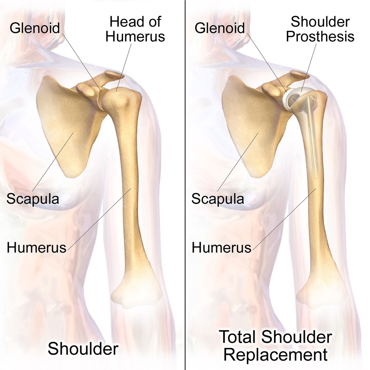
Osteoarthritis: mechanical failure, not systemic inflammation
Osteoarthritis (OA) is progressive loss of articular cartilage with reactive change in the underlying bone. Cartilage thins and fissures, the joint space narrows, subchondral bone becomes sclerotic and forms cysts, and the joint margins throw out osteophytes. The result is pain that is worse with use and better with rest, morning stiffness that resolves in under thirty minutes, crepitus, bony rather than boggy enlargement, and an asymmetric distribution in the weight-bearing joints — knees, hips, lumbar and cervical spine — plus the hands, where Heberden nodes at the distal interphalangeal joints and Bouchard nodes at the proximal interphalangeal joints are characteristic. Systemic features are absent, and ESR and CRP are normal. That last point is the examination discriminator: rheumatoid arthritis is symmetric, involves the metacarpophalangeal and proximal interphalangeal joints while sparing the distal interphalangeal joints, produces morning stiffness lasting longer than an hour with fatigue and low-grade fever, and raises inflammatory markers. Rheumatoid and other inflammatory arthritides are covered in Chapter 9.
Management is a ladder. Non-pharmacologic first and always: weight loss (each pound lost removes roughly four pounds of force from the knee), low-impact exercise and quadriceps strengthening, physical and occupational therapy, assistive devices, heat before activity and cold after. Pharmacologic: topical NSAIDs are first-line for knee and hand OA in older adults because systemic absorption is minimal; oral acetaminophen up to 3 g/day (and no more than 2 g/day in the older adult, in liver disease or with regular alcohol use); oral NSAIDs at the lowest effective dose with attention to gastrointestinal bleeding, hypertension, kidney injury and heart failure — a genuinely risky combination in the frail patient already taking an anticoagulant and an ACE inhibitor. Intra-articular corticosteroid injections give weeks to a few months of relief and are limited to about three or four per joint per year because repeated injection damages cartilage. Opioids are not a maintenance therapy for osteoarthritis. When pain limits sleep and daily function despite the ladder, joint replacement is offered.
Total hip and total knee arthroplasty
Pre-operatively, teach before the anaesthetic rather than after it: incentive spirometry, ankle pumps, the transfer technique, the pain plan and the precautions. Screen for infection anywhere in the body, since bacteraemia seeds a prosthesis; confirm that the surgical-site antibiotic (usually cefazolin, vancomycin for MRSA colonisation) is given within sixty minutes of incision — vancomycin is the exception and is started within one hundred and twenty minutes because it must be infused slowly; and verify the surgical site marking with the patient awake. Post-operatively the priorities are airway and haemodynamics first, then neurovascular checks distal to the joint, then pain control adequate to permit the physiotherapist to work, then VTE prophylaxis, then mobilisation — usually on the day of surgery or the first post-operative morning.
Hip precautions depend on the surgical approach and you must know which one your patient had. After a posterior approach (the most common), the prohibited movements are the ones that lever the head out the back of the socket: no hip flexion beyond ninety degrees, no adduction past midline (no crossing legs or ankles), and no internal rotation of the operative leg. After an anterior approach, the risk is the opposite: avoid hyperextension, external rotation and bridging. Precautions typically last six to twelve weeks. Equip the home accordingly: elevated toilet seat, chairs with arms and a firm high seat, a reacher, a long-handled shoe horn and sponge, sock aid, and no low soft couches or deep recliners. An abduction pillow or wedge is used in bed after posterior approaches, and the patient is turned toward the unoperated side with the pillow between the legs unless the surgeon specifies otherwise.
Prosthetic hip dislocation is an emergency you must recognise: the patient reports a pop and sudden severe pain, cannot move the leg, and the leg is shortened and internally rotated (posterior dislocation, the usual direction). Keep the patient still and supine, support the limb, do not attempt to reposition or ambulate them, notify the surgeon and keep them nil by mouth for closed reduction under sedation. After total knee arthroplasty, the priorities differ: the enemy is a flexion contracture, so the knee is kept in extension at rest and a pillow is never placed under the knee — elevate the whole leg on a pillow under the calf instead. Goals are typically ninety degrees of flexion by discharge and full extension, progressing toward one hundred and ten to one hundred and twenty degrees. Expect knee swelling, use cold therapy, and remember that new fever with wound drainage after post-operative day five, escalating rather than improving pain, or drainage that persists beyond seventy-two hours suggests a prosthetic joint infection and must be reported rather than simply redressed.
| Task on the orthopaedic unit | RN | LPN/LVN | UAP |
|---|---|---|---|
| Initial and post-operative neurovascular assessment; any change in status | Yes — RN only | No | No |
| Teaching hip precautions, crutch gait, bisphosphonate administration | Yes — RN only | Reinforce established teaching | No |
| Evaluating response to analgesia or to a new mobility plan | Yes — RN only | No | No |
| First post-operative ambulation; ambulating an unstable patient | Yes — RN (with therapy) | Per policy once stable | No |
| Pin-site care and dressing changes | Yes | Yes, per protocol | No |
| Applying sequential compression devices, ice packs, repositioning, assisting a stable patient to ambulate with a taught device, intake and output, vital signs | Supervises | Yes | Yes — report findings to the RN |
Amputation and the painful back
Amputation is most often performed for peripheral arterial disease and diabetic limb infection, and less often for trauma, tumour or congenital deformity. Immediately post-operatively the priorities are haemorrhage and pain: keep a large tourniquet at the bedside for the first twenty-four to forty-eight hours, monitor the dressing and the drain, and record the amount of drainage rather than describing it. Elevate the residual limb on a pillow for the first twenty-four hours only to control oedema; after that, elevation risks a hip or knee flexion contracture that will make prosthetic fitting impossible. From day two, position the patient prone for twenty to thirty minutes three to four times daily after a lower-limb amputation, keep the residual limb flat when supine, avoid sitting in a chair for prolonged periods with the knee flexed, and never place a pillow between the thighs (that promotes abduction contracture). Teach residual-limb wrapping in a figure-of-eight pattern with more pressure distally to shape the limb into a cone.
Phantom limb sensation — the feeling that the limb is still present — is nearly universal and is normal. Phantom limb pain is burning, cramping or shooting pain perceived in the missing limb, is real neuropathic pain, and is treated with gabapentin or pregabalin, tricyclic antidepressants such as amitriptyline, mirror therapy, desensitisation, transcutaneous electrical nerve stimulation and early prosthetic use. Never tell a patient the pain cannot be real. Grief, altered body image and depression are expected; ask directly, involve peer support and rehabilitation early, and remember that the contralateral limb must be protected obsessively in the diabetic patient because it is now the only one.
Low back pain is the most common musculoskeletal complaint you will meet. Most acute mechanical back pain resolves in four to six weeks with continued activity (bed rest actually worsens outcomes), heat, NSAIDs, and reassurance; imaging is not indicated in the first six weeks unless red flags exist. A herniated intervertebral disc, usually at L4–L5 or L5–S1, compresses a nerve root and produces radiating pain below the knee, dermatomal numbness, a positive straight-leg raise and myotomal weakness — foot dorsiflexion for L5, plantar flexion and a reduced ankle reflex for S1. The red flags that must never be missed are: cauda equina syndrome (new urinary retention or incontinence, faecal incontinence, saddle anaesthesia, bilateral leg weakness) which is a surgical emergency requiring decompression within hours; fever with back pain and intravenous drug use (spinal epidural abscess or vertebral osteomyelitis); a history of cancer with new unrelenting night pain (metastasis with cord compression); and significant trauma or unexplained weight loss. After lumbar fusion, teach the “no BLT” rule — no bending, no lifting more than five to ten pounds, no twisting — and log-roll the patient in bed with the shoulders and hips moving as a unit.
After joint replacement and amputation, most of what protects the patient is positioning — approach-specific hip precautions, the knee kept in extension, and the residual limb kept flat with scheduled prone lying — and positioning is teaching, which belongs to the RN.
Section 5Bone Integrity: Osteoporosis, Metabolic Bone Disease and Bone Tumours
Osteoporosis: the silent disease until it is not
Bone is continuously remodelled: osteoclasts resorb, osteoblasts rebuild. Osteoporosis is the state in which resorption outpaces formation, so bone mass falls and microarchitecture deteriorates while the bone that remains is chemically normal — which is why serum calcium, phosphate and ALP are all typically normal in uncomplicated osteoporosis. Oestrogen restrains osteoclasts, so bone loss accelerates sharply for five to ten years after menopause. Other major risks: age, low body weight, white or Asian ancestry, family history of hip fracture, smoking, more than two or three alcoholic drinks daily, sedentary lifestyle, low calcium and vitamin D intake, hyperthyroidism and hyperparathyroidism, malabsorption, chronic kidney disease, and drugs — particularly glucocorticoids (prednisone 5 mg daily or more for three months or longer), anticonvulsants, proton pump inhibitors, aromatase inhibitors, androgen deprivation therapy and long-term heparin.
Osteoporosis produces no symptoms until it produces a fracture. The three classic sites are the vertebrae (compression fractures causing acute back pain, progressive height loss of more than 1.5 inches and thoracic kyphosis, the “dowager hump”), the hip and the distal radius (Colles fracture from a fall on an outstretched hand). Screening with DEXA of the hip and lumbar spine is recommended for all women aged 65 and older, men aged 70 and older, and younger post-menopausal women or men over 50 with risk factors or a fragility fracture. The T-score compares the patient to a healthy young adult: above −1.0 is normal, −1.0 to −2.5 is low bone mass (osteopenia), and −2.5 or below is osteoporosis; a T-score of −2.5 or below plus a fragility fracture is severe osteoporosis. The Z-score compares to age-matched peers and is the score used in premenopausal women, men under 50 and children; a Z-score of −2.0 or lower prompts a search for a secondary cause. FRAX estimates ten-year fracture probability and helps decide whether to treat osteopenia.
Every patient gets the foundation: 1,000 to 1,200 mg of elemental calcium daily from diet where possible (dairy, fortified plant milks, sardines with bones, kale, broccoli, tofu set with calcium) and 800 to 1,000 international units of vitamin D daily; weight-bearing and resistance exercise; smoking cessation; alcohol moderation; and rigorous fall prevention. Calcium supplements are absorbed only about 500 to 600 mg at a time, so divide the dose. Calcium carbonate is cheap but needs gastric acid, so take it with food and expect constipation; calcium citrate is absorbed without acid and is the correct choice for a patient on a proton pump inhibitor or with achlorhydria. Warn about constipation and, in the patient with a history of calcium stones or hypercalciuria, keep supplementation conservative and push fluids.
The drugs, and the teaching that makes them safe
| Drug class | Mechanism | Representative agent | What the nurse monitors and teaches |
|---|---|---|---|
| Bisphosphonate (oral) | Binds hydroxyapatite and inhibits osteoclast resorption | Alendronate 70 mg weekly; risedronate; ibandronate | Take first thing in the morning on an empty stomach with a full 6 to 8 oz glass of plain water; remain upright for 30 minutes (60 for ibandronate) and take nothing else by mouth in that time; report new dysphagia, heartburn or retrosternal pain (oesophagitis and ulceration); dental examination before starting (osteonecrosis of the jaw); report new thigh or groin pain (atypical femoral fracture); avoid if creatinine clearance is under about 30–35 mL/min; correct hypocalcaemia and vitamin D deficiency first |
| Bisphosphonate (intravenous) | Same | Zoledronic acid 5 mg IV once yearly | Hydrate well before and after; expect a flu-like acute-phase reaction for 24–72 hours after the first dose, treated with acetaminophen; check calcium and renal function first |
| RANK ligand inhibitor | Monoclonal antibody that blocks osteoclast formation and survival | Denosumab 60 mg subcutaneously every 6 months | Usable in renal impairment but check calcium first — hypocalcaemia risk is real, especially in CKD; osteonecrosis of the jaw; never simply stop it without transitioning to another agent, because rebound vertebral fractures follow discontinuation; must be given on schedule |
| Selective oestrogen receptor modulator | Oestrogen agonist at bone, antagonist at breast | Raloxifene 60 mg daily | Reduces vertebral fracture and breast cancer risk; increases VTE risk — stop before prolonged immobility or surgery; causes hot flashes and leg cramps |
| Anabolic PTH analogue | Intermittent dosing stimulates osteoblasts — builds new bone | Teriparatide, abaloparatide, daily subcutaneous | For severe osteoporosis; generally limited to about 2 years of lifetime use; teach self-injection and pen refrigeration; watch for transient hypercalcaemia and orthostatic dizziness after the first doses (sit or lie down) |
| Sclerostin inhibitor | Increases formation and decreases resorption | Romosozumab monthly for 12 months | Boxed warning for myocardial infarction, stroke and cardiovascular death — avoid within one year of MI or stroke; follow with an antiresorptive agent |
Two safety themes cut across the whole table. First, correct the calcium and vitamin D before the antiresorptive, or you will cause symptomatic hypocalcaemia — perioral numbness, tingling fingers, cramps, Trousseau and Chvostek signs, and in severe cases tetany, laryngospasm and a prolonged QT interval. Second, every antiresorptive carries a small risk of osteonecrosis of the jaw and atypical femoral fracture, so a dental examination before starting and a low threshold for investigating new thigh, groin or jaw pain are standing nursing responsibilities, not optional extras.
Osteomalacia, Paget disease and tumours of bone
Osteomalacia is defective mineralisation of the bone matrix in adults, almost always from vitamin D deficiency (poor intake, minimal sun exposure, dark skin, malabsorption after bariatric surgery or in coeliac disease, chronic kidney disease or certain anticonvulsants). Unlike osteoporosis, the mineral chemistry is abnormal: calcium is low or low-normal, phosphate is low, ALP is elevated, PTH rises in compensation, and 25-hydroxy vitamin D is low. The patient reports diffuse aching bone pain and tenderness, proximal muscle weakness and a waddling gait, and radiographs may show Looser zones (pseudofractures). Treatment is vitamin D repletion with calcium, and the symptoms improve within weeks. The paediatric equivalent is rickets, with bowed legs and growth-plate widening.
Paget disease of bone is a focal disorder of wildly accelerated, disorganised remodelling: osteoclasts resorb at high speed and osteoblasts lay down structurally chaotic bone that is larger but weaker. Calcium and phosphate are normal and ALP is markedly elevated — often two to several times the upper limit — which is the classic laboratory fingerprint. Many patients are asymptomatic and are found on an incidental radiograph or ALP; others report deep bone pain, bowing of the tibia or femur, skull enlargement (a hat that no longer fits), hearing loss from compression of the eighth cranial nerve, headache, pathologic fracture, secondary osteoarthritis of adjacent joints, and rarely high-output heart failure from hypervascular bone or malignant transformation to osteosarcoma. Treatment is a bisphosphonate, most often intravenous zoledronic acid, with ALP monitored as the marker of response, plus calcium and vitamin D, analgesia and fall precautions.
Bone tumours. Benign lesions (osteochondroma, osteoid osteoma, giant cell tumour) are far more common than malignant ones and are often incidental. Among primary malignancies, osteosarcoma is the most common, occurs mostly in adolescents and young adults around the knee (distal femur, proximal tibia) and in older adults with Paget disease, and presents with deep pain worse at night, a palpable mass and elevated ALP and LDH; radiographs show a “sunburst” periosteal reaction and Codman triangle. Ewing sarcoma occurs in children and adolescents and may mimic infection with fever and elevated inflammatory markers, with an “onion-skin” periosteal reaction. Chondrosarcoma occurs in middle-aged and older adults. Treatment is neoadjuvant chemotherapy, limb-salvage resection or amputation, and further chemotherapy. But by far the commonest malignant bone lesion is metastasis — breast, prostate, lung, kidney and thyroid primaries spread to the vertebrae, pelvis, ribs, proximal femur and skull. Watch for pathologic fracture from trivial force, and for hypercalcaemia of malignancy presenting as the classic quartet of bones, stones, abdominal groans and psychiatric overtones: nausea, constipation, polyuria, dehydration, confusion and lethargy. Treatment is aggressive isotonic saline followed by calcitonin and an intravenous bisphosphonate or denosumab. New back pain with leg weakness in a patient with known cancer is spinal cord compression until proven otherwise — MRI, high-dose corticosteroids and urgent radiation oncology or neurosurgery consultation.
Osteoporosis is normal bone in too small a quantity and its labs are normal, whereas osteomalacia and Paget disease announce themselves in the chemistry — and in all three the nursing win is the fracture that never happens.
The chapter in one breath
Musculoskeletal nursing rests on one repeated assessment and a handful of unforgiving timelines. The neurovascular check — pain, paraesthesia, pallor, pulselessness, paralysis, poikilothermia, compared side to side and entry to entry — is what protects a limb, and its most important finding is pain out of proportion with pain on passive stretch, because that is acute compartment syndrome and the tissue has four to six hours. Fractures heal through haematoma, soft callus, hard callus and remodelling, supported by casts that must be elevated and iced early and watched for constriction, by traction whose weights hang free and are never removed, and increasingly by early surgical fixation because early fixation reduces the risk of fat embolism and lets the older adult stand up. The complications keep a calendar: compartment syndrome in the first two days, fat embolism at one to three days with hypoxaemia, confusion and petechiae, thromboembolism from day two onward, infection from day five. Joint replacement care is largely positioning — approach-specific hip precautions, the knee in extension — and amputation care is contracture prevention with scheduled prone lying and a tourniquet at the bedside. Finally, bone integrity: normal labs and a T-score of −2.5 or lower mean osteoporosis, high ALP with normal calcium means Paget disease, low phosphate with high ALP means osteomalacia. The best orthopaedic outcome is always the fracture that never happens.
Words worth knowing
| Term | What it means |
|---|---|
| Acute compartment syndrome | Pressure within a closed fascial compartment exceeds capillary perfusion pressure, causing ischaemia of muscle and nerve; irreversible after roughly 4 to 6 hours; treated by fasciotomy |
| Delta pressure | Diastolic blood pressure minus measured compartment pressure; a value below 30 mmHg indicates fasciotomy |
| Fat embolism syndrome | Marrow fat entering the circulation 24 to 72 hours after long-bone or pelvic fracture, producing hypoxaemia, neurological change and a petechial rash over the chest and axillae |
| Volkmann ischaemic contracture | Permanent flexion deformity of the hand and wrist following untreated forearm compartment syndrome, classically after a supracondylar humerus fracture |
| Fragility fracture | A fracture caused by a fall from standing height or less; clinically establishes osteoporosis regardless of DEXA result |
| Bivalve | Cutting a cast lengthwise into two halves to relieve pressure; the underlying padding must also be cut to relieve pressure fully |
| Counter-traction | The opposing force — usually the patient’s body weight and bed position — that allows traction to pull effectively |
| External fixation | Pins driven into bone and connected to an external frame; used for open, comminuted or contaminated fractures and unstable patients |
| ORIF | Open reduction with internal fixation — surgical realignment with plates, screws, rods or intramedullary nails, allowing earlier mobilisation |
| Sequestrum | A fragment of dead, avascular bone in chronic osteomyelitis that antibiotics cannot penetrate; requires surgical debridement |
| Involucrum | New bone laid down around a sequestrum in chronic osteomyelitis |
| Avascular necrosis | Bone death from loss of blood supply, classically the femoral head after displaced femoral neck fracture, the scaphoid and the talus |
| Heberden and Bouchard nodes | Bony enlargements of the distal (Heberden) and proximal (Bouchard) interphalangeal joints characteristic of osteoarthritis |
| Osteophyte | Bony outgrowth at joint margins in osteoarthritis, visible on radiograph along with joint-space narrowing and subchondral sclerosis |
| Flexion contracture | Permanent shortening of soft tissue holding a joint in flexion; prevented after knee replacement by resting in extension and after amputation by prone positioning |
| Phantom limb pain | Genuine neuropathic pain perceived in an amputated limb, treated with gabapentinoids, tricyclics, mirror therapy and early prosthetic use |
| Cauda equina syndrome | Compression of the lumbosacral nerve roots causing saddle anaesthesia, urinary retention or incontinence and bilateral leg weakness; a surgical emergency |
| T-score | DEXA comparison with healthy young adults: above −1.0 normal, −1.0 to −2.5 low bone mass, −2.5 or lower osteoporosis |
| Looser zones | Radiolucent pseudofracture lines seen in osteomalacia |
| Rhabdomyolysis | Breakdown of skeletal muscle releasing myoglobin and potassium after crush or prolonged compression; tea-coloured urine, CK above five times the upper limit of normal (roughly over 1,000 U/L), hyperkalaemia, and a high risk of acute kidney injury once CK exceeds about 5,000 U/L |
Check yourself
Integumentary Disorders and Burns
Skin is the only organ you assess with your eyes and hands every single shift, and it is the organ students most often under-report. It is a barrier, a thermostat, an immune sentinel and a fluid container — and when it fails, the failure is systemic. A cellulitic leg becomes bacteraemia. A sacral pressure injury becomes osteomyelitis. A forty-percent flame burn becomes distributive-plus-hypovolaemic shock inside an hour, and then, three days later, becomes sepsis. This chapter teaches the integument the way it is practised on a busy floor: recognise the lesion, name the depth, escalate the airway, calculate the fluid, protect the tissue that is still salvageable, and feed the patient enough protein to close the wound. Burns get the most space here because burns are where nursing judgement is measured in millilitres per hour and where the wrong priority in the first sixty minutes cannot be undone later.
By the end of this chapter you can
- Describe the layers and functions of the integument, name primary and secondary lesions, and document a wound using length × width × depth, undermining and tunnelling in clock positions.
- Differentiate cellulitis, erysipelas and necrotising fasciitis, and state the finding that makes necrotising fasciitis a surgical emergency.
- Recognise Stevens–Johnson syndrome and toxic epidermal necrolysis, identify the high-risk drug classes, and name the single most important intervention.
- Stage a pressure injury using current NPIAP criteria, apply the Braden Scale, and build a prevention plan that includes what may and may not be delegated.
- Distinguish venous, arterial and neuropathic ulcers by location and appearance, and explain why the ankle–brachial index must precede compression.
- Classify burn depth, estimate extent with the Rule of Nines and the rule of palm, and calculate resuscitation volume with the Parkland/consensus formula, titrating to urine output.
- Identify the airway red flags of inhalation injury, carbon monoxide and cyanide toxicity, and escalate using SBAR before the airway closes.
- Explain why the first 24 hours are about fluid and the period after 48–72 hours is about infection and hypermetabolism, and describe topical antimicrobials, grafting and anti-contracture positioning.
- Modify assessment and management for the older adult, whose skin is thinner, whose burns are deeper and whose infection may present only as confusion.
Section 1The Integument, Skin Assessment and Wound Healing
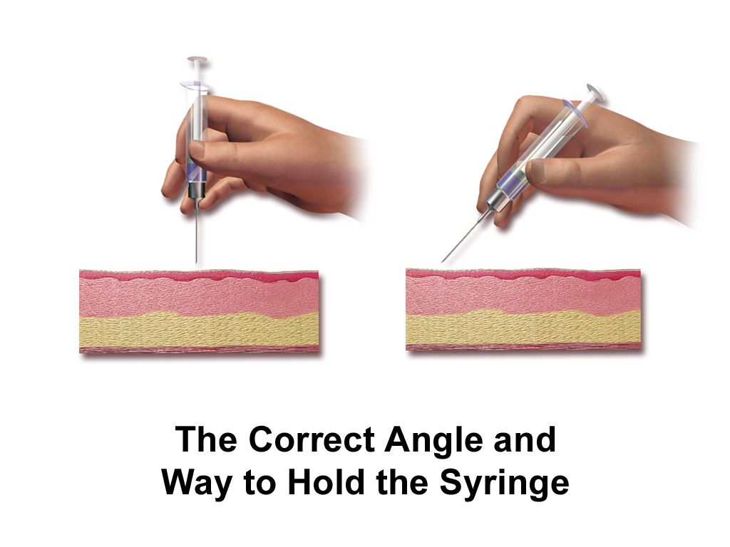
Structure that explains the clinical picture
The epidermis is avascular. It is a stratified sheet of keratinocytes that migrate from the basal layer to the surface over roughly 28 days, seeded with melanocytes for pigment and Langerhans cells for immune surveillance. Because it has no vessels of its own, an injury confined to epidermis cannot bleed and cannot scar — it peels and resolves. That single fact is why a sunburn is painful and red but heals in a week without a mark, and why it is not counted in burn surface-area calculations.
The dermis is where the clinically interesting structures live: capillary loops, sensory nerve endings, sebaceous and sweat glands, and hair follicles. Those appendages dip down from the epidermis into the dermis, and they carry epithelial cells with them. When a wound destroys the surface but spares the follicles, new epithelium crawls out of each follicle like grass seeding a field, and the wound closes on its own. When the wound destroys the deep dermis, the seed is gone, and closure requires a graft. Every burn-depth decision you will make is really a question about follicles.
The hypodermis is subcutaneous fat — insulation, energy store and mechanical cushion over bony prominences. Collectively the integument provides barrier, thermoregulation, sensation, vitamin D synthesis, immune defence and fluid containment. Lose enough of it and the patient loses heat, loses plasma, loses the ability to keep organisms out, and loses the sensory feedback that would normally make them shift their weight off an ischaemic heel.
Looking, and looking in every skin tone
Describe what you see in the standard vocabulary, because that is how dermatology and the medical record talk. Primary lesions arise on previously normal skin; secondary lesions are what happens to a primary lesion after time, scratching, infection or treatment. Add configuration (annular, linear, grouped, dermatomal) and distribution (flexural, extensor, sun-exposed, dermatomal, intertriginous), because distribution is often more diagnostic than morphology.
| Lesion | Definition | Typical example |
|---|---|---|
| Macule | Flat, non-palpable colour change under 1 cm | Freckle, early drug rash |
| Papule | Raised, solid, under 1 cm | Acne, early varicella |
| Plaque | Raised, flat-topped, over 1 cm | Psoriasis |
| Nodule | Solid, deeper, 1–2 cm | Lipoma, erythema nodosum |
| Vesicle / bulla | Fluid-filled under 1 cm / over 1 cm | Herpes zoster / burn blister, TEN |
| Pustule | Pus-filled | Folliculitis, pustular psoriasis |
| Wheal | Transient oedematous plaque | Urticaria |
| Erosion / ulcer | Loss of epidermis / loss through dermis | Ruptured blister / pressure injury |
| Fissure, lichenification, excoriation | Linear crack, leathery thickening, scratch-induced loss | Chronic eczema |
Erythema in deeply pigmented skin does not look red. It looks violaceous, darker than the surrounding skin, or simply different in a way that is easy to miss under fluorescent light. This is not a cosmetic point — it is the single largest source of missed early pressure injury and missed cellulitis. Use natural or high-quality light, compare the area with adjacent skin and with the contralateral limb, and lead with your hands: warmth, coolness, boggy softness and induration are palpable regardless of pigment. Ask about pain and itch, because a patient will report tenderness over a stage 1 injury before you can see it. For pallor, cyanosis and jaundice, look at conjunctivae, hard palate, palms, soles and nail beds.
How wounds heal, and what stops them
Healing runs in overlapping phases. Haemostasis is immediate: vasoconstriction, platelet plug, fibrin. The inflammatory phase (roughly days 0–4) brings neutrophils then macrophages; the wound looks red, warm and swollen, and this is normal, not infection. The proliferative phase (days 4–21) builds beefy red granulation tissue, contracts the edges and re-epithelialises from the margins and remaining appendages. The maturation phase runs from three weeks to two years, remodelling collagen; a healed wound reaches only about 80 percent of original tensile strength, which is why you keep teaching lifting restrictions long after the incision looks fine. Wounds closed surgically heal by primary intention; open wounds granulating in heal by secondary intention; a contaminated wound left open and closed later heals by tertiary intention.
Healing fails for predictable reasons, and each one is a nursing intervention. Perfusion and oxygenation come first — tissue cannot build collagen without oxygen, which is why smoking, peripheral arterial disease and hypotension are wound problems. Nutrition is second: protein for collagen, vitamin C for cross-linking, zinc and arginine for proliferation. Hyperglycaemia impairs neutrophil function, so glycaemic control is wound care. Add corticosteroids and other immunosuppressants, chemotherapy, infection, repeated trauma, moisture imbalance (too wet macerates, too dry desiccates) and simple age. A moist, warm, protected wound bed heals faster than a dry scab — that is the entire rationale behind modern dressings.
Document so the next nurse can tell whether the wound is improving without ever having seen it: anatomical location; measurements in centimetres as length (head to toe) × width (side to side) × depth; undermining and tunnelling described in clock positions with 12 o’clock toward the head; percentage of the bed that is granulation, slough or eschar; exudate amount, colour and odour after cleansing; wound edges (attached, rolled/epibole, macerated); and the periwound skin. Photograph per policy. “Wound looks better” is not documentation.
Whether a wound can close itself depends on whether epithelial cells survive in the dermal appendages — and whether it will close depends on perfusion, protein and glucose.
Section 2Inflammatory, Infectious and Life-Threatening Skin Disorders
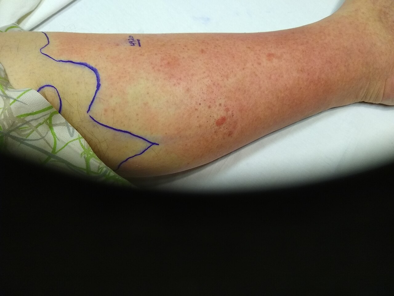
Bacterial infection: from cellulitis to a surgical emergency
Cellulitis is infection of the deep dermis and subcutaneous tissue, usually Streptococcus pyogenes or Staphylococcus aureus, entering through a break you may have to hunt for — tinea pedis between the toes, a venous ulcer, an IV site, a shaving nick. It presents as warm, tender, oedematous erythema with indistinct borders. Erysipelas is the more superficial cousin: sharply demarcated, raised, fiery red, often on the face or shin, classically streptococcal and more likely to spike fever early. Risk is stacked in patients with diabetes, lymphoedema, chronic venous insufficiency, obesity and prior episodes.
Nursing management is unglamorous and effective. Outline the erythema with a skin marker and write the date and time on the skin, then reassess each shift — expansion beyond the mark on appropriate antibiotics is an escalation trigger. Elevate the limb above heart level, because gravity does more for the oedema than any drug. Warm compresses for comfort. Mark and monitor temperature, heart rate and white count. Treatment for non-purulent cellulitis targets streptococci: cephalexin orally, cefazolin intravenously. Purulent cellulitis with an abscess needs incision and drainage — drainage is the treatment, antibiotics are the adjunct — plus MRSA coverage with trimethoprim–sulfamethoxazole, doxycycline or clindamycin orally, or vancomycin intravenously with trough or AUC monitoring and attention to renal function. Send cultures before the first dose when you can.
Necrotising fasciitis is the diagnosis you must not miss. Infection tracks along fascial planes, thromboses the perforating vessels and kills the overlying skin from below, so the surface can look deceptively unimpressive while the patient is dying. The cardinal early sign is pain out of proportion to the visible findings. Then come rapid hourly progression, oedema extending past the erythema, dusky or grey mottled skin, haemorrhagic bullae, crepitus, foul “dishwater” drainage, and later anaesthesia over the area as the cutaneous nerves die — a numb, agonising limb is a catastrophic sign. Systemically the patient is toxic: fever, tachycardia, hypotension, altered mentation, rising lactate, marked leukocytosis. Management is emergent surgical debridement plus broad-spectrum antibiotics (typically vancomycin with piperacillin–tazobactam, plus clindamycin for its antitoxin effect on streptococcal exotoxin). Imaging must never delay the operating room.
Viral, fungal and parasitic disease
Herpes zoster is reactivation of varicella-zoster virus latent in a dorsal root ganglion. Patients report two to three days of burning, itching or stabbing pain in a band before anything appears; then grouped vesicles on an erythematous base in a unilateral dermatomal distribution that stops at the midline. Antivirals — acyclovir, valacyclovir or famciclovir — shorten the course and reduce postherpetic neuralgia, but only if started within 72 hours of rash onset, so a patient reporting a new one-sided rash needs to be seen today, not next week. Pain is neuropathic: gabapentin or pregabalin, tricyclics, lidocaine 5 percent patches, opioids as a bridge. Postherpetic neuralgia may persist for months and is far more common in older adults. Lesions are infectious to varicella-naive contacts until crusted: cover localised lesions and use standard plus contact precautions; disseminated zoster or zoster in an immunocompromised patient requires airborne plus contact precautions. Involvement of the ophthalmic branch of the trigeminal nerve — especially vesicles on the tip of the nose, the Hutchinson sign — is a same-day ophthalmology referral. Recombinant zoster vaccine, two doses, is recommended from age 50 and for immunocompromised adults from 19.
Fungal disease is common and usually simple. Tinea corporis is an annular plaque with a scaly, advancing, raised border and central clearing; tinea pedis macerates the web spaces and is a frequent portal for cellulitis; tinea capitis needs an oral agent because topicals cannot reach the follicle. Candidiasis favours warm moist folds — inframammary, panniculus, groin — and produces beefy red plaques with satellite lesions, the finding that distinguishes it from simple intertrigo. Treat with topical antifungals and, more importantly, keep the fold dry and separated; barrier products and moisture-wicking textiles do more than any cream.
Scabies is Sarcoptes scabiei burrowing in the stratum corneum, and it should be suspected in any patient with relentless nocturnal itch and burrows or papules in the finger web spaces, flexor wrists, axillae, waistline, umbilicus or genitalia. Treat with permethrin 5 percent cream applied from the neck down (including under nails), left on 8–14 hours, repeated in seven days; treat all household and intimate contacts simultaneously even if asymptomatic; launder linens and clothing in hot water and dry on high heat, or bag non-washables for 72 hours. Oral ivermectin is used for crusted (Norwegian) scabies, which is highly contagious and warrants contact precautions. Teach explicitly that itching commonly persists two to four weeks after successful treatment — that is a hypersensitivity reaction to dead mite antigen, not failure, and re-treating repeatedly causes dermatitis.
Chronic inflammatory disease and severe drug reactions
Psoriasis is T-cell–driven keratinocyte hyperproliferation: turnover drops from 28 days to about four, so cells pile up as well-demarcated erythematous plaques with adherent silvery scale over extensor surfaces, scalp, umbilicus and sacrum. Removing scale causes pinpoint bleeding (Auspitz sign); new plaques appear at sites of trauma (Koebner phenomenon); nails show pitting and onycholysis. Treat mild disease topically with corticosteroids, vitamin D analogues such as calcipotriene, tar and keratolytics; moderate-to-severe disease with narrowband UVB phototherapy, methotrexate, cyclosporine, apremilast or biologics targeting TNF-alpha, IL-17 and IL-23. Nursing implications matter here: methotrexate is weekly, not daily — a daily dosing error is lethal — requires folic acid supplementation, CBC and liver function monitoring, alcohol avoidance and contraception, since it is teratogenic. Biologics require screening for latent tuberculosis and hepatitis B before the first dose and holding for active infection. Psoriasis is not contagious and it is not a hygiene failure; it travels with psoriatic arthritis, metabolic syndrome, cardiovascular disease and depression, so screen for joint pain and mood.
Atopic dermatitis is a skin-barrier disease with an itch–scratch cycle on top. Teach “soak and seal”: short lukewarm bath, gentle non-soap cleanser, pat dry, then a thick ointment-based emollient within three minutes, with topical corticosteroid applied to active lesions first. Use the lowest effective steroid potency on the face, neck and skin folds, teach the fingertip-unit measure, and warn about atrophy, striae and telangiectasia with prolonged high-potency use. Topical calcineurin inhibitors (tacrolimus, pimecrolimus) are steroid-sparing for the face; dupilumab is available for moderate-to-severe disease. Sudden worsening with punched-out erosions and fever suggests eczema herpeticum — a same-day referral.
Stevens–Johnson syndrome and toxic epidermal necrolysis are a single spectrum of keratinocyte apoptosis, almost always drug-induced, separated by the percentage of body surface with epidermal detachment: SJS under 10 percent, overlap 10–30 percent, TEN over 30 percent. The usual culprits are allopurinol, sulfonamide antibiotics, aromatic anticonvulsants (lamotrigine, carbamazepine, phenytoin), nevirapine and oxicam NSAIDs, typically 4–28 days after starting. Suspect it when a flu-like prodrome is followed by painful skin, dusky targetoid macules, and erosions of at least two mucosal surfaces — eyes, mouth, genitalia. Lateral pressure sloughs the epidermis (Nikolsky sign positive). The single most important intervention is to stop the offending drug immediately; every hour of continued exposure worsens mortality. After that, care is burn care: transfer to a burn or intensive care unit, fluid and electrolyte replacement, meticulous non-adherent wound care with no tape on skin, temperature regulation, aggressive analgesia, nutrition, and an early ophthalmology consult, because ocular scarring and blindness are the leading long-term morbidity. Document the culprit drug as a true allergy in every system, and teach the patient and family to name that drug and its whole class for the rest of their life.
In skin infection, pain out of proportion to appearance means necrotising fasciitis until a surgeon says otherwise; in a drug rash, mucosal involvement plus a positive Nikolsky sign means stop the drug now and call the burn unit.
Section 3Pressure Injuries, Chronic Wounds and Skin Cancer
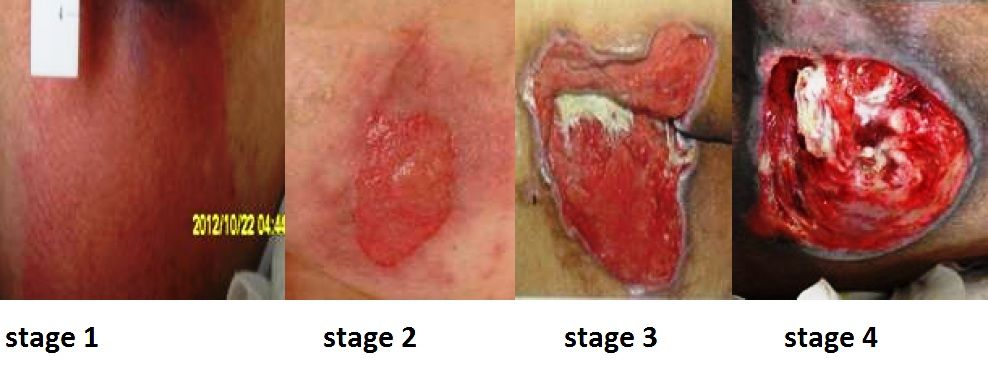
Pressure injury: mechanism, staging and prevention
Pressure injury develops when sustained pressure over a bony prominence exceeds capillary closing pressure and occludes perfusion; ischaemia, reperfusion injury and cell death follow. Shear makes it far worse by tearing the deeper tissue planes while the skin stays fixed — which is exactly what happens when the head of the bed is elevated and the patient slides down. Deep muscle over bone dies before the skin does, which is why a small surface lesion can sit over a large cavity, and why deep tissue pressure injury exists as its own category. Add moisture, malnutrition, impaired sensation, immobility, hypotension and friction and you have every risk factor on the Braden Scale.
| Category | Defining findings | Practice implication |
|---|---|---|
| Stage 1 | Intact skin, non-blanchable erythema; in darker tones may be purple or maroon, with warmth, firmness or pain | Off-load immediately; fully reversible |
| Stage 2 | Partial-thickness loss of dermis: shallow pink-red bed, or intact/ruptured serum-filled blister. No slough, granulation or eschar | Not a skin tear, moisture-associated damage or burn — those are staged separately |
| Stage 3 | Full-thickness loss; fat may be visible; slough or eschar may be present; undermining and tunnelling possible; no exposed bone, tendon or muscle | Depth varies by site — the ear and bridge of the nose have no fat, so stage 3 there is shallow |
| Stage 4 | Full-thickness with exposed or directly palpable bone, tendon, muscle, ligament or cartilage | Assume osteomyelitis risk; surgical consult |
| Unstageable | Base obscured by slough or eschar | Cannot stage until debrided — except stable, dry, intact eschar on an ischaemic heel, which is left alone as a biological cover |
| Deep tissue pressure injury | Persistent non-blanchable deep red, maroon or purple discoloration, or a blood-filled blister; skin may be intact | May declare itself as a stage 3 or 4 within days; document evolution |
| Medical device–related | Conforms to the pattern of the device — oxygen tubing, cervical collar, SCD, endotracheal tube tie | Assess under and around every device at least twice daily |
The Braden Scale scores six subscales — sensory perception, moisture, activity, mobility, nutrition, and friction/shear — for a total of 6 to 23. Lower is worse: 15–18 mild risk, 13–14 moderate, 10–12 high, 9 or below severe. Score on admission, at every change in condition and per unit policy, and treat the low subscale, not just the total: a patient scoring low only on moisture needs a barrier and a continence plan, not a specialty mattress.
Prevention is a bundle and every element is testable. Reposition on an individualised schedule, conventionally every two hours in bed and hourly in a chair — or teach the alert patient to do a 15-second weight shift every 15 minutes. Use the 30-degree lateral tilt rather than a full 90-degree side-lying position, which loads the trochanter. Keep the head of bed at or below 30 degrees unless contraindicated, and use the knee gatch or a lift sheet to stop sliding. Float the heels completely off the mattress with a pillow lengthwise under the calves or an offloading boot — the heel is the second most common site and the hardest to heal. Never massage over a bony prominence, never use ring or donut cushions, and never turn a patient onto an existing injury. Manage incontinence with a structured barrier regimen, keep linens dry and wrinkle-free, optimise protein and calorie intake, and choose a support surface matched to risk. And never reverse-stage: a healing stage 4 is a “healing stage 4,” not a stage 2, because muscle and fat are replaced by scar, not by the original tissue.
Lower-extremity ulcers: three diseases that look alike
A leg ulcer is not a diagnosis, it is a finding, and the treatment for one type actively harms another. Compression heals a venous ulcer and can infarct an arterial one, so you must know which you are looking at before the wrap goes on.
| Feature | Venous | Arterial | Neuropathic (diabetic) |
|---|---|---|---|
| Location | Medial malleolus, gaiter area | Lateral malleolus, toes, pressure points | Plantar surface, metatarsal heads, heel |
| Appearance | Shallow, irregular border, ruddy granulation | “Punched out,” pale or necrotic dry base | Round, callused rolled rim |
| Exudate | Moderate to heavy | Minimal | Variable; purulent if infected |
| Pain | Aching, better with elevation | Severe, worse with elevation, better dangling | Absent — that is the danger |
| Surrounding skin | Haemosiderin staining, oedema, lipodermatosclerosis, weeping | Thin, shiny, hairless, cool, pale on elevation | Dry, cracked, callused; deformity, Charcot changes |
| Pulses / ABI | Present; ABI usually normal | Diminished or absent; ABI under 0.9 | Often present but ABI may be falsely high (over 1.3) from calcification |
| Treatment core | Compression, elevation, moist dressing | Revascularisation; never compress | Offloading (total contact cast), debride callus, glycaemic control |
The ankle–brachial index is the gatekeeper: 0.91–1.30 is normal, 0.90 or below indicates arterial disease, 0.40 or below is severe ischaemia with rest pain, and above 1.30 means non-compressible calcified vessels that make the number unreliable. Full multilayer compression is generally reserved for an ABI above 0.8; modified or no compression below that, pending vascular assessment. Every diabetic patient gets daily foot inspection with a mirror, well-fitted closed shoes, no barefoot walking, no heating pads, straight-across nail trimming and an annual monofilament and pulse exam — teaching that prevents amputations far more effectively than any dressing.
Skin cancer and photoprotection
Basal cell carcinoma is the most common human cancer: a pearly, translucent papule with telangiectasias and a rolled border, often on the nose, ear or eyelid, sometimes with a central ulcer that scabs and re-bleeds. It invades locally and rarely metastasises. Squamous cell carcinoma is a firm, scaly, hyperkeratotic or ulcerated nodule on sun-exposed skin, arising from actinic keratoses; it can metastasise, and its risk is markedly higher in solid-organ transplant recipients and in chronic wounds and old burn scars — a non-healing ulcer within a mature burn scar is a Marjolin ulcer until biopsied. Melanoma is the one that kills. Teach ABCDE: Asymmetry, Border irregularity, Colour variegation, Diameter over 6 mm, and Evolving — plus the “ugly duckling” lesion that simply does not match the patient’s other moles. Diagnosis is by full-thickness biopsy; Breslow depth in millimetres is the dominant prognostic factor and drives sentinel lymph node biopsy. Prevention teaching is concrete: broad-spectrum SPF 30 or higher applied 15–30 minutes before exposure and reapplied every two hours and after swimming, protective clothing and wide-brimmed hats, shade between 10:00 and 16:00, no tanning beds at any age, and a monthly self-examination including scalp, soles and between the toes.
Pressure injuries are prevented by offloading and staged only when the base is visible; leg ulcers are treated by their cause — compression for venous, revascularisation for arterial, offloading for neuropathic — and any non-healing lesion or changing mole gets a biopsy, not another dressing.
Section 4Burn Injury: Depth, Extent and the First 24 Hours
Mechanism: why a skin injury becomes a whole-body problem
Heat coagulates protein. At the centre of the burn is the zone of coagulation — irreversibly dead tissue. Surrounding it is the zone of stasis, where perfusion is marginal and the tissue is alive but threatened; and outside that the zone of hyperaemia, which recovers. Everything you do in the first 24 hours — fluid, warmth, avoiding pressure and vasoconstrictors — exists to keep the zone of stasis from converting to necrosis. Under-resuscitate and the burn gets deeper. Over-resuscitate and you drown the patient. Both errors are nursing-visible before they are physician-visible.
Once a burn exceeds roughly 20–25 percent total body surface area (TBSA), the inflammatory response stops being local. Histamine, prostaglandins, bradykinin and thromboxane flood the circulation and make capillaries everywhere leak. Plasma, protein and electrolytes shift into the interstitium — massive third-spacing — producing generalised oedema and intravascular volume loss. This is burn shock: hypovolaemic and distributive at once, with an early depression of cardiac output out of proportion to the volume lost. Haematocrit rises from haemoconcentration even as the patient bleeds volume into the tissues. Potassium rises from cell lysis. Sodium falls as it follows water into the interstitium. Metabolic acidosis develops from hypoperfusion. Glucose rises with the catecholamine surge. The gut becomes ileus.
Then, at roughly 48–72 hours, capillary integrity is restored and the process reverses. Interstitial fluid remobilises into the vasculature, the patient diureses briskly, haematocrit falls by dilution, and potassium now drops — back into cells and out through the urine. The nurse who keeps running the resuscitation rate through this transition causes pulmonary oedema. The transition point is the boundary between the emergent and acute phases, and recognising it is a bedside judgement.
| Parameter | Emergent phase (0–48 h) | Acute phase (after fluid remobilisation) |
|---|---|---|
| Fluid shift | Intravascular to interstitial (third spacing) | Interstitial back to intravascular |
| Urine output | Low — the resuscitation target | Diuresis |
| Haematocrit | Elevated (haemoconcentration) | Falls (dilution, plus anaemia of injury) |
| Potassium | Hyperkalaemia from cell lysis | Hypokalaemia from shift and urinary loss |
| Sodium | Hyponatraemia (sodium follows water out) | Hyponatraemia (dilutional) or hypernatraemia with evaporative loss |
| Acid–base | Metabolic acidosis, base deficit, elevated lactate | Corrects with perfusion; watch for sepsis-driven acidosis |
| Dominant threat | Airway and hypovolaemic shock | Infection, hypermetabolism, contracture |
Depth and extent: naming the burn correctly
| Depth | Tissue involved | Appearance and sensation | Healing |
|---|---|---|---|
| Superficial (first-degree) | Epidermis only | Dry, red, blanches, no blisters, painful | 3–7 days, no scar. Not counted in TBSA |
| Superficial partial-thickness | Epidermis + papillary dermis | Moist, weeping, blisters, blanches with brisk refill, very painful | 7–21 days, minimal scarring |
| Deep partial-thickness | Into reticular dermis | Mottled red and waxy white, drier, sluggish or absent blanching, decreased light touch but pressure is painful | Over 21 days; hypertrophic scarring; usually excised and grafted |
| Full-thickness (third-degree) | Entire dermis | Dry, leathery eschar — waxy white, tan, brown or charred; does not blanch; insensate; thrombosed vessels visible | Cannot re-epithelialise; requires grafting |
| Fourth-degree | Fat, fascia, muscle, tendon, bone | Charred, exposed deep structures | Debridement, flaps, often amputation |
Two cautions students get wrong. First, a full-thickness burn is painless in its centre, but the patient still hurts badly, because full-thickness areas are ringed by partial-thickness tissue with intact nerves — never withhold analgesia because “third-degree burns do not hurt.” Second, burn depth evolves over 48–72 hours; the wound you chart on admission may be deeper on day three, and that is expected, not a documentation error.
For extent, the Rule of Nines gives a fast field estimate in adults: head and neck 9 percent; each arm 9; anterior trunk 18; posterior trunk 18; each leg 18; perineum 1. In infants and small children the head is proportionally much larger — about 18 percent — and each leg smaller, about 13.5 percent, so the adult rule overestimates the legs and underestimates the head. The Lund and Browder chart adjusts by age and is the accurate tool used in burn centres. For scattered or irregular burns, use the rule of palm: the patient’s own palm including the fingers is approximately 1 percent TBSA. Count only partial- and full-thickness burns.
The American Burn Association transfer criteria decide destination: partial-thickness burns over 10 percent TBSA; any full-thickness burn; burns of the face, hands, feet, genitalia, perineum or over major joints; electrical injury including lightning; chemical burns; inhalation injury; burns in patients with significant comorbidity; burns with concomitant trauma; and children in facilities without paediatric burn capability.
What you do first, in order
Stop the burning process — remove smouldering clothing and all jewellery and constricting items before oedema develops, brush off dry chemical powder before irrigating, and irrigate chemical burns with copious room-temperature water for at least 20 minutes (longer for alkalis, which cause deeper liquefactive necrosis; never attempt to neutralise). Cool small burns briefly with room-temperature water, but never apply ice — it vasoconstricts the zone of stasis and deepens the injury — and never cool a large burn, because hypothermia kills burn patients. Cover with a clean dry sheet and keep the patient warm.
Then run ABC. The airway is the first killer and the one with a closing window. Two lists, and students collapse them into one. Suspect inhalation injury with any of: burns sustained in an enclosed space; loss of consciousness at the scene; facial burns; singed nasal hairs, eyebrows or eyelashes; soot in the mouth or nose; or carbonaceous sputum. Those are exposure findings, and they are poor standalone predictors of obstruction — what they mandate is close serial airway assessment, humidified 100 percent oxygen and a carboxyhaemoglobin level, not a reflex tube. Do not intubate on exposure history alone; an unnecessary tube in a burn patient costs ventilator days, pneumonia, sedation and unplanned extubations. Escalate for immediate airway control when the dynamic signs appear: new or worsening hoarseness or voice change; a brassy cough; stridor; drooling or difficulty swallowing; wheezing; respiratory distress; deep circumferential facial or neck burns; or altered mentation. Those mean intubate now, before the oedema closes the airway. Upper-airway swelling builds over the first 12 hours and peaks at roughly 24 to 48 hours, and the resuscitation fluid you are about to give makes it worse — so vigilance does not relax at the end of your shift. A patient talking comfortably now can be unintubatable in ninety minutes; do not wait for hypoxia to declare itself.
Remember two poisons that pulse oximetry will not show you. Carbon monoxide binds haemoglobin with roughly 200 times the affinity of oxygen, and standard pulse oximetry cannot distinguish carboxyhaemoglobin from oxyhaemoglobin — the SpO2 reads normal while the patient is starving. Diagnose with a carboxyhaemoglobin level by co-oximetry: under 5 percent is normal in a non-smoker, 15–20 percent causes headache and confusion, 20–40 percent nausea and disorientation, 40–60 percent coma and seizures. Treat with 100 percent oxygen by non-rebreather, which shortens the carboxyhaemoglobin half-life from about 4–5 hours on room air to roughly 60–90 minutes; hyperbaric therapy is considered for severe poisoning. Cyanide, released when plastics, wool and silk burn, blocks cellular oxygen use; suspect it with profound persistent lactic acidosis and altered mentation despite adequate oxygen, and treat with intravenous hydroxocobalamin, warning the team that it turns skin, urine and serum deep red and interferes with colorimetric laboratory assays and pulse oximetry.
Fluid resuscitation, worked through
Two large-bore IVs go through unburned skin if possible, through burned skin if not — access matters more than aesthetics. Lactated Ringer’s is the crystalloid of choice, because it is isotonic and its lactate is metabolised to bicarbonate, helping the acidosis. Fluid is required for burns of roughly 20 percent TBSA or more in adults (10 percent in children and older adults).
The classic Parkland formula is 4 mL × body weight in kg × %TBSA of lactated Ringer’s over the first 24 hours, with half given in the first 8 hours and the remaining half over the next 16 hours. The critical detail: the clock starts at the time of injury, not the time of arrival. Current American Burn Association consensus starts lower — 2 mL/kg/%TBSA for thermal injury in adults, 3 mL for high-voltage electrical injury — and titrates upward, because decades of over-resuscitation (“fluid creep”) produced compartment syndromes and pulmonary oedema. Know both; the formula is a starting point, not a prescription.
Worked example. An 80 kg man sustains 45 percent TBSA partial- and full-thickness flame burns at 20:00. He reaches your emergency department at 22:00.
Parkland total: 4 mL × 80 kg × 45 = 14,400 mL lactated Ringer’s in the first 24 hours. First half = 7,200 mL, due in the first 8 hours from injury — that is by 04:00. But two hours have already elapsed, so those 7,200 mL must run over the remaining 6 hours: 7,200 ÷ 6 = 1,200 mL/hr. The second half, 7,200 mL, runs over the following 16 hours (04:00 to 20:00): 7,200 ÷ 16 = 450 mL/hr. Had he arrived immediately, the first rate would have been 7,200 ÷ 8 = 900 mL/hr. Using the ABA 2 mL starting point instead: 2 × 80 × 45 = 7,200 mL total, 3,600 mL in the first 8 hours = 450 mL/hr, then 225 mL/hr.
Now the part that actually matters at the bedside: the formula is titrated to urine output every hour. Insert an indwelling catheter and target 0.5 mL/kg/hr in adults — about 30–50 mL/hr for most adults, so roughly 40 mL/hr for this 80 kg man — and 1 mL/kg/hr in children under 30 kg. In high-voltage electrical injury with tea-coloured or port-wine urine from myoglobin, target 75–100 mL/hr until the urine clears, because myoglobin precipitating in tubules causes acute kidney injury. Falling urine output, rising heart rate, worsening base deficit and rising lactate say increase the rate. Urine output well above target, ballooning oedema, rising peak airway pressures and a tense abdomen say back off — over-resuscitation causes abdominal compartment syndrome, extremity compartment syndrome, ocular compartment syndrome and pulmonary oedema. Colloid, typically 5 percent albumin, is commonly added after the first 12–24 hours once capillaries begin to seal.
Meanwhile: keep the room hot (about 30–33 °C) and use warmed fluids and blankets, because a burn patient cannot thermoregulate and hypothermia worsens coagulopathy and acidosis. Give analgesia as intravenous opioid only — intramuscular and subcutaneous absorption is unreliable during third spacing and will dump a bolus into the circulation when perfusion returns. Insert a nasogastric tube for burns over 20 percent TBSA because ileus is near-universal, and start stress-ulcer prophylaxis to prevent a Curling ulcer. Update tetanus immunisation. Monitor circumferential burns hourly with a Doppler: loss of pulse, deep unrelenting pain, pallor, paraesthesia and delayed capillary refill in an extremity, or rising peak airway pressures with restricted chest excursion, mean the eschar is acting as a tourniquet and an escharotomy is needed — a bedside incision through the insensate eschar; if the deeper compartments are involved, a fasciotomy in theatre.
Estimate depth and TBSA accurately, protect the airway before oedema closes it, start Parkland from the time of injury — and then let hourly urine output, not the formula, decide the rate.
Section 5Acute and Rehabilitative Burn Care: Infection, Nutrition and Function
Infection: the threat after 48 to 72 hours
Once diuresis begins, the emergent phase ends and the acute phase runs until every wound is closed. The burn wound is a warm, moist, protein-rich, avascular surface with no epidermal barrier and no white cells able to reach it — a perfect culture medium. Infection and sepsis are now the leading cause of death. Early colonisation is with gram-positive skin flora; by the end of the first week gram-negatives dominate, especially Pseudomonas aeruginosa, followed by fungi such as Candida in patients on prolonged antibiotics.
Prophylactic systemic antibiotics are not given — they select resistant organisms without reducing wound infection. Control is topical, plus early excision of dead tissue. Recognising infection in a burn patient is a nursing skill, because the usual signs are unreliable. Watch the wound for a change in colour (green or blue-green discoloration suggests Pseudomonas), for rapid or premature separation of eschar, for focal dark brown or black spots in previously clean tissue, for conversion of a partial-thickness wound to full-thickness, and for a new odour. Watch the patient for the burn-specific sepsis picture: hypothermia or hyperthermia, new confusion or agitation, return of ileus or feeding intolerance, unexplained hyperglycaemia and rising insulin requirement, thrombocytopenia, tachypnoea and falling urine output. A burn patient who suddenly stops tolerating tube feeds is septic until proved otherwise.
| Topical agent | How it works / penetration | Nursing implications |
|---|---|---|
| Silver sulfadiazine 1 percent | Broad-spectrum; poor eschar penetration | Contraindicated in sulfa allergy, late pregnancy and infants under 2 months (kernicterus risk); may cause transient leukopenia — monitor CBC; forms a pseudo-eschar that must be cleansed off; painless to apply |
| Mafenide acetate | Penetrates eschar and cartilage well — agent of choice for ear and nose burns | Painful and burning on application — premedicate; carbonic anhydrase inhibitor, so it causes metabolic acidosis — monitor ABG and respiratory rate |
| Silver nitrate 0.5 percent solution | Surface antisepsis; does not penetrate | Stains everything black; dressings must be kept wet; causes hyponatraemia, hypochloraemia and hypokalaemia — monitor electrolytes |
| Silver-impregnated dressings | Sustained silver release | May stay in place 3–7 days — fewer painful changes; do not use with oil-based products |
| Bacitracin / plain petrolatum gauze | Low-toxicity coverage | Common on facial and superficial partial-thickness burns; frequent reapplication |
Wound care itself is hydrotherapy or bedside cleansing, debridement and re-dressing, usually once or twice daily and always after adequate analgesia. Debridement may be mechanical (careful cleansing, forceps), enzymatic (collagenase applied to slough), autolytic (occlusive dressing) or surgical. Early excision and grafting — taking full-thickness eschar to a viable bed within the first several days — is the single biggest advance in burn survival, because it removes the culture medium. Use strict infection-prevention practice: hand hygiene, gowns and gloves for wound contact, dedicated equipment, no fresh flowers or standing water, and cohorting per policy.
Hypermetabolism, nutrition and pain
A major burn produces the most extreme hypermetabolic state in medicine: resting energy expenditure can approach twice normal, peaking around days 5–7 and persisting for months after wound closure. Catecholamines, cortisol and glucagon drive gluconeogenesis, insulin resistance and relentless skeletal-muscle catabolism. If you do not feed the patient, they will burn their own diaphragm and their wounds will not close.
Start enteral nutrition early — within 24 hours, and often within 4–6 hours in major burns — because early feeding preserves gut mucosal integrity and reduces bacterial translocation. Targets are high-calorie and high-protein: commonly 1.5–2 g/kg/day of protein and 25–35 kcal/kg/day plus a burn factor (the older Curreri formula, 25 kcal/kg plus 40 kcal per %TBSA, still illustrates the magnitude). Supplement vitamin C, vitamin A, zinc and often glutamine. Nursing monitoring is daily weights on the same scale at the same time, strict intake and output, calorie counts, glucose control with insulin infusions as needed, and tracking of nitrogen balance and prealbumin with the understanding that prealbumin falls in inflammation regardless of intake. Hold nothing for the sake of routine: burn patients are chronically underfed by NPO orders that nobody cancels.
Pain has two components and both need a plan. Background pain is continuous and is best covered by scheduled or continuous opioid plus adjuvants; procedural pain during dressing changes and physiotherapy is severe, predictable and requires premedication timed to peak effect, often with ketamine or short-acting agents, plus non-pharmacological methods — virtual reality distraction, music, controlled breathing and giving the patient a say in pausing. Anxiety amplifies pain; treat both. Later, pruritus in healing burns is intense and under-treated: use emollients, cool compresses, antihistamines, and gabapentin or pregabalin for the neuropathic component, and keep nails short.
Grafting, contracture prevention and going home
Deep partial- and full-thickness burns are closed with a split-thickness autograft harvested from the patient’s own unburned skin. Meshing the graft expands coverage and lets exudate escape but leaves a permanent lattice pattern. Cultured epithelial autograft is used when donor sites are scarce. Cadaver allograft and porcine xenograft are temporary biological dressings only — they will be rejected, and teaching families otherwise causes real distress. After grafting, immobilise and elevate the site for three to five days, protect absolutely against shear, monitor for haematoma or seroma beneath the graft (which lifts it off its blood supply and is the commonest cause of failure), and keep the dressing intact until the surgeon says otherwise. The donor site is a fresh superficial partial-thickness wound: it is often more painful than the graft, heals in 10–14 days, and may be reharvested.
Rehabilitation begins on day one, not at discharge. The rule to remember is that the position of comfort is the position of contracture: burned tissue contracts into flexion, and a patient left comfortable will fuse into a useless posture. Anterior neck burns are positioned in slight hyperextension with no pillow; axillae are abducted 90–110 degrees on airplane splints; elbows and knees are splinted in extension; hips stay extended and neutrally rotated with no pillow under the knees; hands are splinted in the intrinsic-plus position (wrist slightly extended, MCP joints flexed 70–90 degrees, IP joints straight, thumb abducted). Range-of-motion exercise happens daily, and ambulation starts as soon as haemodynamics allow. Once wounds are closed, custom pressure garments worn 23 hours a day for 12–18 months flatten hypertrophic scar and are the treatment patients most often abandon — make adherence an explicit teaching goal.
Discharge teaching covers wound and graft care with return demonstration; signs of infection to report; the pressure-garment and splinting schedule; daily moisturising of healed skin, which has no sebaceous glands and cracks; strict sun avoidance and SPF 30 or higher for at least a year because new skin hyperpigments permanently; itch management; a high-protein diet with weight monitoring; and realistic expectations about fatigue, cold intolerance and the two-year timeline of scar maturation. Screen for post-traumatic stress, depression and altered body image, and connect the patient with burn survivor peer support — psychosocial recovery is slower than wound closure and predicts return to work better than TBSA does.
After the first 48–72 hours the burn patient no longer dies of shock — they die of infection and catabolism, so the acute-phase priorities are topical antimicrobials with early excision, aggressive enteral protein, and splinting that protects function before the scar decides for you.
The chapter in one breath
Skin is a barrier, a thermostat and a fluid container, and every integumentary disorder is a version of one of those failing. Assess it in every skin tone using light, comparison and your hands, name lesions in standard morphology, and document wounds in centimetres with clock-position tunnelling. In infection, the finding that changes everything is pain out of proportion to appearance — that is necrotising fasciitis and it goes to theatre, not to a dressing cart. In drug eruptions, mucosal involvement with a positive Nikolsky sign is Stevens–Johnson syndrome or toxic epidermal necrolysis, and stopping the drug is the intervention that saves the patient. Pressure injuries are prevented by offloading, floated heels, 30-degree tilts and moisture control, staged only when the base is visible, and never reverse-staged. Leg ulcers are treated by cause: compress venous, revascularise arterial, offload neuropathic, and check the ankle–brachial index first. In burns, classify depth by whether dermal appendages survive, estimate extent with the Rule of Nines, protect the airway before oedema closes it, and start Parkland from the time of injury while titrating hourly to a urine output of 0.5 mL/kg/hr. Past 48–72 hours, the enemy changes from shock to infection and catabolism — topical antimicrobials, early excision and grafting, high-protein enteral feeding, and splinting that preserves the function the scar would otherwise take.
Words worth knowing
| Term | What it means |
|---|---|
| Eschar | Dry, leathery, devitalised tissue over a full-thickness burn or wound; acts as a tourniquet when circumferential and as a culture medium when retained |
| Escharotomy | Bedside incision through full-thickness eschar to relieve constriction of a limb or the chest; the eschar itself is insensate |
| Zone of stasis | The salvageable ring of marginally perfused tissue around a burn; adequate resuscitation and warmth prevent it converting to necrosis |
| Parkland formula | 4 mL × kg × %TBSA of lactated Ringer’s in 24 hours, half in the first 8 hours from the time of injury; ABA consensus starts at 2 mL and titrates |
| Rule of Nines | Rapid adult TBSA estimate in multiples of 9 percent; the paediatric head is 18 percent and each leg 13.5 percent |
| Rule of palm | The patient’s palm including fingers approximates 1 percent TBSA; used for scattered burns |
| Carboxyhaemoglobin | Haemoglobin bound to carbon monoxide; requires co-oximetry because pulse oximetry reads it as saturated |
| Curling ulcer | Stress-related gastroduodenal ulceration following major burns; the reason for early acid suppression |
| Autograft / allograft / xenograft | Permanent graft from the patient’s own skin / temporary cadaveric skin / temporary porcine skin |
| Intrinsic-plus position | Anti-contracture hand splinting: wrist extended, MCP joints flexed 70–90 degrees, IP joints straight, thumb abducted |
| Nikolsky sign | Epidermis shears off with gentle lateral pressure; positive in TEN and pemphigus |
| Deep tissue pressure injury | Persistent non-blanchable deep red, maroon or purple discoloration, or a blood-filled blister, over intact or non-intact skin |
| Braden Scale | Six-subscale pressure-injury risk tool scored 6–23; 18 or below indicates risk, 9 or below severe risk |
| Ankle–brachial index (ABI) | Ankle-to-arm systolic pressure ratio; 0.91–1.30 normal, 0.90 or less arterial disease, above 1.30 non-compressible calcified vessels |
| Marjolin ulcer | Squamous cell carcinoma arising in a chronic wound or old burn scar; any non-healing scar ulcer needs biopsy |
| Auspitz sign | Pinpoint bleeding after removing psoriatic scale, from the dilated capillaries of elongated dermal papillae |
| Erysipelas | Superficial dermal and lymphatic streptococcal infection with a sharply demarcated raised border, unlike the indistinct margin of cellulitis |
| Hypermetabolic response | The post-burn catabolic surge that can double resting energy expenditure for months; managed with early high-protein enteral feeding |
| Undermining / tunnelling | Tissue destruction under intact wound edges / a narrow sinus tract; both charted in clock positions with 12 o’clock toward the head |
| Pressure garment | Custom compression worn 23 hours daily for 12–18 months to flatten hypertrophic burn scar |
Check yourself
Sensory Disorders: Eye and Ear
Sensory nursing is quiet work until it suddenly is not. Most of what you do for eyes and ears is patient teaching, drop technique, fall prevention and the unglamorous business of making sure a frightened person can hear the plan of care — and then, once or twice a year, a patient tells you their vision went dark in one eye an hour ago, or that a splash of drain cleaner hit their face, or that they woke up deaf on one side, and the difference between a good outcome and permanent loss is measured in minutes. This chapter teaches the mechanism first, because the mechanism predicts the finding: pressure that cannot escape gives you a rock-hard painful eye with a fixed mid-dilated pupil, a retina starved of blood gives you painless blackout, endolymph under pressure gives you vertigo with fluctuating hearing loss. Learn the mechanism and you will never have to memorise a list of symptoms again.
By the end of this chapter you can
- Perform and document a focused bedside eye and ear assessment including visual acuity, confrontation fields, pupil testing, the whisper test and the Weber and Rinne tuning-fork tests.
- Explain aqueous humour dynamics and distinguish primary open-angle glaucoma from acute angle-closure glaucoma, including which one is a same-hour emergency and why.
- Compare the drug classes that lower intraocular pressure by mechanism, and state the systemic monitoring each one demands.
- Triage sudden painless vision loss among central retinal artery occlusion, retinal detachment, vitreous haemorrhage and giant cell arteritis, and act on the correct time window.
- Manage diabetic retinopathy and age-related macular degeneration, including anti-VEGF therapy, the Amsler grid and AREDS2 supplementation.
- Differentiate the red eye and initiate immediate irrigation for a chemical ocular burn before any other assessment.
- Distinguish conductive from sensorineural hearing loss, recognise sudden sensorineural hearing loss as an emergency, and identify common ototoxic drugs.
- Differentiate benign paroxysmal positional vertigo, vestibular neuritis, labyrinthitis and Ménière disease, and teach safety for each.
- Escalate ocular and otologic red flags using SBAR and delegate appropriately, keeping assessment, teaching, evaluation and unstable patients with the registered nurse.
Section 1Assessing Vision, Hearing and the Safety They Buy
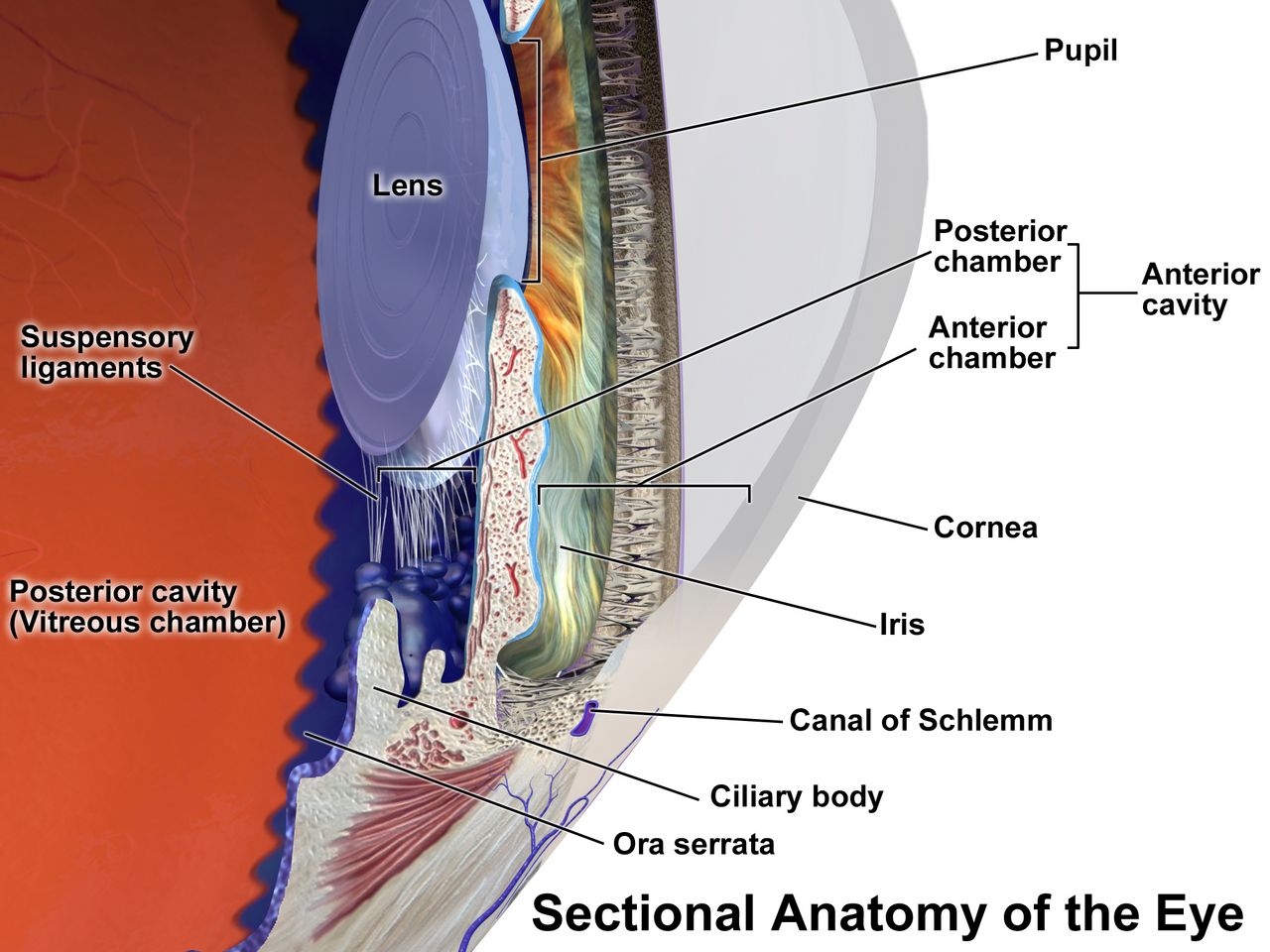
The mechanism you need before anything else
The eye is a pressurised sphere with a one-way plumbing problem. The ciliary body, sitting behind the iris, secretes aqueous humour continuously at roughly 2 to 3 microlitres per minute. That fluid flows from the posterior chamber, through the pupil, into the anterior chamber, and drains out through the trabecular meshwork at the angle where the iris meets the cornea. Production never stops. If drainage slows or the angle closes, pressure rises, and pressure is transmitted to the one structure that cannot tolerate it: the axons of the optic nerve as they turn and exit at the optic disc. Everything you will learn about glaucoma is a variation on that sentence.
Light that survives the trip lands on the retina, and the retina is metabolically the hungriest tissue in the body per gram. It is fed by two independent systems — the central retinal artery for the inner layers and the choroidal circulation for the outer layers — and neither has meaningful collateral supply. That is why retinal ischaemia behaves like brain ischaemia rather than like a bruised muscle: the tissue dies on a clock measured in minutes to a couple of hours, not days. The macula, the small central zone packed with cones, provides reading vision and face recognition; lose the macula and you keep peripheral navigation but lose the ability to live independently.
The ear divides the same way into a mechanical half and a neural half, and that division is the whole of hearing-loss diagnosis. The external canal and the middle ear ossicles are a mechanical amplifier: anything that blocks or stiffens them — cerumen, fluid, a perforated drum, a fixed stapes — produces conductive loss. The cochlea and the eighth cranial nerve are the transducer and the cable: anything that damages hair cells or the nerve — noise, ageing, aminoglycosides, a schwannoma — produces sensorineural loss. Sitting alongside the cochlea, sharing its fluid, is the vestibular labyrinth, which is why so many patients arrive with hearing and balance complaints in the same breath.
The vision assessment you can do at the bedside
Always test and document visual acuity before you do anything else to an eye, with the single exception of a chemical splash. Acuity is the vital sign of the eye. Use a Snellen chart at 20 feet or a handheld near card at 14 inches, test each eye separately with the other occluded, and let the patient wear their usual glasses or contacts — you want corrected acuity, not a refraction study. Record it exactly as measured: 20/40 means the patient sees at 20 feet what a normal eye sees at 40. Normal is 20/20; legal blindness in the United States is 20/200 or worse in the better eye with correction, or a visual field no wider than 20 degrees. If a patient cannot read the largest letter, step down through counting fingers, hand motion, and light perception, and document which.
Then check the pupils, the fields and the external eye. Test direct and consensual reaction and swing the light rapidly between eyes: if the pupil dilates when the light arrives on it, that is a relative afferent pupillary defect and it means the optic nerve or a large area of retina on that side is not conducting. That single finding separates true optic-nerve or retinal disease from cataract, refractive error and functional complaints. Confrontation fields are crude but free — sit at eye level, cover the opposite eyes, and bring a wiggling finger in from each quadrant. Inspect for redness, discharge, lid position, proptosis and corneal clarity, and test extraocular movements in the six cardinal directions, noting pain on movement.
Diagnostics you will see ordered follow logically. Tonometry measures intraocular pressure; normal is roughly 10 to 21 mm Hg. Slit-lamp examination with fluorescein stain shows corneal abrasions and ulcers as green uptake under cobalt-blue light. Gonioscopy visualises the drainage angle and tells the ophthalmologist whether a glaucoma is open-angle or closed. Automated perimetry maps visual fields and is how open-angle glaucoma is followed over years. Optical coherence tomography images retinal and optic-nerve layers in cross-section, and fluorescein angiography demonstrates leaking or non-perfused vessels in diabetic retinopathy and wet macular degeneration.
The hearing and balance assessment you can do at the bedside
Start with the otoscope and with observation. Pull the pinna up and back in an adult to straighten the canal — down and back in a child under three — and inspect the canal for cerumen, oedema, discharge and foreign bodies before judging the drum. A normal tympanic membrane is pearly grey, translucent, and shows a cone of light; a bulging erythematous drum with loss of landmarks means acute otitis media, an amber immobile drum means effusion, and a retraction pocket packed with white debris suggests cholesteatoma. Note whether pulling the pinna or pressing the tragus hurts — that pain is external otitis, not middle-ear disease.
The whisper test is a legitimate screen: stand about two feet behind the patient, occlude and rub the tragus of the non-tested ear, exhale fully, and whisper a combination of three numbers and letters. Inability to repeat at least three of six items is a positive screen. Then use a 512 Hz tuning fork. In the Weber test you place the fork on the midline forehead or vertex; sound should be heard equally in both ears. In the Rinne test you compare air conduction at the meatus with bone conduction on the mastoid; normally air conduction lasts about twice as long as bone conduction. The pattern of the two tests, not either alone, tells you which half of the ear is broken.
| Test | Normal | Conductive loss (left ear) | Sensorineural loss (left ear) |
|---|---|---|---|
| Weber (fork on midline) | Heard equally, midline | Lateralises TO the left (bad) ear — blocked ear is shielded from room noise | Lateralises AWAY, to the right (good) ear — left cochlea cannot transduce |
| Rinne (air vs bone) | Air conduction greater than bone conduction, both ears | Bone conduction greater than air conduction on the left | Air conduction still greater than bone conduction, but both are reduced on the left |
| Typical causes | — | Cerumen, effusion, perforation, otosclerosis, cholesteatoma | Presbycusis, noise, ototoxicity, Ménière, acoustic neuroma |
Formal audiometry quantifies what the fork suggests. Normal adult thresholds are 0 to 25 decibels; 26 to 40 dB is mild loss, 41 to 55 dB moderate, 56 to 70 dB moderately severe, 71 to 90 dB severe, and above 90 dB profound. Tympanometry measures drum mobility — a type A tracing is normal, a flat type B curve means effusion or perforation, and a type C curve means negative middle-ear pressure from eustachian tube dysfunction. Otoacoustic emissions and auditory brainstem response testing are used when the patient cannot cooperate, including newborn screening.
Sensory loss is a safety diagnosis
A patient who cannot see the call light or hear the alarm is a fall risk, an aspiration risk, a medication-error risk and a delirium risk, and none of that shows up on a lab report. Older adults arrive with layered baseline changes: presbyopia from a stiffened lens, slowed dark adaptation, glare sensitivity, reduced tear production, arcus senilis, lens yellowing that dulls blues and greens, drier and more impacted cerumen, and presbycusis that eats the high frequencies first. Because consonants live in the high frequencies, presbycusis does not make speech quieter so much as mushy — which is why shouting fails and why the patient is so often mislabelled confused when they are simply not receiving the sentence.
Communicate the way the deficit requires. Face the patient at eye level in good light without covering your mouth, lower your pitch rather than raising your volume, rephrase instead of repeating the identical sentence, reduce background noise by turning off the television, and confirm hearing aids are in and switched on with working batteries before you conclude anything about cognition. For low vision, announce yourself on entering and before touching, keep the room arrangement constant, orient the patient to the tray using clock positions, and use the sighted-guide technique — the patient holds your elbow and walks half a step behind rather than being pushed from in front.
Delegation follows the usual rule with no sensory exception. Unlicensed assistive personnel may reinforce teaching you have already delivered, position the patient, assist with ambulation and hygiene, clean eyeglasses and hearing aids, gather irrigation supplies, and report what they observe. They do not perform the initial or ongoing assessment, do not test acuity as a nursing judgement, do not teach a newly diagnosed patient, do not evaluate response to therapy, and do not take the unstable patient. Instillation of eye and ear medication is medication administration — it belongs to the licensed nurse within scope and facility policy, and a licensed practical nurse giving drops still reports abnormal findings to the registered nurse.
Document visual acuity and a hearing screen on every patient with an eye or ear complaint, because acuity is the eye’s vital sign and unrecognised sensory loss masquerades as confusion, non-adherence and falls.
Section 2Pressure and the Lens: Glaucoma and Cataract
Two glaucomas, two tempos
Primary open-angle glaucoma is the common form and accounts for roughly nine in ten cases in the United States. The drainage angle is anatomically open, but the trabecular meshwork offers increasing outflow resistance, so pressure creeps up over years. The optic nerve dies from the outside in: peripheral vision is lost first, in arcuate patterns the patient does not notice because the brain fills in the gaps and the other eye compensates. By the time a patient reports tunnel vision, a large fraction of the retinal ganglion cells are gone and cannot be recovered. Glaucomatous vision loss is permanent, which reframes the entire nursing goal — you are not restoring sight, you are protecting what remains.
Risk factors matter because screening depends on them: age over 40, African ancestry (earlier onset and more aggressive course), Hispanic ancestry in older age groups, family history, high myopia, diabetes, chronic corticosteroid use in any form including inhaled and topical, and prior ocular trauma. The disease is defined by optic-nerve damage and field loss, not by pressure alone — normal-tension glaucoma damages the nerve at pressures under 21 mm Hg, and ocular hypertension raises pressure without damaging the nerve. Screening is a dilated examination with tonometry, optic-disc assessment for an enlarged cup-to-disc ratio, and perimetry.
Because the disease is silent, adherence is the entire nursing intervention. Patients stop drops because the drops sting, because nothing feels different when they skip them, because a bottle costs money, and because arthritic hands cannot squeeze a small bottle over an eye they cannot aim at. Assess each of those explicitly. Teach the drop schedule attached to existing routines, demonstrate and then watch a return demonstration, offer drop-guide devices, and warn that missing doses causes no symptom at all until the field is gone. Tell the patient to inform every clinician they see that they have glaucoma, especially before any procedure involving anticholinergic or sympathomimetic drugs.
Acute angle-closure glaucoma: the one that cannot wait
In a predisposed eye — short axial length, hyperopia, shallow anterior chamber, thick or forward-sitting lens, often an older adult of East Asian or Inuit ancestry — the peripheral iris can bunch forward and occlude the trabecular meshwork completely. Production continues into a closed compartment, and intraocular pressure rockets from a normal 10 to 21 mm Hg to 40, 60, even 80 mm Hg within hours. The trigger is anything that mid-dilates the pupil: walking into a dark room or cinema, emotional stress, and above all drugs — anticholinergics, antihistamines, tricyclic antidepressants, some antipsychotics, scopolamine, topical mydriatics, high-dose sympathomimetics, and even nebulised ipratropium that leaks around a loose mask.
The presentation is dramatic and easy to miss because the loudest complaints are systemic. The patient reports sudden severe unilateral eye pain and a frontal headache, blurred vision with coloured haloes around lights, and nausea and vomiting that can be severe enough for them to land on a medical floor with a presumed gastrointestinal problem or migraine. On examination the eye is injected with a ciliary flush, the cornea is hazy or steamy from oedema, the pupil is mid-dilated and fixed or minimally reactive, and the globe feels rock-hard compared with the other eye. Vision is markedly reduced. This is an emergency — untreated, optic-nerve damage begins within hours and can be irreversible within a day or two.
What you do first: call the ophthalmologist immediately and do not wait for rounds, keep the patient supine because that lets the lens settle backward and deepens the anterior chamber, keep the room quiet but normally lit rather than darkened — darkness mid-dilates the pupil and worsens the block, which is the same reason a dark cinema can trigger an attack — hold every anticholinergic and every mydriatic drop, give the prescribed analgesic and antiemetic because vomiting and Valsalva raise pressure further, and start nothing by mouth in case an intravenous osmotic or a procedure is coming. Medical therapy layers agents that cut production and pull fluid out: a topical beta-blocker, a topical alpha-2 agonist, a topical or oral carbonic anhydrase inhibitor, and intravenous mannitol as an osmotic. Once pressure begins to fall, pilocarpine constricts the pupil and pulls the peripheral iris off the meshwork — it does not work at extremely high pressure because the sphincter is ischaemic. Definitive treatment is laser peripheral iridotomy, and the fellow eye is treated prophylactically because it shares the anatomy.
Drugs that lower intraocular pressure
Every agent works by one of two levers: make less aqueous or drain more of it. Knowing which lever a drug pulls tells you its systemic risk. Anything that suppresses production by blocking beta receptors or carbonic anhydrase carries systemic cardiopulmonary or metabolic consequences; anything that improves outflow tends to cause local cosmetic and comfort effects. Teach punctal occlusion with every drop — press the inner canthus for one to two minutes after instillation — because it both increases ocular contact time and dramatically reduces systemic absorption through the nasolacrimal duct.
| Class and example | Mechanism | What the nurse watches and teaches |
|---|---|---|
| Prostaglandin analogue — latanoprost, bimatoprost, travoprost | Increases uveoscleral outflow | First-line; once daily at bedtime. Permanent brown iris darkening, darker and longer lashes, periorbital fat atrophy, conjunctival hyperaemia. Refrigerate latanoprost until opened. Warn about the cosmetic changes before the patient discovers them. |
| Beta-blocker — timolol, betaxolol | Decreases aqueous production | Systemic absorption is real: bradycardia, hypotension, bronchospasm, fatigue, masked hypoglycaemia. Contraindicated or used with great caution in asthma, chronic obstructive pulmonary disease, heart block and decompensated heart failure. Check apical pulse before instilling. Punctal occlusion is essential. |
| Alpha-2 agonist — brimonidine, apraclonidine | Decreases production and increases uveoscleral outflow | Dry mouth, allergic conjunctivitis, fatigue, hypotension. Contraindicated in infants and young children and in patients on monoamine oxidase inhibitors; can cause central nervous system depression and apnoea in the very young. |
| Topical carbonic anhydrase inhibitor — dorzolamide, brinzolamide | Decreases production | Bitter taste, stinging, blurred vision. Sulfonamide-derived — ask about sulfa allergy. |
| Systemic carbonic anhydrase inhibitor — acetazolamide | Decreases production sharply | Reserved for acute angle closure and refractory cases. Monitor potassium, sodium bicarbonate and creatinine; expect paraesthesias, polyuria, metabolic acidosis, malaise. Avoid in sulfa allergy, renal impairment and marked electrolyte derangement. |
| Rho-kinase inhibitor — netarsudil | Increases trabecular outflow | Conjunctival hyperaemia is very common, plus corneal verticillata and small conjunctival haemorrhages. Reassure but report pain or vision change. |
| Cholinergic miotic — pilocarpine | Constricts pupil, pulls iris off the meshwork, increases trabecular outflow | Mainly used in angle closure. Brow ache, dim vision especially at night, accommodative spasm. Teach fall precautions and no night driving. |
When drops fail, laser trabeculoplasty is offered for open-angle disease and surgical drainage follows: trabeculectomy creates a filtering bleb, and tube-shunt devices divert aqueous to an external plate. Minimally invasive glaucoma surgery stents are increasingly combined with cataract extraction. After any filtering procedure, teach the patient to avoid bending below the waist, heavy lifting, straining at stool, vigorous coughing and sexual activity for the period the surgeon specifies, to sleep with an eye shield, and to report immediately any increase in pain, decrease in vision, purulent discharge or flashes and floaters — a bleb infection can seed the inside of the eye.
Cataract and the post-operative eye
A cataract is opacification of the crystalline lens, and it is the leading treatable cause of blindness worldwide. Age is the dominant cause, with lens proteins denaturing and aggregating over decades, but the accelerators are worth teaching: ultraviolet exposure, smoking, diabetes, chronic corticosteroids, blunt or penetrating trauma, and prior intraocular surgery or radiation. Symptoms are painless and gradual — progressive blurring, glare and haloes around headlights that make night driving frightening, colours that look faded or yellowed, and, characteristically, improved near vision in some patients as the lens becomes more refractive, the so-called second sight. The red reflex dims and the pupil may look cloudy or white in advanced disease.
There is no drug that clears a cataract. Surgery is elective and driven by function, not by an appearance on examination: when the patient can no longer read, drive safely or work, phacoemulsification with intraocular lens implantation is offered. It is usually a same-day procedure under local or topical anaesthesia with light sedation, one eye at a time. Preoperatively you confirm the correct eye is marked, review anticoagulants and alpha-blockers such as tamsulosin with the surgical team because the latter causes intraoperative floppy iris syndrome, instil the ordered dilating and anti-inflammatory drops on schedule, and confirm the patient has a ride home and someone to help with drops.
Postoperative teaching is where nurses prevent the real complications. The patient wears a protective shield at night and sunglasses in daylight, sleeps on the non-operative side, and does not rub or press the eye. They avoid bending at the waist, lifting more than about ten pounds, straining at stool, and vigorous coughing or vomiting for the period ordered — a stool softener is a legitimate nursing suggestion. Mild scratchiness, itching and a bloodshot appearance are expected; what is not expected is increasing pain, decreasing vision, a sudden shower of floaters or flashing lights, or purulent discharge. Those mean endophthalmitis or retinal detachment and require the surgeon the same hour, not at the next scheduled visit.
Open-angle glaucoma steals vision silently over years and is a war of adherence, while acute angle-closure glaucoma produces a painful red eye with a mid-dilated fixed pupil and rock-hard globe and must be treated within hours — and any anticholinergic can precipitate it.
Section 3The Retina: Sudden Loss and Slow Loss
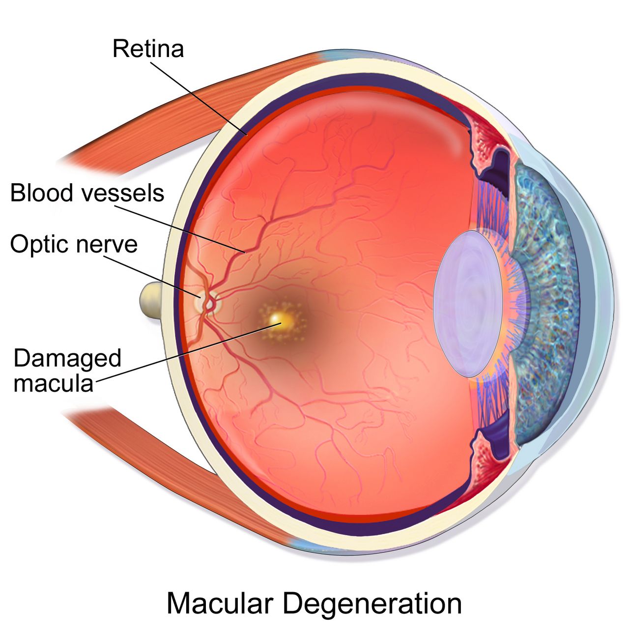
Sudden painless vision loss: sorting it in five minutes
Sudden vision loss is one of the few complaints where the correct nursing action changes depending on which of four diagnoses it is, so learn to sort them by pain, pattern and preceding symptoms. Central retinal artery occlusion is the retinal stroke: profound painless loss over seconds in one eye, an afferent pupillary defect, a pale retina with a cherry-red spot at the fovea on fundoscopy. The retina tolerates roughly ninety minutes to a few hours of complete ischaemia before infarction, so this is a hyperacute emergency handled like a cerebrovascular event — activate the stroke pathway, obtain urgent imaging, and expect a carotid and cardiac embolic workup, because the next embolus may go to the brain. Amaurosis fugax, a transient monocular blackout that resolves, is the same warning in a reversible package.
Retinal detachment announces itself differently. The patient describes a sudden shower of new floaters, flashes of light called photopsia, and then a curtain, shadow or veil advancing across the field from one side. There is no pain and no redness. Risk factors are high myopia, prior cataract or vitreoretinal surgery, blunt trauma, proliferative diabetic retinopathy and a previous detachment in either eye. It is urgent rather than hyperacute, but the clock still matters because a detachment that reaches the macula converts a good visual prognosis into a poor one. Keep the patient at rest, restrict activity and head movement, avoid Valsalva, keep them nothing by mouth pending surgery, and position as the ophthalmologist directs so that gravity holds the detached area against the wall of the eye.
Two more belong on the list. Vitreous haemorrhage — common in proliferative diabetic retinopathy and in anticoagulated patients — produces sudden floaters, cobwebs or a red or black haze, with a lost red reflex; it is managed with head elevation, activity restriction and observation, or vitrectomy if it does not clear. Giant cell arteritis is the one you must not miss in anyone over 50: monocular vision loss with a new temporal headache, scalp tenderness, jaw claudication when chewing, fever, weight loss and proximal muscle aching from polymyalgia rheumatica. Erythrocyte sedimentation rate is typically above 50 mm per hour and C-reactive protein is elevated. High-dose corticosteroid is started immediately on suspicion, before the temporal artery biopsy, because the whole purpose of treatment is to save the second eye.
| Presentation | Likely cause | First nursing action |
|---|---|---|
| Instant, total, painless monocular loss; afferent pupillary defect | Central retinal artery occlusion | Emergency — treat as a stroke, notify ophthalmology and the stroke team now, note the exact time of onset |
| Flashes, new floaters, then an advancing curtain; painless | Retinal detachment | Urgent ophthalmology, bed rest, restrict head movement, nothing by mouth, position as directed |
| Sudden floaters or red haze, lost red reflex, diabetic or anticoagulated | Vitreous haemorrhage | Head of bed elevated, limit activity, ophthalmology referral, review anticoagulation |
| Vision loss plus temporal headache, jaw claudication, age over 50 | Giant cell arteritis | Draw sedimentation rate and C-reactive protein, expect high-dose steroids before biopsy — do not delay the steroid |
| Severe pain, haloes, vomiting, hard eye, fixed mid-dilated pupil | Acute angle-closure glaucoma | Ophthalmology now, supine in a quiet room with the lights left on (darkness dilates the pupil), hold anticholinergics, antiemetic and analgesia |
Diabetic retinopathy
Chronic hyperglycaemia damages the retinal microvasculature through glycation, pericyte loss and capillary basement-membrane thickening. The result is leaky, weak capillaries and, eventually, capillaries that close entirely. Non-proliferative retinopathy shows microaneurysms, dot-and-blot haemorrhages, hard exudates and cotton-wool spots, and it becomes vision-threatening when fluid collects at the macula — diabetic macular oedema is the most common cause of vision loss in diabetes. When enough capillary bed has closed, the ischaemic retina releases vascular endothelial growth factor and grows fragile new vessels: this is proliferative retinopathy, and those vessels bleed into the vitreous and contract into fibrous bands that pull the retina off, causing tractional detachment.
Screening schedules are examination questions and clinic reality. A patient with type 1 diabetes needs a dilated eye examination within five years of diagnosis, then annually. A patient with type 2 diabetes needs one at the time of diagnosis, because they have often been hyperglycaemic for years already, then annually. Pregnancy accelerates retinopathy, so a woman with pre-existing diabetes should be examined before conception or in the first trimester and followed closely. The most powerful interventions remain metabolic: glycaemic control with an individualised haemoglobin A1c target commonly around less than 7 percent, blood pressure control generally below 130/80 mm Hg, lipid management and smoking cessation.
Treatment of established disease is intravitreal anti-VEGF injection — ranibizumab, aflibercept, or bevacizumab used off-label — given at intervals of roughly four to eight weeks initially, which is a substantial adherence burden and a common reason patients drop out of care. Panretinal photocoagulation ablates ischaemic peripheral retina to remove the VEGF stimulus, at the cost of some peripheral and night vision, and vitrectomy clears non-resolving haemorrhage and relieves traction. After any intravitreal injection, teach the patient that mild grittiness and a subconjunctival red patch are normal but that increasing pain, worsening vision, a shower of floaters or discharge over the next several days means endophthalmitis and requires the retina service immediately.
Age-related macular degeneration
Macular degeneration is the leading cause of irreversible central vision loss in adults over 60 in high-income countries. The dry, or non-exudative, form accounts for roughly 85 to 90 percent of cases: metabolic waste accumulates as drusen beneath the retinal pigment epithelium, cells atrophy, and central vision fades gradually over years, ending in geographic atrophy. The wet, or exudative, form is less common but far more aggressive: abnormal choroidal vessels grow up under the macula and leak blood and fluid, and central vision can distort or drop out over days to weeks. The hallmark early symptom is metamorphopsia — straight lines such as door frames and window blinds appear wavy or bent — followed by a central grey or blank spot.
Teach every at-risk patient to use an Amsler grid daily: tape it at reading distance, wear reading glasses, cover one eye, fix on the central dot, and report on the same day any new wavy, blurred, missing or dark area. That single home test is what converts wet macular degeneration from a catastrophe into a treatable problem, because anti-VEGF injections preserve and often improve vision when started early. Modifiable risk factors are smoking, which roughly doubles risk and is the strongest one you can change, hypertension, obesity and low dietary intake of leafy greens and fish.
For intermediate or advanced dry disease, the AREDS2 formulation slows progression: vitamin C 500 mg, vitamin E 400 international units, zinc oxide 80 mg, cupric oxide 2 mg, lutein 10 mg and zeaxanthin 2 mg. The critical safety point is that beta-carotene was removed because it increased lung-cancer risk in current and former smokers — never hand a smoker an older beta-carotene formulation. AREDS2 does not prevent macular degeneration in people who do not have it and does not help early disease. Beyond drugs, nursing care is low-vision rehabilitation: magnifiers, high-contrast large-print materials, task lighting, screen-reader and text-to-speech settings on the phone the patient already owns, referral to occupational therapy and a state low-vision service, and an honest conversation about driving.
Sudden painless monocular vision loss is a retinal emergency until proved otherwise — note the exact time of onset, and remember that a curtain means detachment, instant blackout means arterial occlusion, and vision loss with a temporal headache over age 50 means steroids before biopsy.
Section 4The Red Eye: Infection, Inflammation and Trauma
Sorting the red eye
Most red eyes are conjunctivitis and most conjunctivitis is self-limited, so the clinical skill is finding the minority that will blind the patient. Four findings should stop you every time: significant pain rather than irritation, reduced visual acuity, photophobia, and a ciliary flush — redness concentrated in a ring around the cornea rather than spread diffusely toward the lids. Add a hazy cornea, a fixed or irregular pupil, or a history of contact-lens wear or trauma, and the eye needs to be seen the same day. Simple conjunctivitis itches or feels gritty, spares acuity, and does not hurt in the deep aching way that uveitis and keratitis do.
Bacterial conjunctivitis produces thick purulent discharge with lids crusted shut on waking, usually starting in one eye; it is treated with topical antibiotic drops and scrupulous hand hygiene. Viral conjunctivitis, usually adenoviral, produces watery discharge, a gritty foreign-body sensation, a tender preauricular lymph node and frequent bilateral spread; it is exquisitely contagious for up to two weeks, so teach separate towels, no shared pillows and no eye makeup, and it needs supportive care rather than antibiotics. Allergic conjunctivitis is bilateral, intensely itchy, with stringy mucus and often rhinitis; it responds to antihistamine or mast-cell-stabiliser drops and cool compresses.
The dangerous inflammations look different. Bacterial keratitis, most often in a contact-lens wearer who slept in lenses or used tap water, gives pain, photophobia, a white corneal infiltrate and reduced acuity, and Pseudomonas can perforate a cornea in a day or two — fortified antibiotic drops are given as often as hourly around the clock, and the eye is never patched. Herpes simplex keratitis produces a branching dendritic ulcer on fluorescein staining and is treated with antivirals; topical corticosteroid alone can melt that cornea, which is why unlabelled steroid drops must never be borrowed from a family member. Anterior uveitis gives deep aching pain, photophobia, a small or irregular pupil and ciliary flush, and is treated with steroid and cycloplegic drops while the team looks for an associated systemic inflammatory disease.
Chemical splash and ocular trauma
A chemical ocular burn is the single ophthalmic situation where you act before you assess. Begin copious irrigation immediately with normal saline or lactated Ringer solution — or with plain water if that is what is within reach at the moment of injury — and irrigate continuously for at least fifteen to thirty minutes, holding the lids open, sweeping the fornices, and directing flow from the inner canthus outward so runoff does not cross the other eye. Remove contact lenses. Only after irrigation do you test pH with litmus paper in the lower fornix, aiming for a normal 7.0 to 7.4, and if pH remains abnormal you keep irrigating and recheck every twenty to thirty minutes. Do not neutralise an acid with a base or a base with an acid; the exothermic reaction adds a thermal burn.
Alkali burns — drain cleaner, oven cleaner, lye, ammonia, wet cement and plaster — are far worse than acid burns because alkali causes liquefactive necrosis and penetrates deep into the anterior chamber, while acid causes coagulative necrosis that partly seals the surface. A deceptively white, quiet-looking eye after an alkali splash is an ominous sign of limbal ischaemia, not a reassuring one. All chemical burns need urgent ophthalmology, and cement burns need particulate lime removed mechanically from the fornices.
Blunt and penetrating trauma follow different rules. A hyphema is blood layered in the anterior chamber after blunt injury: elevate the head of the bed 30 to 45 degrees so blood settles inferiorly and clears the visual axis, enforce rest, shield the eye, avoid aspirin and other antiplatelet or anticoagulant agents when possible, and watch for a pressure spike and for rebleeding around days two to five. A suspected globe rupture or penetrating foreign body demands the opposite of instinct: do not remove an impaled object, do not irrigate, do not instil drops, do not measure pressure, and do not press on the eye. Place a rigid shield — not a pressure patch — over the eye, keep the patient nothing by mouth with the head elevated, control pain and nausea aggressively because vomiting can extrude intraocular contents, give tetanus prophylaxis and antibiotics as ordered, and get the patient to the operating room. A corneal abrasion, by contrast, is common and benign: fluorescein confirms it, topical antibiotic prophylaxis and oral analgesia manage it, and topical anaesthetics are never sent home because repeated use prevents epithelial healing.
Periocular infection, drops, and the technique that matters
Distinguishing preseptal from orbital cellulitis is a high-yield bedside judgement. Preseptal (periorbital) cellulitis involves only the tissue anterior to the orbital septum: the lid is red and swollen, but the eye moves fully and painlessly, acuity is normal and there is no proptosis, and many patients are treated with oral antibiotics. Orbital cellulitis involves the orbit itself and typically follows sinusitis: it adds pain with eye movement, restricted or painful extraocular motion, proptosis, reduced acuity, an afferent pupillary defect and systemic toxicity. That patient is admitted for intravenous antibiotics with computed tomography of the orbits and sinuses, and can progress to abscess, cavernous sinus thrombosis or intracranial extension. Herpes zoster ophthalmicus — a vesicular dermatomal rash in the first trigeminal division, especially with vesicles on the tip of the nose, the Hutchinson sign — needs systemic antiviral therapy and an ophthalmology examination because corneal involvement is common.
Endophthalmitis is infection inside the eye, usually after intraocular surgery or an intravitreal injection, occasionally from a penetrating injury or haematogenous seeding. It presents within days as increasing pain, rapidly worsening vision, marked redness, a hypopyon of layered white cells in the anterior chamber, and lid oedema. It is a same-hour emergency requiring vitreous sampling and intravitreal antibiotics. This is precisely why every post-operative and post-injection teaching sheet says the same three things: increasing pain, decreasing vision, and increasing redness or discharge mean call today.
Drop technique is a nursing skill that changes drug delivery more than most dose adjustments do. Perform hand hygiene, verify the correct eye, have the patient tilt the head back and look up, pull the lower lid down to form a conjunctival sac, and drop the medication into that sac without letting the bottle tip touch lashes, lid or eye. One drop is the full dose — the conjunctival sac holds far less than a drop, and a second drop mostly washes the first away. Have the patient close the eye gently, without squeezing, and occlude the punctum at the inner canthus for one to two minutes. Wait at least five minutes between different drops so the second does not rinse out the first, give all drops before any ointment because ointment blocks absorption, and if a suspension such as a steroid or brinzolamide is ordered, shake it first. For ear drops the parallel rules are: warm the bottle to body temperature in your hands to avoid provoking vertigo, position the affected ear up, straighten the canal, instil along the canal wall rather than onto the drum, and keep the patient on their side for about five minutes.
For a chemical splash, irrigation begins before assessment, before history and before the ophthalmology call — and for any other red eye, pain, reduced acuity, photophobia or a ciliary flush moves the patient from routine to same-day.
Section 5The Ear: Infection, Hearing Loss and Vertigo
External and middle ear disease
Otitis externa is infection of the canal skin, classically after swimming or after a cotton swab has stripped the protective cerumen layer. The distinguishing sign is pain on traction of the pinna or pressure on the tragus, with a swollen weeping canal and often debris that obscures the drum. Treatment is topical antibiotic drops with or without a steroid — ofloxacin or ciprofloxacin with dexamethasone — sometimes with a wick placed to carry drops down a swollen canal. Teach the patient to keep the ear dry, to stop using swabs entirely, and to use a drying agent after swimming once the infection clears. The dangerous variant is necrotising (malignant) otitis externa in an older adult with diabetes or immunocompromise: it is a Pseudomonas osteomyelitis of the skull base with severe deep pain, granulation tissue in the canal and possible cranial nerve palsies, and it needs admission and prolonged systemic antipseudomonal therapy.
Acute otitis media is a middle-ear infection behind an intact drum, driven by eustachian tube dysfunction after a viral upper respiratory infection. Adults present with ear pain, muffled hearing, fever and a bulging erythematous immobile drum; sudden relief of pain with purulent drainage means the drum has perforated. Amoxicillin remains first-line, with amoxicillin-clavulanate for treatment failure or recent antibiotic exposure. Otitis media with effusion is fluid without acute infection: muffled hearing, an amber or retracted drum, a flat type B tympanogram, and usually watchful waiting. Persistent unilateral effusion in an adult is not casual — it can signal a nasopharyngeal mass obstructing the eustachian tube and warrants referral.
Two chronic middle-ear problems deserve recognition. Mastoiditis follows untreated or inadequately treated otitis media: postauricular pain, swelling and erythema with the pinna pushed outward and downward, fever and toxicity, requiring intravenous antibiotics and sometimes mastoidectomy. Cholesteatoma is a keratin-filled sac that erodes bone: chronic painless foul-smelling drainage, progressive conductive hearing loss and, if it erodes further, facial nerve palsy or vertigo — surgical removal is the only cure. Finally, cerumen impaction is the most common and most easily fixed cause of conductive loss in an older adult. Irrigate only after confirming there is no perforation, no tympanostomy tube, no previous mastoid surgery and no active infection; use body-temperature solution and direct the stream along the canal wall, and stop for pain or vertigo.
Hearing loss, including the emergency version
Sort every hearing complaint into conductive or sensorineural first, because the pathways diverge completely. Conductive loss is mechanical and usually fixable — cerumen, effusion, perforation, ossicular disruption, otosclerosis where the stapes footplate fixes to the oval window and which is corrected by stapedectomy. Sensorineural loss is cochlear or neural: presbycusis from cumulative hair-cell loss, noise-induced loss that classically notches around 4000 Hz, ototoxic injury, Ménière disease, and vestibular schwannoma (acoustic neuroma). Unilateral sensorineural hearing loss with tinnitus and imbalance is a schwannoma until magnetic resonance imaging with gadolinium says otherwise — asymmetry is the finding that must never be shrugged off as ageing, because ageing is symmetric.
Sudden sensorineural hearing loss is the otologic emergency students routinely miss. It is defined as a loss of 30 decibels or more across three contiguous frequencies developing within 72 hours, usually in one ear, often noticed on waking, and often accompanied by aural fullness and tinnitus. Patients and even clinicians attribute it to wax or fluid and wait a week — and the treatment window closes. If the Weber lateralises away from the affected ear and the Rinne shows air conduction still greater than bone conduction, the loss is sensorineural and the patient needs urgent audiometry and ear-nose-throat evaluation. High-dose oral corticosteroid, sometimes with intratympanic steroid injection, gives the best chance of recovery when started within 72 hours and diminishing benefit after two weeks.
| Ototoxic agent | Injury pattern | Monitoring and mitigation |
|---|---|---|
| Aminoglycosides — gentamicin, tobramycin, amikacin | Cochlear and vestibular hair cells; often permanent, may progress after the drug stops | Trough levels and renal function, shortest effective course, baseline and serial audiometry, ask daily about tinnitus, fullness and unsteadiness |
| Loop diuretics — furosemide, bumetanide | Stria vascularis; usually transient but permanent with rapid high-dose intravenous administration | Do not exceed the recommended intravenous push rate (furosemide about 4 mg per minute in adults); risk rises sharply in renal failure and when combined with aminoglycosides |
| Cisplatin and other platinum agents | Outer hair cells, dose-cumulative, high-frequency first, usually permanent | Baseline and pre-cycle audiometry, report new tinnitus promptly, dose modification decisions belong to oncology |
| High-dose salicylates and quinine | Tinnitus and reversible high-frequency loss | Tinnitus is an early warning of salicylate toxicity — report it rather than dismissing it |
| Vancomycin, macrolides at high dose | Uncommon, usually reversible; risk increases with concurrent ototoxins | Levels, renal function, and audiometric surveillance in prolonged therapy |
Vertigo: telling the peripheral causes apart
Start by clarifying what the patient means. Vertigo is the illusion of movement — the room spins, or the patient spins — and it points to the vestibular system. Lightheadedness or near-syncope points to perfusion: orthostatic hypotension, arrhythmia, anaemia, hypoglycaemia, medication. Disequilibrium without spinning points to the legs, the joints, the eyes and the cerebellum. Once true vertigo is established, the useful questions are duration of each episode, what triggers it, and whether hearing is involved. Then screen for central causes, which are the ones that kill: sudden severe headache, diplopia, dysarthria, dysphagia, focal weakness or numbness, ataxia out of proportion to the vertigo, or vertical or direction-changing nystagmus all mean posterior-circulation stroke until imaging says otherwise.
| Disorder | Episode length and trigger | Hearing | Management |
|---|---|---|---|
| Benign paroxysmal positional vertigo | Seconds to under a minute, triggered by rolling over, lying down, looking up | Normal | Dix-Hallpike confirms; Epley canalith-repositioning manoeuvre treats it, often in one visit; fall precautions |
| Ménière disease | 20 minutes to 12 hours, spontaneous, clustered then quiescent | Fluctuating low-frequency sensorineural loss, tinnitus, aural fullness | Sodium restriction 1500 to 2000 mg daily, thiazide or thiazide-plus-potassium-sparing diuretic, limit caffeine, alcohol and nicotine; acute attacks treated with meclizine, a benzodiazepine and an antiemetic; refractory cases get intratympanic steroid or gentamicin, endolymphatic sac surgery or labyrinthectomy |
| Vestibular neuritis | Days of continuous vertigo, worse with head motion, after a viral illness | Normal | Short-course vestibular suppressants and antiemetics, then early vestibular rehabilitation; prolonged suppressant use delays compensation |
| Labyrinthitis | Days of continuous vertigo | Hearing loss and tinnitus present — this is what separates it from neuritis | As for neuritis, plus urgent audiology; consider steroids and evaluate for a suppurative source |
| Central (stroke, tumour, multiple sclerosis) | Variable, often with headache or focal signs | Usually spared | Emergency neuroimaging and stroke pathway — do not treat as peripheral vertigo |
Ménière disease deserves its mechanism spelled out because the teaching follows from it. Excess endolymph distends the membranous labyrinth — endolymphatic hydrops — and when the membrane ruptures, potassium-rich endolymph floods the perilymphatic space and paralyses the hair cells, producing the attack. That is why sodium and fluid load matter, why diuretics help some patients, and why hearing fluctuates early and becomes permanently worse over years. Practically, teach the patient to sit or lie down at the first hint of aural fullness rather than trying to walk it off, to keep the environment free of clutter, to avoid ladders, swimming alone and driving during clusters, to keep a rescue antiemetic accessible, and to keep a symptom and salt diary to find their own triggers.
Otologic surgery, devices and the older adult
After tympanoplasty, stapedectomy or mastoidectomy, teaching prevents the complications. The patient does not blow the nose, sneezes with the mouth open, avoids heavy lifting, straining and vigorous coughing, keeps water out of the ear, and does not fly until cleared — all because pressure changes displace a graft or a prosthesis. Warn them that hearing in the operated ear will be worse initially because of packing and oedema, and that popping and crackling are expected. What they must report is vertigo that is new or worsening, facial droop or weakness on the operated side because the facial nerve runs through the temporal bone, clear watery drainage suggesting cerebrospinal fluid leak, fever, or a sudden increase in pain. Keep the operated ear up when lying down unless the surgeon says otherwise, and use antiemetics liberally since vomiting raises middle-ear pressure.
Hearing devices are nursing territory because patients are discharged with them and readmitted without them. For hearing aids, teach daily cleaning of the earmould, opening the battery door at night to extend battery life and dry the compartment, keeping them out of the bathroom and away from hairspray, checking for feedback whistle that usually means poor fit or wax, and always labelling and storing them in a container rather than a napkin on a meal tray. Cochlear implants bypass dead hair cells and stimulate the cochlear nerve directly and are indicated for severe to profound sensorineural loss with poor benefit from hearing aids; recipients need pneumococcal vaccination because of meningitis risk, must have the external processor removed for certain imaging, and require months of auditory rehabilitation to interpret the new signal. An implant does not restore normal hearing; set that expectation before surgery.
Older adults concentrate all of this. Presbycusis plus impacted cerumen plus a noisy unit plus a mask over your mouth equals a patient who looks confused and is not. Vestibular function declines with age, so vertigo in an older adult is a fall risk before it is anything else — and meclizine and benzodiazepines, the very drugs that relieve it, are on the Beers criteria for causing sedation, anticholinergic effects and falls, so use the smallest effective dose for the shortest time and pair it with a bed alarm, non-skid footwear, a clear path to the bathroom and a physical-therapy referral. Older adults with an ear infection may present with confusion and anorexia rather than fever and pain. And the same anticholinergic burden that we so casually add for nausea, sleep or bladder spasm is what precipitates acute angle-closure glaucoma, so review the medication list of every older adult with a red painful eye or a new balance complaint.
Match the tempo to the diagnosis — seconds of positional spinning is benign paroxysmal positional vertigo, hours with fluctuating hearing loss and fullness is Ménière, days without hearing loss is vestibular neuritis, and a 30-decibel drop within 72 hours is a steroid emergency.
The chapter in one breath
Eyes and ears reward mechanism-based thinking. Aqueous humour is made continuously behind the iris and must drain through the angle in front of it, so open-angle glaucoma silently strangles the optic nerve over years while acute angle-closure glaucoma slams the door in hours and produces a painful red eye, a steamy cornea, a mid-dilated fixed pupil and a rock-hard globe — commonly after an anticholinergic. The retina has no collateral supply, so sudden painless monocular loss is an emergency: instant blackout is arterial occlusion handled as a stroke, flashes and an advancing curtain are detachment, and vision loss with a temporal headache after 50 is giant cell arteritis that gets steroids before the biopsy. Diabetic retinopathy and macular degeneration are slower losses managed with glycaemic and blood-pressure control, dilated screening, anti-VEGF injections, an Amsler grid and AREDS2 without beta-carotene for smokers. A red eye with pain, reduced acuity, photophobia or ciliary flush is seen the same day, and a chemical splash is irrigated before anything else is done. In the ear, conductive loss is mechanical and usually fixable, sensorineural loss is cochlear or neural, sudden sensorineural loss is a 72-hour steroid emergency, and vertigo is sorted by how long each episode lasts and whether hearing is involved. Throughout, undetected sensory loss looks like confusion, non-adherence and falls.
Words worth knowing
| Term | What it means |
|---|---|
| Aqueous humour | Clear fluid made continuously by the ciliary body that nourishes the lens and cornea and must drain through the trabecular meshwork; its outflow determines intraocular pressure. |
| Tonometry | Measurement of intraocular pressure; normal is roughly 10 to 21 mm Hg. |
| Ciliary flush | Redness concentrated in a ring at the limbus around the cornea, suggesting keratitis, uveitis or acute glaucoma rather than simple conjunctivitis. |
| Miotic | A drug such as pilocarpine that constricts the pupil, pulling the peripheral iris away from the drainage angle. |
| Mydriatic and cycloplegic | Drugs that dilate the pupil and paralyse accommodation; they relieve uveitis pain but can precipitate angle closure in a susceptible eye. |
| Afferent pupillary defect | Pupil dilates rather than constricts when light swings to it, indicating optic-nerve or extensive retinal dysfunction on that side. |
| Photopsia | Perceived flashes of light, classically from vitreoretinal traction preceding a retinal detachment. |
| Metamorphopsia | Straight lines appear wavy or bent; the earliest symptom of wet macular degeneration, detected on the Amsler grid. |
| Drusen | Yellow deposits of metabolic waste beneath the retinal pigment epithelium, the hallmark of dry macular degeneration. |
| Anti-VEGF therapy | Intravitreal injection of ranibizumab, aflibercept or bevacizumab that blocks vascular endothelial growth factor to stop abnormal vessel growth and leakage. |
| Hyphema | Blood layered in the anterior chamber after blunt trauma; managed with head elevation, rest, a shield and pressure monitoring. |
| Endophthalmitis | Infection inside the globe after surgery, injection or penetrating injury; increasing pain with decreasing vision is the warning pair. |
| Hutchinson sign | Herpes zoster vesicles on the tip of the nose, predicting corneal involvement in herpes zoster ophthalmicus. |
| Presbyopia | Age-related stiffening of the lens causing loss of near focus, usually noticed after age 40. |
| Presbycusis | Age-related, symmetric, high-frequency sensorineural hearing loss that blurs consonants and makes speech sound mumbled. |
| Otosclerosis | Fixation of the stapes footplate producing progressive conductive hearing loss, correctable by stapedectomy. |
| Cholesteatoma | Keratin-filled sac in the middle ear that erodes bone, causing chronic foul painless drainage and conductive loss; requires surgery. |
| Endolymphatic hydrops | Excess endolymph distending the membranous labyrinth, the mechanism underlying Ménière disease. |
| Nystagmus | Involuntary rhythmic eye movement; vertical or direction-changing nystagmus suggests a central rather than peripheral cause of vertigo. |
| Tympanometry | Test of tympanic membrane mobility; type A is normal, flat type B suggests effusion or perforation, type C indicates negative middle-ear pressure. |
Check yourself
IMMUNE AND RHEUMATOLOGIC DISORDERS
The immune system fails in only three interesting ways: it overreacts, it underperforms, or it mistakes the patient for the enemy. Everything in this chapter falls into one of those three buckets, and knowing which bucket you are in tells you what to do. The patient whose lips are swelling twelve minutes after a first dose of cefazolin needs intramuscular epinephrine in the next sixty seconds, not an antihistamine and a reassessment. The patient with a CD4 count of 140 and a dry cough needs an oxygen saturation on ambulation, not on the pillow. The woman with lupus whose complement is falling and whose urine is suddenly frothy is losing a kidney quietly while her joints get the attention. And the patient on a tumour necrosis factor inhibitor who feels “a little off” without a fever may be septic, because you have chemically removed her ability to mount one. Immunology at the bedside is pattern recognition plus ruthless prioritisation.
By the end of this chapter you can
- Distinguish the four hypersensitivity types by mechanism and name a clinical example of each, including Type I IgE-mediated and Type IV delayed cell-mediated reactions.
- Recognise anaphylaxis within seconds and state the first drug, the correct dose, the correct route and the correct site without hesitation.
- Interpret a CD4 count and HIV viral load, identify the threshold that defines AIDS, and match CD4 strata to opportunistic infection prophylaxis.
- Explain the antibody profile, organ involvement and flare markers of systemic lupus erythematosus, including anti-dsDNA and falling complement (C3, C4).
- Compare rheumatoid arthritis, osteoarthritis and gout by joint distribution, stiffness pattern, laboratory findings and treatment.
- Describe the monitoring, screening and teaching that must precede and accompany methotrexate, biologic DMARDs and JAK inhibitors.
- Differentiate hyperacute, acute and chronic transplant rejection and identify the nursing assessments that detect each.
- Apply SBAR escalation to red flags including anaphylaxis, giant cell arteritis with visual change, scleroderma renal crisis and neutropenic fever.
- Delegate appropriately in the immunosuppressed patient, retaining assessment, teaching, evaluation and unstable patients for the registered nurse.
Section 1When the Immune System Overreacts: Hypersensitivity, Allergy and Anaphylaxis
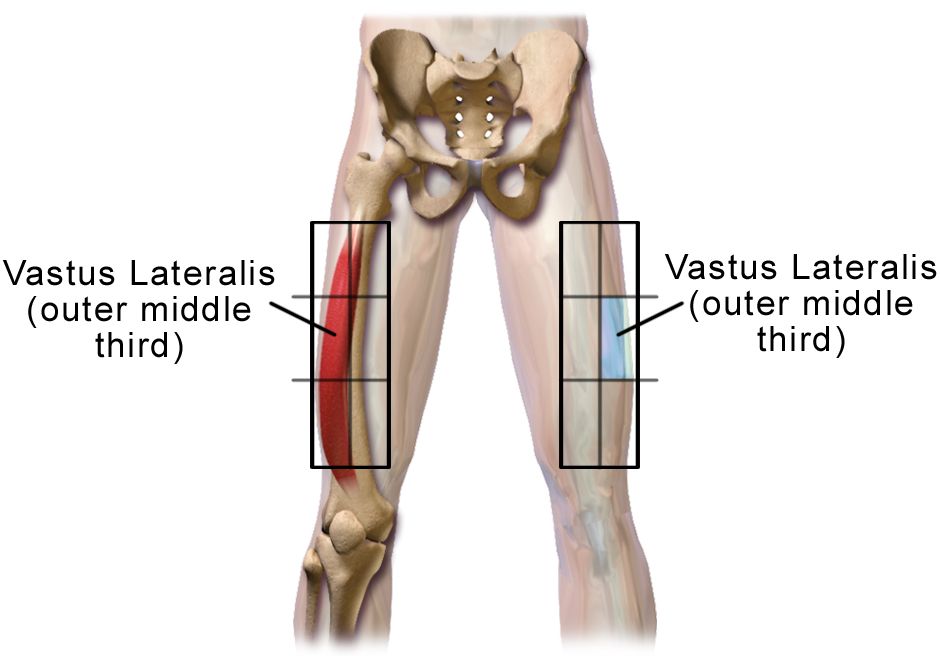
The four hypersensitivity patterns
Hypersensitivity means the immune response causes more damage than the antigen would have. Four mechanisms are described, and the mechanism predicts both the timing and the treatment. Type I is immediate and IgE-mediated: a previously sensitised patient re-encounters the antigen, IgE bound to mast cells and basophils cross-links, and the cells degranulate within minutes, dumping histamine, tryptase, leukotrienes and prostaglandins. The result is vasodilation, capillary leak, bronchoconstriction and urticaria — anaphylaxis, allergic rhinitis, atopic asthma, food and insect sting allergy. Because it requires prior sensitisation, the first exposure is often uneventful and the second is the catastrophe. Students remember this as “the second dose is the dangerous one,” which is why you never relax during a second antibiotic dose.
Type II is antibody-mediated cytotoxicity: IgG or IgM binds antigen on the surface of the patient’s own cells and marks them for complement lysis or phagocytosis. Acute haemolytic transfusion reaction from ABO incompatibility is the classic, along with immune thrombocytopenia, heparin-induced thrombocytopenia, Graves disease and Goodpasture syndrome. Type III is immune complex disease: antigen and antibody form soluble complexes that deposit in vessel walls, glomeruli, skin and synovium, fixing complement and drawing neutrophils. Serum sickness, post-streptococcal glomerulonephritis and much of the organ damage in systemic lupus erythematosus are Type III. This is why serum complement falls in a lupus flare — it is being consumed by the complexes.
Type IV is delayed and cell-mediated, driven by sensitised T lymphocytes rather than antibody, and it takes 24 to 72 hours to appear. The tuberculin skin test read at 48 to 72 hours is a deliberately induced Type IV reaction. So is contact dermatitis to nickel or poison ivy, most solid-organ graft rejection, and the severe cutaneous drug reactions Stevens–Johnson syndrome and toxic epidermal necrolysis. The nursing implication is timing: a Type IV reaction will not be caught in a thirty-minute post-dose observation, so patients started on a new sulfonamide, allopurinol, lamotrigine or antiepileptic must be taught to report a spreading rash, mucosal blistering or fever days into therapy.
| Type | Mechanism | Onset | Clinical examples |
|---|---|---|---|
| I — Immediate | IgE on mast cells, degranulation | Seconds to 30 minutes | Anaphylaxis, food and sting allergy, allergic rhinitis, atopic asthma |
| II — Cytotoxic | IgG or IgM against cell-surface antigen, complement lysis | Minutes to hours | ABO haemolytic transfusion reaction, immune thrombocytopenia, heparin-induced thrombocytopenia, Goodpasture |
| III — Immune complex | Antigen–antibody complexes deposit in tissue, complement consumed | Hours to weeks | Serum sickness, post-streptococcal glomerulonephritis, lupus nephritis and vasculitis |
| IV — Delayed | Sensitised T cells and macrophages, no antibody | 24 to 72 hours or longer | Tuberculin skin test, contact dermatitis, graft rejection, Stevens–Johnson syndrome |
Anaphylaxis: recognise it in seconds, treat it in one
Anaphylaxis is a clinical diagnosis, not a laboratory one, and it does not require hives. Suspect it when a patient develops acute onset of illness involving skin or mucosa (urticaria, flushing, swollen lips or tongue) plus either respiratory compromise (stridor, wheeze, hypoxaemia, throat tightness) or hypotension and end-organ dysfunction. It should also be suspected when two or more body systems light up quickly after a likely allergen — skin, respiratory, cardiovascular, or persistent gastrointestinal cramping and vomiting — or when a known allergen is followed by hypotension alone. In hospital the usual culprits are beta-lactam antibiotics, vancomycin, contrast media, neuromuscular blockers, latex and blood products. Onset is typically within five to thirty minutes of an intravenous agent, and faster is worse.
The first action is epinephrine 0.3 to 0.5 mg of the 1 mg/mL concentration, intramuscularly into the anterolateral thigh (vastus lateralis), repeated every five to fifteen minutes as needed. Not subcutaneous, not the deltoid, not intravenous unless the patient is in arrest or a physician is titrating an infusion in a monitored setting, and not after you have tried an antihistamine. Epinephrine works because alpha-1 stimulation reverses vasodilation and mucosal oedema while beta-2 stimulation bronchodilates and beta-1 supports cardiac output. Antihistamines relieve itch and hives; they do nothing for airway or blood pressure. Corticosteroids take hours. The paediatric dose is 0.01 mg/kg to a maximum of 0.3 mg (0.5 mg in adolescents). At the same time, stop the infusion, call for help, apply high-flow oxygen, and place the patient supine with the legs elevated unless vomiting or severe respiratory distress demands otherwise — abruptly sitting a hypotensive anaphylactic patient upright can empty the vena cava and cause cardiac arrest.
Adjuncts follow: rapid crystalloid boluses of 1 to 2 L for hypotension (capillary leak can shift a third of the plasma volume in ten minutes), an H1 blocker such as diphenhydramine 25 to 50 mg IV, an H2 blocker such as famotidine 20 mg IV, nebulised albuterol for persistent bronchospasm, and methylprednisolone 125 mg IV to blunt a late-phase reaction. If the patient takes a beta blocker and is refractory to epinephrine, glucagon 1 to 5 mg IV over five minutes bypasses the beta receptor. Serum tryptase drawn between fifteen minutes and three hours after onset can confirm mast cell degranulation retrospectively, but it never delays treatment. Because a biphasic reaction can recur one to eight hours later (occasionally up to seventy-two hours) without re-exposure, observe for a minimum of four to six hours — longer after severe reactions, repeat epinephrine doses, or asthma history.
Allergy, latex and drug reaction on a busy floor
Every admission assessment includes allergies, but a name alone is useless documentation. Ask what happened and when: nausea after codeine is an intolerance, hives and throat swelling after penicillin is an allergy, and the distinction changes what the patient may safely receive for the rest of their life. Roughly ninety percent of patients labelled penicillin-allergic tolerate penicillin on formal evaluation, and that mislabel pushes them toward broader, more toxic and less effective antibiotics. Document the reaction, the timing and the severity, and refer for allergy evaluation rather than perpetuating a forty-year-old label.
Latex allergy deserves its own line. Natural rubber latex proteins cause a Type I reaction; the accelerators in gloves cause a Type IV contact dermatitis with delayed erythema and fissuring. Highest risk are patients with spina bifida, urogenital anomalies, and anyone with repeated procedures or occupational exposure. Cross-reactivity with banana, avocado, kiwi, chestnut, papaya and passion fruit is real, so a patient who reports lip tingling with kiwi is telling you something about their glove tolerance. A latex-allergic patient needs a documented latex-safe environment: synthetic gloves, latex-free tourniquets, blood pressure cuff barriers, medication vials with non-latex stoppers, and a band and chart flag. Powdered latex gloves aerosolise protein, so the exposure risk is airborne, not just contact.
Two other reactions on the floor are commonly mislabelled as allergy. Vancomycin infusion reaction (formerly “red man syndrome”) is a direct, non-IgE mast cell degranulation from rapid infusion: flushing of the face, neck and upper torso with pruritus, sometimes hypotension. It is managed by stopping the infusion, giving an antihistamine, and restarting at a slower rate — typically no faster than 1 g per hour — not by permanently banning vancomycin. Contrast-associated reactions are also usually non-IgE, and premedication with corticosteroids and antihistamines is often used for a documented prior reaction. Finally, teach the discharged patient with a true anaphylaxis history to carry two epinephrine autoinjectors, demonstrate the technique back to you, replace them before expiry, and call emergency services after using one even if symptoms resolve.
In suspected anaphylaxis, intramuscular epinephrine into the anterolateral thigh is the first and non-negotiable action — every other drug is an adjunct that treats symptoms rather than the shock.
Section 2When the Immune System Fails: Immunodeficiency and HIV
Primary and secondary immunodeficiency
Primary immunodeficiencies are inherited defects in a specific arm of immunity and usually declare themselves in childhood: recurrent sinopulmonary infection with encapsulated organisms in antibody deficiencies such as common variable immunodeficiency, or opportunistic and fungal infection in T cell defects. Adults reach the medical–surgical floor more often with secondary immunodeficiency, and the causes are the ordinary furniture of hospital medicine: corticosteroids and other immunosuppressants, chemotherapy, malignancy (particularly haematologic), chronic kidney disease and dialysis, cirrhosis, poorly controlled diabetes, malnutrition, splenectomy, advanced age and HIV infection.
Two practical consequences follow. First, an immunocompromised patient may not mount the signs you rely on: no fever, no leucocytosis, minimal infiltrate on chest radiograph, no purulence at a wound because there are too few neutrophils to make pus. In a neutropenic patient a single temperature of 38.3 degrees C, or 38.0 degrees C sustained for one hour, with an absolute neutrophil count under 500 cells/mm3, is an emergency requiring blood cultures and antibiotics within one hour. Second, asplenic patients — surgical or functional, as in sickle cell disease — are at lifelong risk of overwhelming sepsis from encapsulated organisms and must have pneumococcal, meningococcal and Haemophilus influenzae type b vaccination plus a standing plan for febrile illness.
HIV: virology, testing and staging
HIV is a retrovirus that binds the CD4 receptor and a co-receptor on helper T lymphocytes, reverse-transcribes its RNA into DNA, integrates into the host genome and turns the cell into a virus factory. The progressive loss of CD4 T cells dismantles cell-mediated immunity, which is precisely the arm that controls intracellular pathogens, fungi and certain cancers. Transmission is through blood, semen, vaginal and rectal fluids, and breast milk — not through saliva, tears, sweat, casual contact or shared utensils. Standard precautions protect the nurse; the occupational risk from a hollow-bore needlestick with infected blood is roughly 0.3 percent, and lower still when the source patient is virally suppressed.
Testing follows a defined algorithm. The screening test is a fourth-generation antigen/antibody combination immunoassay that detects HIV-1 and HIV-2 antibodies plus the p24 antigen, shortening the window period to roughly eighteen to forty-five days. A reactive screen is followed by an HIV-1/HIV-2 antibody differentiation immunoassay; if that is negative or indeterminate, an HIV-1 nucleic acid test resolves it. Never tell a patient the screening result is a diagnosis before confirmation. Acute retroviral syndrome — fever, pharyngitis, lymphadenopathy, rash, myalgia two to four weeks after exposure — occurs when antibody may still be negative but viral load is enormous and transmissibility is highest, so a nucleic acid test is the right test in that window.
Two numbers drive care. The CD4 count (normal roughly 500 to 1,600 cells/mm3) measures immune damage and predicts opportunistic infection. The viral load measures how well treatment is working; the goal is undetectable, usually reported as fewer than 20 to 50 copies/mL. A diagnosis of AIDS is made when the CD4 count falls below 200 cells/mm3, when the CD4 percentage is below 14, or when an AIDS-defining illness occurs — Pneumocystis jirovecii pneumonia, oesophageal candidiasis, disseminated Mycobacterium avium complex, cryptococcal meningitis, cytomegalovirus retinitis, Kaposi sarcoma, central nervous system lymphoma, or HIV wasting. Once the AIDS stage is reached the diagnosis does not revert even if the CD4 recovers on therapy.
| CD4 count (cells/mm3) | Risk | Standard prophylaxis |
|---|---|---|
| Above 500 | Near-normal immunity | None; routine vaccination and ART |
| 200 to 500 | Bacterial pneumonia, tuberculosis, herpes zoster, oral candidiasis | None specific; treat and vaccinate |
| Below 200 | Pneumocystis jirovecii pneumonia | Trimethoprim–sulfamethoxazole one double-strength tablet daily |
| Below 100 | Toxoplasma encephalitis, cryptococcal disease | Trimethoprim–sulfamethoxazole (covers toxoplasma if IgG positive) |
| Below 50 | Disseminated Mycobacterium avium complex, cytomegalovirus retinitis | Azithromycin 1,200 mg weekly if ART cannot be started promptly; dilated eye exams |
Antiretroviral therapy, adherence and prevention
Antiretroviral therapy is started as soon as possible after diagnosis, at any CD4 count, ideally the same day. Regimens combine agents from different classes so the virus cannot mutate around a single pressure point, and most first-line regimens are a single daily tablet built on an integrase strand transfer inhibitor: bictegravir with emtricitabine and tenofovir alafenamide, or dolutegravir paired with two nucleoside analogues. The other classes are nucleoside and nucleotide reverse transcriptase inhibitors (tenofovir, emtricitabine, lamivudine, abacavir, zidovudine), non-nucleoside reverse transcriptase inhibitors (efavirenz, rilpivirine, doravirine), protease inhibitors (darunavir, atazanavir, usually boosted with ritonavir or cobicistat), a CCR5 antagonist (maraviroc), a fusion inhibitor (enfuvirtide) and a capsid inhibitor (lenacapavir).
The nursing implications are concrete. Abacavir requires HLA-B*5701 testing before the first dose; a positive result predicts a potentially fatal hypersensitivity reaction and the drug is never rechallenged. Tenofovir disoproxil fumarate is nephrotoxic and reduces bone density, so monitor creatinine, estimated glomerular filtration rate and urine protein; the alafenamide form is gentler on kidney and bone. Integrase inhibitors are well tolerated but cause weight gain and chelate with polyvalent cations — calcium, magnesium, aluminium, iron — so the integrase inhibitor is given at least two hours before, or six hours after, any antacid, calcium, magnesium, iron or multivitamin product. Protease inhibitors are potent cytochrome P450 inhibitors, making drug interaction review mandatory at every admission; statins, rifampin, some anticoagulants and inhaled fluticasone are common collisions. Adherence must exceed roughly ninety-five percent to prevent resistance, so assess barriers honestly: cost, stigma, housing, mental health, substance use, pill burden, and simple forgetting.
Immune reconstitution inflammatory syndrome can appear in the first weeks to months after starting therapy in patients with very low CD4 counts. As immunity returns, the body mounts a florid inflammatory response to a previously silent organism, and the patient looks worse on effective treatment — new fever, enlarging nodes, worsening infiltrates. It is not treatment failure and therapy is usually continued while the underlying infection is treated. For prevention, pre-exposure prophylaxis requires a documented negative HIV test before the first dose, and the retesting interval follows the formulation: daily oral tenofovir/emtricitabine is retested at least every three months, while long-acting injectable cabotegravir — given every two months — requires an HIV test before every injection, because a breakthrough infection left on a long-acting agent is exposed to prolonged effective monotherapy and selects integrase-inhibitor resistance. Post-exposure prophylaxis after occupational or sexual exposure is a twenty-eight-day three-drug regimen started as soon as possible and within seventy-two hours. Finally, teach and reinforce U=U: a person with sustained undetectable viral load does not transmit HIV sexually. That fact is both accurate and therapeutic.
The CD4 count tells you what infection the patient is vulnerable to and therefore what to prophylax and monitor; the viral load tells you whether the antiretroviral regimen and the patient’s adherence are working.
Section 3When the Immune System Turns Inward: Lupus and Connective Tissue Disease
Systemic lupus erythematosus
Lupus is a chronic multisystem autoimmune disease in which autoantibodies against nuclear antigens form immune complexes that deposit in skin, joints, serosal membranes, kidney, blood vessels and brain. It affects women about nine times as often as men, most commonly between fifteen and forty-five years of age, and is more prevalent and more severe in Black, Hispanic, Asian and Indigenous populations — a disparity driven by both biology and access to care. The course is relapsing and remitting, and flares are frequently triggered by ultraviolet light, infection, physical or emotional stress, pregnancy and the postpartum period, and certain drugs.
The classic findings are worth memorising because they map to the diagnostic criteria. Constitutional: fatigue that is out of proportion to activity, low-grade fever, weight loss. Skin: the malar rash across the cheeks and nasal bridge sparing the nasolabial folds, discoid plaques that scar, photosensitivity, non-scarring alopecia and painless oral or nasal ulcers. Joints: a symmetric, non-erosive, non-deforming polyarthritis of the small joints. Serosa: pleuritis with pleuritic chest pain, pericarditis with a friction rub. Renal: proteinuria and cellular casts. Neurologic: seizures, psychosis, cognitive dysfunction, stroke. Haematologic: haemolytic anaemia, leucopenia below 4,000/mm3, lymphopenia below 1,000/mm3, thrombocytopenia below 100,000/mm3. Raynaud phenomenon is common. Drug-induced lupus — from hydralazine, procainamide, isoniazid, minocycline or tumour necrosis factor inhibitors — produces arthralgia, serositis and anti-histone antibodies, spares the kidney and brain, and resolves when the drug is stopped.
Serologically, the antinuclear antibody is positive in roughly ninety-eight percent of patients: extremely sensitive, poorly specific, and positive in many healthy older adults, so it screens but never diagnoses. Anti-double-stranded DNA is specific and its titre tracks disease activity, particularly nephritis. Anti-Smith is the most specific antibody but is present in only about a third. Complement components C3 and C4 fall during an active flare because circulating immune complexes consume them, which is the opposite of the intuition that inflammation raises everything. The erythrocyte sedimentation rate rises with activity while C-reactive protein often stays relatively low — a markedly elevated CRP in a lupus patient should raise suspicion for infection or serositis rather than assuming a flare.
Lupus nephritis, antiphospholipid syndrome and managing the flare
Kidney involvement occurs in roughly half of patients and is the leading determinant of long-term outcome, yet it is nearly silent early. That makes urinalysis the highest-yield cheap test you will order: look for proteinuria, red cells and cellular casts, and follow the spot urine protein-to-creatinine ratio. Rising blood pressure, new peripheral oedema, weight gain and foamy urine in a lupus patient are nephritis until proven otherwise. Renal biopsy classifies the lesion, and proliferative disease (Class III or IV) requires induction immunosuppression — glucocorticoids with mycophenolate mofetil or cyclophosphamide, increasingly combined with belimumab or voclosporin — followed by prolonged maintenance.
Antiphospholipid antibodies (lupus anticoagulant, anticardiolipin, anti-beta-2 glycoprotein I) occur in about a third of lupus patients and, when accompanied by thrombosis or recurrent pregnancy loss, define antiphospholipid syndrome. The name is a trap: the lupus anticoagulant prolongs the activated partial thromboplastin time in the test tube but causes clotting in the patient. These patients require long-term anticoagulation with warfarin after an event — direct oral anticoagulants perform worse in triple-positive disease — and their contraception, surgical and immobility risk must be managed aggressively.
Baseline therapy for essentially every lupus patient is hydroxychloroquine, which reduces flares, protects the kidney, lowers thrombosis risk and improves survival. Dose it at or below 5 mg/kg of actual body weight, obtain a baseline retinal examination, and screen annually after five years of use because retinal toxicity is dose- and duration-dependent and irreversible. Glucocorticoids control flares but the goal is always the lowest dose for the shortest time. Nursing care is largely teaching: rigorous photoprotection with broad-spectrum sunscreen of SPF 30 or higher, hats and long sleeves; energy conservation and paced activity for fatigue; prompt reporting of fever because infection is a leading cause of death and immunosuppression masks it; pregnancy planning with a rheumatologist, since conception should occur during six months of quiescent disease and mycophenolate, cyclophosphamide and methotrexate are teratogenic while hydroxychloroquine is continued.
Scleroderma, Sjögren syndrome and giant cell arteritis
Systemic sclerosis is fibrosis driven by immune activation and vascular injury. Limited cutaneous disease presents as the CREST pattern: calcinosis, Raynaud phenomenon, oesophageal dysmotility, sclerodactyly and telangiectasia, associated with anticentromere antibody and a risk of pulmonary arterial hypertension. Diffuse disease spreads proximally and carries anti-Scl-70 (topoisomerase I) antibody with interstitial lung disease, or anti-RNA polymerase III with scleroderma renal crisis — abrupt severe hypertension, headache, rising creatinine and microangiopathic haemolysis. This is a medical emergency: a new blood pressure rise in a scleroderma patient is escalated the same hour, because delay costs the kidney permanently. Renal crisis is treated with an angiotensin-converting enzyme inhibitor, captopril being the traditional choice, and it is one of the few situations where high-dose corticosteroids increase the risk and are avoided. Nursing priorities are warmth and vasospasm avoidance (layered clothing, gloves in the frozen-food aisle, no smoking, calcium channel blockers such as nifedipine for Raynaud), small frequent meals with upright positioning for ninety minutes after eating and a proton pump inhibitor for reflux, meticulous skin care and range of motion to preserve hand function, and daily blood pressure checks at home.
Sjögren syndrome is lymphocytic destruction of exocrine glands producing dry eyes (keratoconjunctivitis sicca) and dry mouth (xerostomia), with anti-SSA/Ro and anti-SSB/La antibodies and a positive Schirmer test. It is not a nuisance diagnosis: xerostomia causes rampant dental caries and candidiasis, dryness causes corneal damage, and there is a substantially increased lifetime risk of non-Hodgkin lymphoma, so a new persistent parotid mass or lymphadenopathy must be evaluated. Teach preservative-free artificial tears, sugar-free gum or lozenges, frequent water sips, meticulous oral hygiene with fluoride and quarterly dental review, humidified air, and avoidance of anticholinergic drugs that worsen dryness. Pilocarpine or cevimeline stimulate residual glandular secretion.
Giant cell arteritis is the rheumatologic emergency of the older adult. In a patient over fifty with a new temporal or occipital headache, scalp tenderness (the pillow or the comb hurts), jaw claudication when chewing, constitutional symptoms and a sedimentation rate often above 50 mm/h with elevated C-reactive protein, the threat is irreversible monocular blindness from anterior ischaemic optic neuropathy. Transient visual loss or diplopia is the warning shot. Start high-dose glucocorticoids immediately — do not wait for the temporal artery biopsy, which remains positive for one to two weeks after steroids are begun. Prednisone 40 to 60 mg daily for uncomplicated disease; intravenous methylprednisolone 500 to 1,000 mg daily for three days when vision is threatened or lost. Tocilizumab is used as a steroid-sparing agent. Its cousin, polymyalgia rheumatica, causes bilateral shoulder and hip girdle aching and morning stiffness with a dramatic response to low-dose prednisone of 12.5 to 25 mg daily; up to a fifth of these patients also have arteritis, so screen every one of them for headache and visual symptoms at each contact.
| Antibody | Points toward | Bedside meaning |
|---|---|---|
| Antinuclear antibody | Lupus and most connective tissue disease | Sensitive screen only; positive in healthy older adults |
| Anti-double-stranded DNA | Systemic lupus erythematosus | Specific; titre tracks activity and nephritis |
| Anti-Smith | Systemic lupus erythematosus | Most specific for lupus; present in about one third |
| Anti-histone | Drug-induced lupus | Look for hydralazine, procainamide, isoniazid, minocycline |
| Anticentromere | Limited systemic sclerosis (CREST) | Screen for pulmonary arterial hypertension |
| Anti-Scl-70 (topoisomerase I) | Diffuse systemic sclerosis | Interstitial lung disease risk; monitor lung function |
| Anti-RNA polymerase III | Diffuse systemic sclerosis | Renal crisis risk; daily blood pressure at home |
| Anti-SSA/Ro, anti-SSB/La | Sjögren syndrome, neonatal lupus | Dryness care; fetal heart block risk in pregnancy |
| Anti-cyclic citrullinated peptide | Rheumatoid arthritis | Highly specific; predicts erosive disease |
In lupus a rising anti-dsDNA titre with falling C3 and C4 signals an active flare, while a new headache with jaw claudication or visual change in an adult over fifty means starting steroids for giant cell arteritis before any biopsy is arranged.
Section 4Inflammatory Joint Disease: Rheumatoid Arthritis, Spondyloarthritis and Gout
Rheumatoid arthritis
Rheumatoid arthritis is a chronic systemic autoimmune disease in which synovial inflammation forms an invasive pannus that erodes cartilage and bone. The joint pattern is the diagnosis: symmetric involvement of the small joints — metacarpophalangeal, proximal interphalangeal, wrists and metatarsophalangeal — with characteristic sparing of the distal interphalangeal joints. Morning stiffness lasts longer than sixty minutes and improves with use, the exact inverse of osteoarthritis, where brief stiffness worsens through the day with activity. Late deformities include ulnar deviation, swan-neck and boutonnière fingers, and subluxation. Systemic features are prominent: fatigue, low-grade fever, weight loss, rheumatoid nodules over pressure points, interstitial lung disease, pericarditis, scleritis, secondary Sjögren syndrome, and Felty syndrome (rheumatoid arthritis with splenomegaly and neutropenia). Cardiovascular risk is elevated roughly like that of diabetes, so lipids, blood pressure and smoking cessation are part of arthritis care.
Laboratory support comes from rheumatoid factor, positive in seventy to eighty percent but also positive in hepatitis C, Sjögren and healthy older adults, and anti-cyclic citrullinated peptide antibody, which is roughly ninety-five percent specific, appears years before symptoms and predicts erosive disease. Sedimentation rate and C-reactive protein track activity, and a normocytic anaemia of chronic inflammation is typical. Radiographs show periarticular osteopenia and marginal erosions; ultrasound and magnetic resonance imaging detect synovitis earlier. One finding must never be forgotten on a medical–surgical unit: atlantoaxial subluxation. Erosion of the transverse ligament at C1–C2 lets the odontoid impinge on the spinal cord during neck flexion, so any patient with long-standing rheumatoid arthritis needs cervical spine evaluation before intubation, positioning under anaesthesia or aggressive neck manipulation, and new neck pain with paraesthesia or gait change is escalated immediately.
Treatment is treat-to-target and time-sensitive: a conventional disease-modifying antirheumatic drug should start within three months of symptom onset because erosions that occur are permanent. Methotrexate is the anchor, 7.5 to 25 mg once weekly with folic acid 1 mg daily to reduce mucositis and cytopenias. Daily dosing of methotrexate is a classic fatal medication error; when you take that order or reconcile that home list, verify the day of the week and that the patient can say it back. Monitor complete blood count, liver enzymes and creatinine every two to three months, avoid alcohol, hold with active infection, and remember it is a potent teratogen requiring reliable contraception. Alternatives and add-ons include hydroxychloroquine, sulfasalazine (avoid with sulfa allergy or G6PD deficiency; warn about orange discolouration of urine and skin) and leflunomide (teratogenic, with a cholestyramine washout protocol). Non-steroidal anti-inflammatory drugs and short steroid bursts control symptoms but do not stop erosion. Nursing care emphasises heat before activity and cold for acute swelling, joint protection and larger-joint techniques, splints, paced activity with rest periods, occupational therapy for adaptive devices, and structured low-impact exercise.
| Feature | Rheumatoid arthritis | Osteoarthritis | Gout |
|---|---|---|---|
| Pattern | Symmetric, small joints, spares distal interphalangeal | Asymmetric, weight-bearing, distal interphalangeal and first carpometacarpal | Monoarticular, first metatarsophalangeal, midfoot, ankle, knee |
| Stiffness | Over 60 minutes, improves with use | Under 30 minutes, worsens with use | Sudden severe pain, often overnight, peaks in 12 to 24 hours |
| Systemic signs | Fatigue, fever, nodules, lung and eye disease | None | Occasional low-grade fever; tophi in chronic disease |
| Key tests | Anti-CCP, rheumatoid factor, elevated ESR and CRP | Clinical, radiographic joint space narrowing and osteophytes | Arthrocentesis: needle-shaped negatively birefringent crystals |
| Core treatment | Methotrexate and biologic DMARDs | Exercise, weight loss, acetaminophen, topical NSAIDs | Acute: NSAID, colchicine, steroid. Long term: allopurinol |
Spondyloarthritis and the non-inflammatory mimics
Axial spondyloarthritis, including ankylosing spondylitis, is inflammation of the sacroiliac joints and spinal entheses, typically beginning before age forty-five in a patient carrying HLA-B27. The history is the giveaway: inflammatory back pain that is worse at rest and in the second half of the night, better with movement and exercise, with morning stiffness over thirty minutes and insidious onset over months. Over years, syndesmophytes bridge the vertebrae to produce the fused “bamboo spine,” restricting chest expansion and creating a restrictive ventilatory defect. Extra-articular features are anterior uveitis (a painful red eye with photophobia that needs same-day ophthalmology), inflammatory bowel disease, psoriasis and aortic regurgitation. Related conditions include psoriatic arthritis with dactylitis and nail pitting, and reactive arthritis following genitourinary or enteric infection.
Management is exercise-forward: daily extension and postural exercises, sleeping supine on a firm mattress with a thin pillow, swimming, and smoking cessation because smoking accelerates fusion and lung restriction. Non-steroidal anti-inflammatory drugs are first-line pharmacotherapy, and tumour necrosis factor or interleukin-17 inhibitors are added for inadequate response. A safety point that catches nurses out: the rigid ankylosed spine fractures with trivial trauma, often at the cervicothoracic junction, and the fracture is unstable. A spondylitic patient who falls from standing height and reports new neck pain gets full spinal precautions and imaging, not reassurance.
Not every aching patient is inflamed. Fibromyalgia is a disorder of central pain processing producing widespread pain, unrefreshing sleep, cognitive fog and fatigue with normal sedimentation rate, C-reactive protein and imaging. It coexists with rheumatoid arthritis and lupus and is frequently mistaken for a flare, leading to unnecessary steroid escalation. Treatment is graded aerobic exercise, sleep hygiene, cognitive behavioural therapy, and medications acting centrally — duloxetine, milnacipran, pregabalin or low-dose amitriptyline — not opioids and not immunosuppression. Validating the pain while explaining that the tests are normal because the problem is amplification, not destruction, is a genuine nursing skill.
Gout and calcium pyrophosphate deposition
Gout is the deposition of monosodium urate crystals in joints and soft tissue when serum urate exceeds its solubility, roughly 6.8 mg/dL. Risk rises with male sex, obesity, chronic kidney disease, alcohol (beer especially), high-fructose beverages, purine-rich foods, and drugs that raise urate — thiazide and loop diuretics, low-dose aspirin, cyclosporine, niacin. The acute attack is a red-hot, exquisitely tender monoarthritis, classically the first metatarsophalangeal joint (podagra), often waking the patient at night and peaking within twelve to twenty-four hours. Chronic untreated disease produces tophi over the helix of the ear, olecranon and finger pads, and erosive joint destruction.
The definitive test is arthrocentesis: urate crystals are needle-shaped and negatively birefringent (yellow when parallel to the polariser), while calcium pyrophosphate crystals in pseudogout are rhomboid and positively birefringent, favouring the knee and wrist in older adults. Aspiration also answers the far more dangerous question — is this septic arthritis? A synovial white cell count above 50,000/mm3 with a neutrophil predominance points to infection, which is a surgical and antibiotic emergency. Note that serum uric acid is often normal or low during an acute attack, so a normal level never excludes gout; measure it two weeks or more after the flare settles.
Acute treatment is anti-inflammatory: a full-dose non-steroidal anti-inflammatory drug, colchicine 1.2 mg followed by 0.6 mg one hour later, or corticosteroids (oral prednisone or intra-articular injection) when kidney disease, anticoagulation or heart failure make the others unsafe. Colchicine causes dose-limiting diarrhoea, needs dose reduction in renal and hepatic impairment, and interacts dangerously with clarithromycin, verapamil, diltiazem and statins. Urate-lowering therapy is indicated for recurrent flares, tophi, erosions or urate nephrolithiasis: allopurinol, a xanthine oxidase inhibitor, started low at 100 mg daily (50 mg in advanced chronic kidney disease) and titrated to a serum urate below 6.0 mg/dL. Screen for HLA-B*5801 in patients of Han Chinese, Thai or Korean ancestry because of severe cutaneous reactions, and never combine full-dose allopurinol with azathioprine or mercaptopurine without a large dose reduction. Prophylactic low-dose colchicine or an NSAID for three to six months prevents the mobilisation flares that occur as tophi dissolve. Classically, urate-lowering therapy is deferred until a flare settles; current guidance permits starting during a flare provided anti-inflammatory prophylaxis is running — but the drug is never stopped mid-flare once established. Teach 2 to 3 L of fluid daily, weight loss, limiting organ meats, sardines, anchovies, shellfish and red meat, avoiding beer and sweetened beverages, and note that low-fat dairy appears protective.
Joint distribution and stiffness pattern separate the arthritides at the bedside — symmetric small joints with prolonged morning stiffness that eases with use is rheumatoid, and a single hot joint always earns an aspiration before it earns a diagnosis.
Section 5Immunosuppression, Biologics and Transplant: Keeping the Treatment From Becoming the Problem
Corticosteroids: the drug that treats everything and complicates everything
Glucocorticoids suppress essentially every inflammatory pathway, which is why they appear in the treatment of every disease in this chapter and why their adverse effects are a syllabus of their own. Expect hyperglycaemia even in non-diabetics, hypertension and fluid retention, hypokalaemia, weight gain with central fat redistribution, mood lability and insomnia, cataract and glaucoma, thin fragile skin with bruising and poor wound healing, proximal myopathy, osteoporosis, avascular necrosis of the femoral head, peptic ulceration when combined with NSAIDs, and blunted fever and inflammatory markers that mask infection. Give the daily dose in the morning to mimic the cortisol rhythm and reduce insomnia, and administer with food.
Two teaching points prevent readmissions. First, never stop systemic steroids abruptly after more than about two to three weeks of therapy; the hypothalamic–pituitary–adrenal axis is suppressed and abrupt withdrawal precipitates adrenal crisis with hypotension, hypoglycaemia, hyponatraemia, vomiting and shock. Doses are tapered, and patients on chronic therapy need stress dosing for surgery, serious illness or trauma plus a medical alert identifier. Second, protect the skeleton from the outset: 1,000 to 1,200 mg of calcium and 800 international units of vitamin D daily, weight-bearing exercise, smoking and alcohol reduction, baseline bone densitometry, and bisphosphonate therapy for patients who will take the equivalent of 2.5 to 5 mg of prednisone or more for three months or longer. Monitor capillary glucose in every hospitalised patient started on high-dose steroids, whether or not they carry a diabetes diagnosis.
Conventional DMARDs, biologics and JAK inhibitors
Biologic disease-modifying agents target single cytokines or cell populations, and their power is matched by their infection risk. Before the first dose of a tumour necrosis factor inhibitor — etanercept, adalimumab, infliximab, golimumab, certolizumab — the patient must be screened for latent tuberculosis with an interferon-gamma release assay or tuberculin skin test plus chest radiograph, and for hepatitis B and C. These drugs reactivate latent tuberculosis, often extrapulmonary and disseminated. They also worsen moderate to severe heart failure and are avoided in New York Heart Association class III and IV disease, may unmask demyelinating disease, and carry a small lymphoma signal. Interleukin-6 inhibitors (tocilizumab, sarilumab) suppress C-reactive protein and fever, so a patient on one of them can be septic with a normal CRP and a normal temperature; they also raise the risk of gastrointestinal perforation in patients with diverticular disease. Rituximab, an anti-CD20 antibody, causes infusion reactions, reactivates hepatitis B, and rarely causes progressive multifocal leukoencephalopathy. JAK inhibitors (tofacitinib, baricitinib, upadacitinib) carry a boxed warning for serious infection, mortality, major adverse cardiovascular events, malignancy and thrombosis, and commonly reactivate herpes zoster.
Across the whole class the nursing rules are the same. Hold the drug during an active infection and before major surgery, per protocol. Give no live vaccines while immunosuppressed — that excludes intranasal influenza, measles-mumps-rubella, varicella, yellow fever and oral typhoid; any live vaccine that is genuinely indicated must be completed at least four weeks before immunosuppression begins. Inactivated and recombinant vaccines are encouraged and are ideally given at least two weeks before starting therapy, though they may be given at any point during it. The recombinant zoster vaccine is not live and is recommended. Teach subcutaneous self-injection with site rotation, storage in the refrigerator with the pen allowed to reach room temperature for about thirty minutes before injection, safe sharps disposal, and never shaking the pen. Report fever, cough, night sweats, unexplained weight loss, new or spreading rash, or a non-healing wound. Screen for skin cancer annually and use sunscreen, since long-term immunosuppression raises non-melanoma skin cancer risk substantially.
| Agent | Screen before starting | Ongoing monitoring | Teach the patient |
|---|---|---|---|
| Methotrexate | CBC, LFTs, creatinine, pregnancy test, hepatitis serology | CBC, LFTs, creatinine every 2 to 3 months | Once weekly only; daily folic acid; no alcohol; report mouth sores, cough, bruising |
| Hydroxychloroquine | Baseline dilated eye exam | Annual retinal screening after 5 years; keep dose at or below 5 mg/kg | Report blurred vision or difficulty reading; takes 2 to 3 months to work |
| Sulfasalazine | CBC, LFTs, G6PD if indicated, sulfa allergy history | CBC and LFTs periodically | Orange discolouration of urine and skin; drink fluids; report sore throat or rash |
| TNF inhibitors | Latent TB test, chest radiograph, hepatitis B and C, heart failure history | Annual TB screening; infection surveillance | Hold when febrile or infected; no live vaccines; refrigerate pens |
| IL-6 inhibitors | TB and hepatitis screen, lipid panel, diverticular history | CBC, LFTs, lipids; neutrophil count | Fever and CRP may be suppressed — report any new symptom, not just fever |
| Rituximab | Hepatitis B surface antigen and core antibody, immunoglobulins | Infusion reaction monitoring, CBC, immunoglobulin levels | Report new neurologic symptoms, infection, or infusion-site reaction |
| JAK inhibitors | TB and hepatitis screen, lipids, CBC, thrombosis and cardiovascular risk | CBC, lipids, LFTs, creatinine; zoster vigilance | Report shingles rash, chest pain, leg swelling or shortness of breath urgently |
Transplant immunosuppression and rejection
Solid-organ rejection is graded by timing and mechanism. Hyperacute rejection occurs within minutes to hours in the operating room from preformed recipient antibodies against donor antigen; the graft thromboses and must be removed, which is why ABO and crossmatch verification is a two-nurse, no-shortcuts process. Acute rejection is T cell mediated and occurs days to months after transplant — the patient presents with fever, tenderness over the graft, weight gain, decreased urine output and rising creatinine in a kidney recipient, or rising transaminases and bilirubin in a liver recipient. It is treatable with pulse methylprednisolone, antithymocyte globulin or intensified maintenance therapy, so early detection matters enormously. Chronic rejection develops over months to years as fibrosis and vasculopathy, is not reversible, and is managed by slowing decline and planning re-transplantation.
Maintenance therapy is typically triple: a calcineurin inhibitor, an antimetabolite and a corticosteroid. Tacrolimus is monitored by trough level drawn immediately before the morning dose — commonly targeted in the range of 5 to 15 ng/mL early after transplant and lower thereafter, per the transplant centre’s protocol. Its toxicities are nephrotoxicity, tremor and neurotoxicity, hyperglycaemia and post-transplant diabetes, hyperkalaemia, hypomagnesaemia and hypertension. It is metabolised by CYP3A4, so grapefruit juice, azole antifungals, macrolides, diltiazem and many others raise levels while rifampin and St John’s wort drop them into rejection range. Cyclosporine is similar with the added signatures of gingival hyperplasia and hirsutism. Mycophenolate causes gastrointestinal upset and cytopenias and is a known teratogen requiring pregnancy testing and effective contraception under a risk management programme. Azathioprine requires thiopurine methyltransferase assessment and interacts catastrophically with allopurinol. Prophylaxis against opportunistic infection — trimethoprim–sulfamethoxazole for Pneumocystis, valganciclovir for cytomegalovirus, antifungal coverage as indicated — runs for months after transplant.
Nursing care of the immunosuppressed inpatient is unglamorous and decisive: strict hand hygiene as the single highest-yield intervention, private room with protective environment precautions where indicated, daily assessment of every line, wound, mucous membrane and perineum, oral care, no fresh flowers or standing water in profound neutropenia, avoidance of undercooked meat and eggs, unpasteurised dairy and unwashed produce, no rectal temperatures, suppositories or enemas in neutropenic patients, and never assume the absence of fever means the absence of infection. Family and visitors with respiratory symptoms are redirected. Daily weight, intake and output, blood pressure and graft-specific parameters are the early warning system.
Immunosuppressive therapy removes the very signs you use to detect infection, so in these patients a subtle change in mental status, blood pressure or function outranks a normal temperature and white count every single time.
The chapter in one breath
Immune disease reduces to overreaction, underperformance and misdirected attack, and each has its own first move. Overreaction is hypersensitivity: Type I IgE reactions arrive in minutes and anaphylaxis is treated with intramuscular epinephrine 0.3 to 0.5 mg in the anterolateral thigh before any antihistamine or steroid, with supine positioning, fluids, oxygen and four to six hours of observation for a biphasic reaction. Underperformance is immunodeficiency, most commonly drug-induced or from HIV, where the CD4 count predicts which opportunistic infection to fear and prophylax, a count below 200 cells/mm3 defines AIDS, and the viral load reports adherence and regimen success. Misdirected attack is autoimmunity: lupus with a positive antinuclear antibody, a specific anti-dsDNA titre and falling complement during flares, silent nephritis found on urinalysis, and hydroxychloroquine for nearly everyone with a baseline retinal examination and annual screening after five years of use; scleroderma with renal crisis treated urgently by an ACE inhibitor and not by high-dose steroids; giant cell arteritis where steroids precede biopsy because blindness is irreversible; rheumatoid arthritis with symmetric small-joint disease, prolonged morning stiffness, anti-CCP antibody and weekly methotrexate; and gout confirmed by needle-shaped negatively birefringent crystals and controlled to a urate below 6.0 mg/dL. Every therapy in this chapter suppresses immunity, so infection surveillance without relying on fever is the through-line of nursing care.
Words worth knowing
| Term | What it means |
|---|---|
| Anaphylaxis | Rapid, life-threatening Type I hypersensitivity with airway, breathing or circulatory compromise; treated first with intramuscular epinephrine. |
| Biphasic reaction | Recurrence of anaphylaxis one to eight hours (occasionally up to seventy-two) after apparent resolution, without re-exposure. |
| Type IV hypersensitivity | Delayed, T cell mediated reaction appearing 24 to 72 hours later; includes contact dermatitis, tuberculin reaction and graft rejection. |
| CD4 count | Helper T lymphocyte count (normal roughly 500 to 1,600 cells/mm3) that gauges immune damage in HIV; below 200 defines AIDS. |
| Viral load | HIV RNA copies per millilitre of plasma; the treatment target is undetectable, usually fewer than 20 to 50 copies/mL. |
| Immune reconstitution inflammatory syndrome | Paradoxical worsening of a latent infection weeks after starting antiretroviral therapy as immunity recovers. |
| U=U | Undetectable equals untransmittable — sustained viral suppression prevents sexual transmission of HIV. |
| Antinuclear antibody | Highly sensitive but non-specific autoantibody screen; positive in about 98 percent of lupus and in many healthy older adults. |
| Anti-double-stranded DNA | Lupus-specific antibody whose titre rises with disease activity, especially nephritis. |
| Complement (C3, C4) | Serum proteins consumed by immune complexes; falling levels signal an active lupus flare. |
| Antiphospholipid syndrome | Autoantibodies causing thrombosis and pregnancy loss despite a prolonged aPTT in the test tube. |
| Scleroderma renal crisis | Abrupt hypertension with rising creatinine in systemic sclerosis; treated with an ACE inhibitor, worsened by high-dose steroids. |
| Jaw claudication | Pain in the masseters while chewing; a hallmark of giant cell arteritis that precedes irreversible vision loss. |
| Pannus | Invasive inflamed synovial tissue in rheumatoid arthritis that erodes cartilage and bone. |
| Anti-cyclic citrullinated peptide | Antibody roughly 95 percent specific for rheumatoid arthritis that predicts erosive disease. |
| Atlantoaxial subluxation | C1–C2 instability in long-standing rheumatoid arthritis risking cord compression during neck flexion or intubation. |
| Podagra | Acute gouty arthritis of the first metatarsophalangeal joint. |
| Negatively birefringent crystals | Needle-shaped monosodium urate crystals seen on polarised microscopy of synovial fluid; diagnostic of gout. |
| Tophus | Firm subcutaneous deposit of urate crystals in chronic gout, typically on the ear helix, olecranon or finger pads. |
| Acute rejection | T cell mediated graft injury days to months after transplant, reversible with pulse steroids or antithymocyte globulin. |
Check yourself
Infectious Disease and Antimicrobial Stewardship
Volume I taught you to recognise sepsis and start the hour-one bundle. This chapter is the rest of the story: the patient who is infected but not yet septic, the culture that comes back at 0300 with a name and a susceptibility panel, and the four to six weeks of therapy that follow. You will learn to build a diagnostic picture before the first dose rather than after it, to read antimicrobials by mechanism so that the monitoring parameters stop being memorised trivia, and to hold the line on stewardship — the discipline of giving the narrowest effective drug for the shortest effective time. You will meet the resistant organisms by name, the device bundles that prevent most of what we treat, and four infections that fill med-surg beds: cellulitis, necrotising soft-tissue infection, osteomyelitis and infective endocarditis. The chapter closes with tuberculosis, HIV and viral hepatitis — chronic communicable illness, where the nursing is adherence, stigma and public health as much as pharmacology.
By the end of this chapter you can
- Explain how pathogen invasion and the host inflammatory response generate fever, leukocytosis, local erythema, induration and organ-specific findings, and why the older adult may present with delirium or hypothermia instead.
- Collect blood cultures and other specimens correctly and interpret WBC, bandemia, absolute neutrophil count, CRP, ESR, procalcitonin and lactate against real reference values.
- Classify antimicrobials by mechanism — beta-lactams, glycopeptides, lipopeptides, oxazolidinones, aminoglycosides, fluoroquinolones, macrolides, tetracyclines, sulfonamides, nitroimidazoles — and state the monitoring each demands.
- Apply the core actions of antimicrobial stewardship: culture before therapy, de-escalation, IV-to-PO conversion, defined duration, penicillin-allergy delabelling and refusal to treat asymptomatic bacteriuria.
- Differentiate MRSA, VRE, ESBL, CRE, Candida auris and Clostridioides difficile, and select the correct transmission-based precautions and hand-hygiene method for each.
- Prioritise care in necrotising fasciitis, neutropenic fever and septic embolisation using ABC, unstable-over-stable and acute-over-chronic reasoning, and escalate with a complete SBAR.
- Teach the patient on RIPE antitubercular therapy, antiretroviral therapy and direct-acting antivirals what to expect, what to report and why adherence is non-negotiable.
- Delegate appropriately, retaining assessment, teaching, evaluation and the unstable patient for the registered nurse.
Section 1Recognising Infection and Building the Diagnostic Picture
Why infection looks the way it does
An infection begins when a pathogen breaches a barrier — skin, mucosa, an intravascular catheter, an endotracheal tube — and multiplies faster than local defences can clear it. Bacterial cell-wall components such as lipopolysaccharide and lipoteichoic acid are recognised by tissue macrophages, which release interleukin-1, interleukin-6 and tumour necrosis factor. Those cytokines do three things you can see at the bedside. They reset the hypothalamic thermostat upward, producing fever and the shivering, vasoconstricted, cold-feeling phase that precedes it. They dilate local capillaries and increase permeability, producing the erythema, warmth, swelling and pain of a cellulitic limb or an inflamed wound. And they drive the liver to make acute-phase reactants, which is why C-reactive protein climbs within hours.
The same cytokines pull neutrophils out of the marrow. A mature neutrophil takes days to build, so when demand outstrips supply the marrow ships immature band forms — the left shift. Bands above roughly 10 percent are a meaningful sign of bacterial stress even when the total white count is normal. In overwhelming infection the marrow can fail to keep pace and the white count falls instead of rising; a white count of 2,800/mm³ in an ill patient is more worrying, not less, than one of 18,000/mm³.
Local findings tell you the compartment. Dysuria and suprapubic tenderness point to bladder; costovertebral angle tenderness adds kidney. Productive cough with focal crackles and bronchial breath sounds points to lung. A new murmur with peripheral emboli points to valve. Pain that is far worse than the skin looks points to fascia. Learning to pair the systemic response with the local sign is what turns “the patient has a fever” into a working hypothesis you can hand to a provider.
The focused infection assessment, and how the older adult differs
Assess in this order: airway and work of breathing, then perfusion, then the source. Take a full set of vital signs including an accurate temperature by a consistent route, and note the respiratory rate yourself — tachypnoea is the earliest and most-ignored sign of deterioration. Look for tachycardia, a widening pulse pressure, a falling systolic pressure, mottled knees, delayed capillary refill and cool extremities. Check mentation against the patient’s documented baseline, not against your impression of “normal for their age.” Then hunt for the source with your hands and eyes: every line site, every drain, every wound dressing, the perineum and sacrum, between the toes, under pendulous skin folds, the mouth. Infections hide where nobody looks.
Fever is defined as a temperature of 38.0 °C (100.4 °F) or higher, but in the older adult that threshold misses a great deal. Baseline temperature falls with age, so a resident whose usual oral temperature is 36.1 °C is febrile at 37.8 °C. Accepted geriatric criteria are a single oral reading above 37.8 °C (100 °F), repeated readings above 37.2 °C (99 °F) oral or 37.5 °C (99.5 °F) rectal, or a rise of 1.1 °C (2 °F) over the patient’s own baseline. Up to a third of older adults with serious bacterial infection are afebrile, and some are frankly hypothermic, which carries a worse prognosis.
More often the presentation is functional rather than febrile. New confusion or a change in the character of established dementia, a fall, incontinence in a previously continent patient, anorexia, or simply “she is not herself” from a family member are legitimate infection findings in this population and should trigger a full assessment. The corollary matters just as much: confusion alone in an older adult with a positive urine culture and no urinary symptoms is not pyelonephritis. Treating that patient’s asymptomatic bacteriuria exposes them to C. difficile and resistance while the real cause — dehydration, a new opioid, hypoxia, pain, constipation — goes unaddressed.
Cultures, markers and what the numbers mean
Cultures come before antimicrobials whenever it can be done without meaningful delay. In suspected sepsis, obtain them within the first minutes and do not let collection push the first antibiotic dose beyond 45 to 60 minutes. Draw two sets of blood cultures from two separate venepuncture sites, 8 to 10 mL into each bottle, using scrupulous skin antisepsis with chlorhexidine-alcohol and a friction scrub with adequate dry time. Volume is the single biggest determinant of yield; a skimpy bottle is a false negative waiting to happen. Do not routinely draw through an existing catheter — the exception is suspected catheter-related bloodstream infection, where one peripheral set plus one set from the line lets the laboratory compare time to positivity.
Sputum should be deep and purulent, not saliva; the laboratory will reject a specimen with abundant squamous epithelial cells. Wound cultures come from cleansed viable tissue or from aspirated pus, never from a swab dragged across old slough, which grows colonisers and earns the patient an unnecessary antibiotic. Stool is tested for C. difficile only when it is unformed — testing a formed stool detects colonisation and mislabels the patient. Urine is collected clean-catch midstream, or from the sampling port of a freshly changed catheter, never from the drainage bag.
| Test | Reference range | What it tells you at the bedside |
|---|---|---|
| White blood cells | 4,500–11,000/mm³ | Elevation suggests bacterial stress; leukopenia in an ill patient is ominous, not reassuring |
| Neutrophils / bands | Segs 55–70 percent; bands under 5 percent | Bands over 10 percent equal a left shift — the marrow is being emptied |
| Absolute neutrophil count | Over 1,500/mm³ | ANC equals WBC times (segs plus bands) divided by 100; under 500/mm³ is severe neutropenia |
| C-reactive protein | Under 10 mg/L (under 1 mg/dL) | Rises within 6–8 hours, falls quickly with response; good for trending therapy |
| Erythrocyte sedimentation rate | Under 15 mm/hr men, under 20 mm/hr women; rises with age (roughly age÷2 in men, [age+10]÷2 in women) | Slow to move; useful for tracking bone and joint infection over weeks |
| Procalcitonin | Under 0.25 ng/mL low; over 0.5 ng/mL supports bacterial sepsis | Helps justify stopping antibiotics; a falling value supports de-escalation |
| Lactate | Under 2.0 mmol/L | Over 2 is tissue hypoperfusion; over 4 is high risk — repeat within 2–4 hours |
| Blood cultures | No growth at 5 days | Two sets, two sites, before antibiotics; single-bottle coagulase-negative staph is usually contaminant |
One combination of numbers outranks everything else on the list. A single oral temperature of 38.3 °C (101 °F), or 38.0 °C (100.4 °F) sustained for an hour, in a patient whose absolute neutrophil count is under 500/mm³ — or under 1,000/mm³ and predicted to fall — is neutropenic fever, and it is an emergency. Without neutrophils the patient cannot make pus, a visible infiltrate or much erythema, so fever may be the only sign while the bloodstream is already seeded. Draw blood cultures, then give a broad-spectrum antipseudomonal beta-lactam — cefepime, piperacillin-tazobactam or meropenem — within 60 minutes. Do not wait for a source, an imaging study or a rising white count. Avoid rectal temperatures, suppositories and enemas in neutropenia, because mucosal trauma seeds the blood.
Imaging follows the compartment: chest radiograph for pulmonary findings, ultrasound for abscess and for gallbladder, computed tomography for intra-abdominal and deep neck and retroperitoneal collections, magnetic resonance imaging for bone and spine, echocardiography for valve. Remember the governing principle of infection management: source control beats drug selection. An abscess that is not drained, a necrotic limb that is not debrided, an obstructed ureter that is not stented and an infected catheter that is not removed will not be cured by any antibiotic, at any dose, for any duration.
Build the diagnostic picture — systemic response, local source, adequate cultures, correct markers — before the first dose, because after the first dose the cultures may never give you a name.
Section 2Antimicrobial Therapy by Mechanism
Cell-wall agents and the penicillin-allergy label
Beta-lactams — penicillins, cephalosporins, carbapenems and the monobactam aztreonam — bind penicillin-binding proteins and stop cross-linking of the bacterial cell wall, so the organism lyses as it divides. They are bactericidal, remarkably safe for the kidney and liver, and remain the backbone of therapy for streptococci, methicillin-susceptible Staphylococcus aureus, most Enterobacterales and anaerobes. Their killing is time-dependent: what matters is the fraction of the dosing interval that the free drug concentration stays above the organism’s minimum inhibitory concentration. That is why piperacillin-tazobactam and cefepime are increasingly given as extended three- to four-hour infusions and why a late dose is a real clinical event, not a documentation problem.
Know the practical hazards. Cefepime and high-dose beta-lactams accumulate in renal impairment and cause encephalopathy, myoclonus and non-convulsive seizures — new confusion on cefepime with a rising creatinine is a drug effect until proven otherwise. Ceftriaxone causes biliary sludging and must never be co-administered with calcium-containing solutions in neonates. Piperacillin-tazobactam combined with vancomycin raises the risk of acute kidney injury above either drug alone. All beta-lactams disrupt colonic flora and predispose to C. difficile.
Roughly one in ten hospitalised patients carries a penicillin-allergy label, yet fewer than one in a hundred has a true IgE-mediated allergy, and about 80 percent of those who once did lose sensitivity after ten years. The label is not harmless: it pushes patients onto vancomycin, fluoroquinolones and clindamycin, which raise resistance, C. difficile, surgical site infection and cost. Your job is to take a real history. What happened, at what age, how soon after the dose, and what was done about it? A childhood rash, gastrointestinal upset, a family history or “my mother told me” is an intolerance or an unverifiable story, not an allergy. Hives within an hour, angio-oedema, bronchospasm, hypotension or anaphylaxis is an allergy. Blistering, mucosal involvement or skin sloughing describes a severe cutaneous reaction — Stevens-Johnson syndrome or toxic epidermal necrolysis — and that patient must never be rechallenged with the class. Cross-reactivity between penicillins and modern cephalosporins is under 2 percent and is driven by side-chain similarity, not by the beta-lactam ring itself.
The gram-positive workhorses: vancomycin, daptomycin, linezolid
Vancomycin is a glycopeptide that blocks cell-wall precursor assembly and remains first-line intravenous therapy for MRSA. It is poorly absorbed orally, which is a defect everywhere except the colon — and that is precisely why oral vancomycin treats C. difficile while intravenous vancomycin does not. Dosing is now guided by an area-under-the-curve target of 400 to 600 mg·h/L; where trough monitoring is still used, the goal is 15 to 20 mcg/mL for serious MRSA infection, with the trough drawn within 30 minutes before the fourth dose. Monitor serum creatinine at least twice weekly, more often with concurrent nephrotoxins, and watch urine output.
Vancomycin infusion reaction — the flushing, pruritus and erythema of the face, neck and upper torso formerly called red man syndrome — is direct histamine release from rapid infusion, not an allergy. It is a rate problem: infuse at no faster than 1 gram per hour, extend the time, and pretreat with an antihistamine. Do not let a documented infusion reaction be miscoded as an allergy and cost the patient the drug.
Daptomycin is a lipopeptide that depolarises the gram-positive cell membrane. It is excellent for MRSA bacteraemia, right-sided endocarditis and skin infection, and it is inactivated by pulmonary surfactant, so it is never used for pneumonia — a favourite examination point and a real prescribing error. Check creatine kinase at baseline and weekly, and report muscle pain or weakness. Linezolid is an oxazolidinone that blocks the 50S ribosome, is nearly 100 percent orally bioavailable, and covers MRSA and VRE. It causes reversible myelosuppression, especially thrombocytopenia, so draw a baseline CBC and then a weekly CBC in every patient regardless of the expected duration, watching more closely in pre-existing myelosuppression, renal impairment or concurrent marrow-suppressive drugs; the risk rises steeply beyond about two weeks. Beyond about four weeks it can cause peripheral and optic neuropathy. Because it is a weak monoamine oxidase inhibitor, combining it with SSRIs, SNRIs, tramadol or triptans risks serotonin syndrome — agitation, hyperthermia, clonus, hyper-reflexia.
Ribosomal, DNA-active and anaerobic agents
Aminoglycosides — gentamicin, tobramycin, amikacin — bind the 30S subunit and are bactericidal against gram-negative aerobes. Their killing is concentration-dependent with a long post-antibiotic effect, which is why extended-interval dosing (about 5 to 7 mg/kg every 24 hours for gentamicin) gives better killing with less toxicity than the old three-times-daily schedule. Nephrotoxicity is usually reversible; ototoxicity is often not. Ask daily about tinnitus, a sense of fullness, hearing change, vertigo and unsteadiness, and document a baseline. Aminoglycosides also potentiate neuromuscular blockade and are avoided in myasthenia gravis.
Fluoroquinolones inhibit DNA gyrase and topoisomerase, have superb oral bioavailability, and carry a boxed warning worth reciting to patients: tendinitis and Achilles tendon rupture, peripheral neuropathy, central nervous system effects from insomnia to psychosis, exacerbation of myasthenia gravis, plus QT prolongation, dysglycaemia, aortic aneurysm and dissection, and a high C. difficile risk. Antacids, sucralfate, calcium, iron, zinc and multivitamins chelate the drug and can abolish absorption entirely — give the fluoroquinolone at least 2 hours before, or 6 hours after, any of them. Macrolides as a class prolong the QT interval — check an ECG when other QT-prolonging drugs are on board. Cytochrome inhibition, however, is not a class property: erythromycin and clarithromycin are potent CYP3A4 inhibitors with clinically important interactions (statins, with a risk of rhabdomyolysis, plus colchicine, tacrolimus, warfarin and carbamazepine), while azithromycin has minimal CYP inhibition and is deliberately chosen over clarithromycin when interaction risk matters — its signature hazard is QT prolongation. Tetracyclines and doxycycline cause photosensitivity and oesophagitis — take with a full glass of water, sit upright for 30 minutes, and use sunscreen. Trimethoprim-sulfamethoxazole causes hyperkalaemia, a creatinine rise that is partly artefactual, rash, and marrow suppression, and it potentiates warfarin. Metronidazole covers anaerobes and protozoa, causes a metallic taste and peripheral neuropathy, and produces a disulfiram-like reaction — teach absolute avoidance of alcohol, including mouthwash and cough syrup, during therapy and for three days after.
| Class / example | Mechanism | Nursing priority and monitoring |
|---|---|---|
| Penicillins (piperacillin-tazobactam) | Cell-wall cross-link inhibition | Time-dependent — give on time, extended infusion; allergy history; renal dosing |
| Cephalosporins (cefazolin, ceftriaxone, cefepime) | Cell-wall | Cefepime neurotoxicity in renal failure; ceftriaxone and calcium incompatibility in neonates |
| Carbapenems (meropenem, ertapenem) | Cell-wall, very broad | Reserve agents; seizure risk (imipenem); stewardship review at 48–72 hours |
| Glycopeptide (vancomycin IV) | Cell-wall precursor binding | AUC or trough monitoring, creatinine; infuse no faster than 1 g/hr (at least 60 min per gram) |
| Lipopeptide (daptomycin) | Membrane depolarisation | Never for pneumonia; weekly creatine kinase; report muscle pain |
| Oxazolidinone (linezolid) | 50S ribosome | Baseline then weekly CBC in every patient — risk rises steeply beyond 2 weeks; serotonin syndrome with SSRIs; oral equals IV |
| Aminoglycosides (gentamicin) | 30S ribosome | Levels, creatinine, hearing and balance; concentration-dependent dosing |
| Fluoroquinolones (levofloxacin) | DNA gyrase | Tendon rupture, QT, neuropathy, C. difficile; separate from cations |
| Macrolides (azithromycin, clarithromycin, erythromycin) | 50S ribosome | QT prolongation across the class; GI upset; erythromycin and clarithromycin — not azithromycin — are strong CYP3A4 inhibitors (statins, colchicine, tacrolimus, warfarin) |
| Tetracycline (doxycycline) | 30S ribosome | Photosensitivity, oesophagitis; upright with water; avoid in pregnancy and under age 8 |
| Sulfonamide (TMP-SMX) | Folate synthesis | Potassium, creatinine, CBC, rash; warfarin interaction; push fluids |
| Nitroimidazole (metronidazole) | DNA strand breakage | No alcohol during and 3 days after; metallic taste; neuropathy |
| Echinocandin (micafungin) | Fungal cell wall | First-line for candidaemia; few interactions; LFTs |
| Amphotericin B (liposomal) | Fungal membrane sterols | Infusion reactions, nephrotoxicity, hypokalaemia and hypomagnesaemia — replace and monitor |
| Acyclovir IV | Viral DNA polymerase | Crystal nephropathy — hydrate generously, infuse over at least 1 hour |
Every antimicrobial class carries one signature toxicity that flows directly from its mechanism — learn the mechanism and the monitoring parameter names itself.
Section 3Stewardship, Resistance and Healthcare-Associated Infection
What stewardship asks of the bedside nurse
Antimicrobial stewardship is the coordinated effort to give the right drug, at the right dose, by the right route, for the right duration — and to give none at all when none is needed. It exists because resistance is a shared resource problem: every unnecessary course selects for organisms that will one day infect someone else. The formal programme belongs to pharmacy, infectious diseases and microbiology, but the levers that actually move are at the bedside, and most of them are nursing actions.
Six of them are yours. Get adequate cultures before the first dose. Get the first dose in on time — delayed administration is the most common failure in an otherwise correct plan. Take an accurate allergy history and push for delabelling when the story is not an allergy. Prompt the 48- to 72-hour antibiotic time-out: once the culture and susceptibilities are back, ask the team whether the broad empiric regimen can be narrowed to a targeted one, which is de-escalation. Prompt IV-to-PO conversion when the patient has been afebrile for 24 to 48 hours, is haemodynamically stable, has a working gut and is improving — levofloxacin, moxifloxacin, linezolid, metronidazole, doxycycline, fluconazole and TMP-SMX are so well absorbed that the oral route is therapeutically equivalent and removes an intravenous line. Finally, know the intended stop date. Modern evidence supports shorter courses than tradition: five days for most community-acquired pneumonia that is responding, seven days for uncomplicated gram-negative bacteraemia and for most hospital-acquired pneumonia, five to seven days for uncomplicated pyelonephritis.
Refuse two temptations. Do not send cultures reflexively from every warm patient — a positive result from a colonised site generates treatment nobody needed. And do not treat asymptomatic bacteriuria, which is common in older adults and universal in long-term catheters; the only accepted exceptions are pregnancy and before a urologic procedure that will traumatise mucosa.
The resistant organisms, by name
MRSA carries the mecA gene and an altered penicillin-binding protein, so every beta-lactam except ceftaroline fails; treat with vancomycin, daptomycin or linezolid. VRE, usually Enterococcus faecium, is treated with linezolid or daptomycin. ESBL-producing E. coli and Klebsiella destroy penicillins and cephalosporins, so serious infection needs a carbapenem. CRE defeats the carbapenems too and requires newer agents such as ceftazidime-avibactam, meropenem-vaborbactam or cefiderocol. Pseudomonas aeruginosa is intrinsically resistant to much of the formulary and thrives in water, ventilator circuits and moist wounds. Candida auris is a yeast that resists multiple antifungal classes, persists on surfaces for weeks, and requires contact precautions plus a sporicidal-grade disinfectant proven against it — ordinary quaternary-ammonium wipes are not adequate.
Placement and equipment matter as much as drugs. Resistant organisms travel on hands, gowns, stethoscopes, blood-pressure cuffs, glucometers and workstations on wheels. Use dedicated single-patient equipment, disinfect anything shared between patients, and observe contact-time on the wipe label — the surface must stay visibly wet for the full stated dwell time or the disinfection did not happen.
Clostridioides difficile: the infection we cause
Antibiotics strip the colonic anaerobes that normally suppress C. difficile; spores germinate and release toxins A and B, which disrupt the cytoskeleton of colonocytes and produce watery diarrhoea, cramping, fever, leukocytosis and, in the classic case, pseudomembranes. Clindamycin, fluoroquinolones, cephalosporins and carbapenems are the highest-risk classes; age over 65, prolonged hospitalisation, proton pump inhibitors and enteral feeding add risk. Test only unformed stool, ideally three or more loose stools in 24 hours with no laxative, and never test for cure — the assay stays positive long after the patient is well.
Treat an initial episode with oral vancomycin 125 mg four times daily for 10 days or fidaxomicin 200 mg twice daily for 10 days; metronidazole is now reserved for when neither is available. Fulminant disease — hypotension, shock, ileus or megacolon — is treated with high-dose oral or nasogastric vancomycin 500 mg four times daily plus intravenous metronidazole, with rectal vancomycin instillation added if there is ileus, and surgical consultation. Watch for the most dangerous transition in the disease: a patient whose diarrhoea suddenly stops while the abdomen becomes distended, tympanitic and quiet has not improved — that is toxic megacolon, and it is an emergency.
Isolation is contact precautions in a private room with dedicated equipment. Wash hands with soap and water; alcohol-based rub does not kill spores. Clean with a sporicidal agent such as a bleach-based product. Continue precautions for at least 48 hours after diarrhoea resolves — many facilities continue until discharge. Antimotility agents such as loperamide are avoided in acute disease because retaining toxin worsens colitis. Review the proton pump inhibitor: many are continued out of habit and can be stopped.
Precautions and the device bundles
Standard precautions apply to every patient, always: hand hygiene at the five moments, gloves for anything wet, and gown, mask and eye protection whenever splash is possible. Transmission-based precautions are layered on top. Contact means gown and gloves for MRSA, VRE, ESBL, CRE, C. difficile, scabies and draining wounds. Droplet means a surgical mask for influenza, pertussis, Neisseria meningitidis, group A streptococcal pharyngitis and mumps, with the patient masked during transport. Airborne means a fit-tested N95 or a powered air-purifying respirator and a negative-pressure airborne infection isolation room with the door closed, for tuberculosis, measles, varicella and disseminated zoster; only immune staff should enter measles and varicella rooms.
Most of what we treat, we introduced. The prevention bundles are short and they work. CLABSI: hand hygiene, maximal sterile barriers at insertion, chlorhexidine-alcohol skin prep with full dry time, avoid the femoral site, scrub the hub for at least 15 seconds with friction, daily chlorhexidine bathing, and above all a daily out-loud review of whether the line is still needed. CAUTI: insert only for a true indication, use aseptic technique, keep a closed system, secure the tubing, keep the bag below the level of the bladder and off the floor, and remove the catheter at the earliest opportunity — duration is the dominant risk factor. VAP: head of bed 30 to 45 degrees, oral care with toothbrushing, subglottic secretion drainage, daily sedation interruption paired with a spontaneous breathing trial, and early mobility. Surgical site infection: prophylactic antibiotic within 60 minutes of incision (120 minutes for vancomycin or a fluoroquinolone), redosing for long cases or major blood loss, discontinuation at wound closure, clippers rather than razors, normothermia, and perioperative glucose under 180 mg/dL.
Stewardship and infection prevention are the same discipline viewed from two ends: the narrowest drug for the shortest time, and the fewest devices for the fewest days.
Section 4Skin, Bone and Endovascular Infection
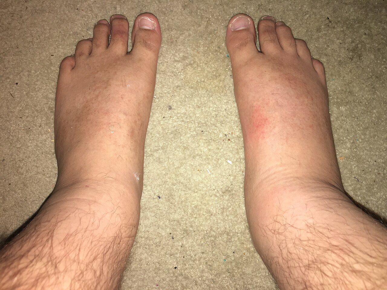
Cellulitis and the abscess that must be opened
Cellulitis is a bacterial infection of the deep dermis and subcutaneous tissue that enters through a fissure, a bite, a venous ulcer, tinea pedis between the toes or an injection site. The presentation is unilateral, warm, tender, poorly demarcated erythema with oedema, often with lymphangitic streaking and regional lymphadenopathy, and sometimes with fever and leukocytosis. Non-purulent cellulitis is usually beta-haemolytic streptococcal and is treated with cefazolin intravenously or cephalexin orally. Purulent cellulitis with an abscess is usually staphylococcal, frequently MRSA, and needs incision and drainage plus TMP-SMX, doxycycline or clindamycin; without drainage, antibiotics alone commonly fail.
Nursing care is simple and effective. Outline the erythema with a skin marker, write the date and time on the skin, and reassess each shift — expansion beyond the mark within the first 24 to 48 hours of therapy is the earliest objective sign of failure, though a modest initial increase can accompany the inflammatory response to bacterial killing. Elevate the limb above the level of the heart, which does more for the oedema than any drug. Treat the portal of entry: an antifungal for macerated toe webs prevents the next episode. Manage pain, control glucose, and assess for venous insufficiency and lymphoedema, which drive recurrence.
Beware the mimics, because both are common and neither needs antibiotics. Bilateral, symmetric, non-tender erythema with scaling in a patient with venous insufficiency is stasis dermatitis, not cellulitis. Acute unilateral calf swelling with pain but little erythema may be deep vein thrombosis. If the leg is red and the patient is well and afebrile with normal inflammatory markers, argue for a second look before committing to a week of antibiotics.
Necrotising soft-tissue infection: the surgical emergency
Necrotising fasciitis spreads along the fascial plane, thrombosing the perforating vessels that feed the skin. Because the destruction is deep and the skin is initially spared, the outside of the limb underestimates the inside of it. The single most important early finding is pain out of proportion to the physical findings — agony in a limb that looks only mildly red. Then come rapid hourly progression, systemic toxicity with fever, tachycardia, hypotension and confusion, tense woody oedema extending beyond the erythema, and later the ominous signs: dusky or violaceous skin, haemorrhagic bullae, crepitus, and skin anaesthesia where cutaneous nerves have infarcted. Risk factors are diabetes, injection drug use, cirrhosis, immunosuppression, obesity, recent surgery and trauma.
Definitive treatment is emergent surgical debridement, and the strongest predictor of survival is time to the operating room. Laboratory scores and imaging support the diagnosis but must never delay surgery; computed tomography showing fascial gas is confirmatory, its absence is not reassuring. Antimicrobial therapy is broad and includes an agent for toxin suppression: vancomycin or linezolid plus piperacillin-tazobactam or a carbapenem, plus clindamycin, which shuts down streptococcal exotoxin production at the ribosome. Resuscitate aggressively — these patients third-space enormous volumes — keep the patient nil by mouth, obtain consent and surgical clearance, type and cross-match, and expect serial debridements.
Osteomyelitis and the diabetic foot
Bone becomes infected by contiguous spread from an overlying ulcer or open fracture, by direct inoculation at surgery or with hardware, or haematogenously — in adults the haematogenous form most often lands in the vertebral bodies. Staphylococcus aureus is the leading organism. Infection raises intra-osseous pressure, thromboses the nutrient vessels, and produces avascular dead bone called sequestrum; because no antibiotic reaches dead bone, chronic osteomyelitis is a surgical disease with antibiotic support rather than the reverse. Acute presentation is deep, constant, unremitting pain with fever, local tenderness and swelling; vertebral disease presents as unrelenting focal back pain that is worse at night and does not ease with rest, and new weakness, numbness, or bladder or bowel change signals epidural abscess with cord compression — an emergency that needs immediate imaging.
Diagnosis rests on inflammatory markers for trending (ESR and CRP), magnetic resonance imaging as the most sensitive imaging modality, and bone biopsy with culture as the gold standard; swab cultures of an overlying ulcer grow colonisers and mislead. In the diabetic foot, a sterile blunt probe that reaches bone through the ulcer strongly predicts osteomyelitis. Treatment is four to six weeks of targeted therapy, historically all intravenous, though high-bioavailability oral regimens are now accepted in selected patients, alongside debridement and, when infection involves hardware, removal or exchange of the implant.
The nursing load is long-haul. If therapy will run through a peripherally inserted central catheter at home, teach line care, flushing, signs of exit-site infection and what to do if the line dislodges, and confirm home-infusion arrangements before discharge. Reinforce adherence for the full course even after the patient feels well — the commonest cause of relapse is stopping early. Protect the limb, offload the diabetic foot with the prescribed device, optimise glucose and nutrition (adequate protein, vitamin C and zinc support healing), and arrange weekly laboratory monitoring appropriate to the drug.
Infective endocarditis
Endocarditis begins where turbulent flow or a foreign surface damages endothelium; platelets and fibrin form a sterile vegetation, and a transient bacteraemia seeds it. Risk factors are prosthetic valves and intracardiac devices, prior endocarditis, congenital heart disease, degenerative valve disease, haemodialysis and injection drug use, which characteristically produces right-sided tricuspid disease with septic pulmonary emboli. Once established, the vegetation is a mass of organisms shielded from immune cells, which is why therapy is intravenous, bactericidal and prolonged — four to six weeks.
Suspect it in anyone with unexplained fever plus a new or changed murmur. Classic peripheral findings are uncommon but worth knowing: splinter haemorrhages under the nails, painless erythematous macules on palms and soles called Janeway lesions, painful nodules on finger and toe pads called Osler nodes (Osler equals ouch), Roth spots on funduscopy, and petechiae. Draw three sets of blood cultures from separate sites before antibiotics; the bacteraemia is continuous, so timing them apart is less important than sampling adequately. Transthoracic echocardiography is the first study; transoesophageal is far more sensitive for vegetations, abscess and prosthetic valves.
Complications drive the nursing surveillance. Valve destruction causes acute regurgitation and heart failure — watch for new dyspnoea, orthopnoea, crackles, an S3 and rising oxygen requirement. Fragments embolise: to brain (sudden focal deficit or altered consciousness), to spleen and kidney (flank or left upper quadrant pain, haematuria), to limbs (a cold pulseless extremity), and from right-sided disease to lung (pleuritic pain, hypoxia). Perform a neurological check every shift and more often if the vegetation is large. Surgical referral is indicated for heart failure from valve destruction, uncontrolled infection or perivalvular abscess, persistently positive cultures on appropriate therapy, and recurrent emboli or a mobile vegetation larger than 10 mm. Antibiotic prophylaxis before dental procedures that manipulate gingival tissue or perforate oral mucosa — typically amoxicillin 2 g orally 30 to 60 minutes prior — is now reserved for the highest-risk patients only: prosthetic valves or prosthetic material, previous endocarditis, specified congenital heart disease and cardiac transplant valvulopathy. Teach meticulous daily oral hygiene, which prevents more bacteraemia than any single prophylactic dose.
In skin, bone and valve infection the drug is only half the treatment — drainage, debridement, hardware removal and valve surgery are the other half, and pain out of proportion to appearance means the operating room, not another dose.
Section 5Tuberculosis, HIV and Viral Hepatitis
Tuberculosis: airborne, chronic, treated for months
Mycobacterium tuberculosis is inhaled in droplet nuclei small enough to stay suspended for hours and to reach the alveoli. Macrophages ingest the bacilli but cannot kill them; the organism survives intracellularly, and cell-mediated immunity walls it into a granuloma. In roughly 90 percent of immunocompetent people that containment holds for life — latent tuberculosis infection: positive test, no symptoms, normal chest film, not infectious. When immunity falters (HIV, tumour necrosis factor inhibitors, chronic steroids, transplantation, diabetes, malnutrition, dialysis, ageing), the granuloma breaks down and caseous material erodes into a bronchus, producing the cavity, the cough and the transmissibility of active disease.
Active pulmonary tuberculosis presents with a cough lasting more than two to three weeks, haemoptysis, drenching night sweats, low-grade afternoon fever, anorexia, fatigue and weight loss. Screening uses the tuberculin skin test, read at 48 to 72 hours by palpating induration rather than measuring redness, or an interferon-gamma release assay, which is not confounded by prior BCG vaccination. Induration of 5 mm is positive in people with HIV, recent close contacts, fibrotic changes on chest film and organ-transplant or otherwise immunosuppressed patients; 10 mm is positive for recent immigrants from high-prevalence countries, injection drug users, healthcare workers, residents and staff of congregate settings, children under four (and any child or adolescent exposed to an adult in a high-risk category) and people with diabetes, silicosis or chronic kidney disease; 15 mm is required in people with no risk factors. A positive screen means infection, not disease — a chest radiograph and symptom review distinguish latent from active, and active disease is confirmed by sputum acid-fast bacilli smear, nucleic acid amplification and culture, which remains the standard for susceptibility.
Suspected active pulmonary tuberculosis goes immediately into a negative-pressure airborne infection isolation room with at least six air changes per hour (twelve in newer construction), door closed. Staff wear a fit-tested N95 or a PAPR; the patient wears a surgical mask — never an N95 with an exhalation valve — whenever they must leave the room, and transport is minimised. Precautions may be lifted only when the patient has clinically improved on effective therapy and has three consecutive negative sputum smears collected 8 to 24 hours apart with at least one early-morning specimen. Standard treatment for drug-susceptible disease is RIPE: rifampin, isoniazid, pyrazinamide and ethambutol for two months, then rifampin and isoniazid for four more, for six months in total, usually given by directly observed therapy. Latent infection is treated with shorter regimens such as weekly isoniazid plus rifapentine for 12 weeks or daily rifampin for four months.
| Drug | Signature toxicity | Teaching and monitoring |
|---|---|---|
| Rifampin | Hepatotoxicity; potent CYP450 inducer | Orange-red urine, sweat and tears; permanently stains soft contact lenses; makes oral contraceptives, warfarin, methadone and many antiretrovirals fail — use a barrier method |
| Isoniazid | Hepatitis; peripheral neuropathy | Give pyridoxine (vitamin B6) 25–50 mg daily; report numbness or tingling; avoid alcohol; baseline and monthly LFTs in at-risk patients |
| Pyrazinamide | Hepatotoxicity; hyperuricaemia | Report joint pain and swelling; push fluids; monitor uric acid and LFTs |
| Ethambutol | Optic neuritis | Baseline and monthly visual acuity and red-green colour discrimination; report any change in vision immediately |
Three of the four are hepatotoxic, so teach every patient to stop the drugs and call for nausea, vomiting, right upper quadrant pain, dark urine, pale stools or jaundice, and to avoid alcohol and unsupervised acetaminophen. The therapeutic problem is time: patients feel well within weeks but must continue for months, and non-adherence is what breeds multidrug-resistant tuberculosis. Directly observed therapy, incentives, transport support, an interpreter and a named public health contact are not niceties — they are the treatment. Tuberculosis is reportable to public health, and contact investigation is a public health function you support with accurate history rather than perform yourself.
HIV: the labs that define stage and the therapy that ends transmission
HIV is a retrovirus that binds CD4 receptors, transcribes its RNA into DNA with reverse transcriptase, integrates into the host genome with integrase, and destroys CD4 T-lymphocytes — the coordinators of cell-mediated immunity. Untreated, the CD4 count falls over years and opportunistic infections appear in a predictable order. Screening uses a fourth-generation antigen and antibody combination immunoassay that detects p24 antigen and shortens the window period to roughly two to three weeks; a reactive result is confirmed with an HIV-1/HIV-2 antibody differentiation assay, and an indeterminate result is resolved with nucleic acid testing. A normal CD4 count is 500 to 1,600 cells/mm³. AIDS is defined by a CD4 count below 200 cells/mm³ or by an AIDS-defining illness regardless of count.
Antiretroviral therapy is started for everyone with HIV, as soon as possible after diagnosis, regardless of CD4 count. Modern first-line regimens are integrase strand transfer inhibitor based — commonly bictegravir with tenofovir alafenamide and emtricitabine as a single daily tablet, or dolutegravir with a nucleoside backbone. The measure of success is the viral load: suppression to undetectable, generally under 50 copies/mL, within three to six months. Teach the two facts that change lives. First, adherence must be near-perfect, because partially suppressive levels select resistance and burn a whole drug class. Second, undetectable equals untransmittable: a person with sustained viral suppression does not transmit HIV sexually. Prevention for those without HIV includes pre-exposure prophylaxis, either a daily oral tenofovir-emtricitabine tablet or long-acting injectable cabotegravir, and post-exposure prophylaxis started within 72 hours — ideally within 2 hours — and continued for 28 days.
Prophylaxis against opportunistic infection is keyed to the CD4 count: TMP-SMX when the count falls below 200 cells/mm³ to prevent Pneumocystis jirovecii pneumonia, which presents insidiously with dry cough, dyspnoea on exertion and marked hypoxia with a near-normal-sounding chest; TMP-SMX also covers toxoplasmosis below 100 cells/mm³ in patients with positive serology; and azithromycin has traditionally been used below 50 cells/mm³ for Mycobacterium avium complex. Watch for immune reconstitution inflammatory syndrome in the first weeks after starting therapy in a severely immunosuppressed patient — a paradoxical worsening of fever and inflammation as returning immunity attacks an existing occult infection. It is generally not a reason to stop antiretrovirals. Finally, remember that HIV status is protected health information: discuss it in private, disclose only to those involved in care, and do not assume the family in the room knows.
Hepatitis B and C, and occupational exposure
Hepatitis B is transmitted percutaneously, sexually and perinatally, and its serology can be read like a sentence. HBsAg positive means the virus is present now; persisting beyond six months defines chronic infection. Anti-HBs alone means immunity from vaccination. Anti-HBs plus anti-HBc (total) means resolved natural infection. Anti-HBc IgM indicates acute infection. HBeAg and a high HBV DNA level indicate high replication and high infectivity. Chronic hepatitis B is suppressed — not usually cured — with tenofovir or entecavir, and it requires lifelong surveillance for cirrhosis and hepatocellular carcinoma. Hepatitis B is vaccine-preventable, and universal adult vaccination through age 59 is now recommended.
Hepatitis C is predominantly bloodborne, most often through injection drug use. Screening is an anti-HCV antibody test; because antibody persists after cure, current infection must be confirmed with HCV RNA. Direct-acting antiviral regimens taken for 8 to 12 weeks cure over 95 percent of patients, measured as an undetectable viral load 12 weeks after therapy ends. Teach that reinfection is possible — cure is not immunity — and screen for hepatitis B before treatment, because HBV can reactivate. Volume I covered the hepatic failure end of this spectrum; here the nursing emphasis is screening, linkage to care, harm reduction and dismantling the stigma that keeps people from returning for the tablets that would cure them.
For an occupational exposure — a needlestick, a splash to mucous membrane or non-intact skin — act in this order. Wash the site with soap and water immediately, or flush mucous membranes with water or saline for several minutes; do not squeeze the wound and do not apply caustics. Report at once to the supervisor and occupational health, because the clock matters. Source and healthcare-worker testing follow institutional protocol. Approximate per-exposure transmission risk from a percutaneous injury with an infected source is up to 30 percent for hepatitis B in an unvaccinated person, roughly 1.8 percent for hepatitis C and about 0.3 percent for HIV. Hepatitis B is managed with vaccine and, when indicated, hepatitis B immune globulin; there is no post-exposure prophylaxis for hepatitis C, so the response is serial testing and immediate treatment if infection occurs; HIV prophylaxis is a three-drug regimen begun within 72 hours and taken for 28 days.
In chronic communicable disease the pharmacology is the easy part — the nursing work is adherence over months, protection of confidentiality, and the isolation and public-health steps that stop the next case.
The chapter in one breath
Infection is a race between a pathogen and a host, and the findings you assess — fever, tachycardia, tachypnoea, leukocytosis with a left shift, local heat and erythema — are the host’s response made visible. Build the picture before the first dose: two sets of blood cultures from two sites, the right specimen from the right compartment, and markers you can actually interpret against reference values. Then remember that source control beats drug selection — drain the abscess, debride the fascia, pull the line, relieve the obstruction. Choose antimicrobials by mechanism, because the mechanism names the monitoring: creatinine and hearing for aminoglycosides and vancomycin, creatine kinase for daptomycin, a CBC for prolonged linezolid, a stool watch for anything that empties the colon of anaerobes. Practise stewardship at the bedside by getting cultures first, giving doses on time, delabelling false penicillin allergies, prompting the 48-hour time-out, converting to oral and stopping on the intended day. Isolate correctly, wash with soap and water for spores, and honour the device bundles that prevent most of what we treat. Escalate immediately for pain out of proportion, a suddenly quiet distended abdomen in C. difficile, a new neurological deficit in endocarditis and fever in neutropenia. In older adults, expect confusion instead of fever — and never treat a urine culture that has no patient attached to it.
Words worth knowing
| Term | What it means |
|---|---|
| Antimicrobial stewardship | Coordinated practice of giving the right drug, dose, route and duration — or no drug — to preserve effectiveness and limit harm |
| De-escalation | Narrowing broad empiric therapy to a targeted agent once culture and susceptibility results return, usually at 48–72 hours |
| Left shift / bandemia | Increased immature band neutrophils (over roughly 10 percent), signalling that the marrow is releasing cells early under bacterial stress |
| Absolute neutrophil count | WBC times (segs plus bands) divided by 100; under 500/mm³ is severe neutropenia and a medical emergency with fever |
| Procalcitonin | Peptide that rises in bacterial infection; used chiefly to support stopping antibiotics rather than starting them |
| Minimum inhibitory concentration | Lowest drug concentration that prevents visible organism growth; the basis of the susceptible, intermediate and resistant report |
| Time-dependent vs concentration-dependent killing | Time-dependent efficacy depends on time above MIC (beta-lactams) — doses must be on time, sometimes as extended infusions; concentration-dependent efficacy depends on peak-to-MIC ratio (aminoglycosides, fluoroquinolones) — the basis of extended-interval dosing |
| Vancomycin infusion reaction | Histamine-mediated flushing and pruritus from rapid infusion; a rate problem, not an allergy |
| MRSA | Methicillin-resistant Staphylococcus aureus; resistant to all beta-lactams except ceftaroline — treat with vancomycin, daptomycin or linezolid |
| ESBL / CRE | Extended-spectrum beta-lactamase organisms (need a carbapenem) and carbapenem-resistant Enterobacterales (need newer agents) |
| Pseudomembranous colitis | Yellow-white mucosal plaques of C. difficile toxin injury; requires soap-and-water hand hygiene and sporicidal cleaning |
| Toxic megacolon | Colonic dilation with systemic toxicity; suspect when diarrhoea suddenly stops and the abdomen becomes distended and quiet |
| Asymptomatic bacteriuria | Bacteria in urine without urinary symptoms; treated only in pregnancy or before mucosa-traumatising urologic procedures |
| Necrotising fasciitis | Fascial-plane infection with pain out of proportion, crepitus, bullae and skin anaesthesia; treated by emergent surgical debridement |
| Sequestrum | Devascularised dead bone in chronic osteomyelitis that no antibiotic can penetrate; must be removed surgically |
| Osler nodes / Janeway lesions | Painful finger and toe nodules versus painless palm and sole macules in infective endocarditis (Osler equals ouch) |
| Latent tuberculosis infection | Positive skin test or IGRA with no symptoms and a normal chest film; not infectious, but treatable to prevent reactivation |
| Induration | The palpable raised firmness measured when reading a tuberculin skin test at 48–72 hours — redness alone is not measured |
| Undetectable equals untransmittable | Sustained HIV viral suppression on antiretroviral therapy prevents sexual transmission |
| Immune reconstitution inflammatory syndrome | Paradoxical inflammatory worsening after starting antiretroviral therapy as immunity returns and attacks an occult infection |
Check yourself
Reproductive and Sexual Health Disorders
Reproductive complaints arrive on a med-surg floor wearing a disguise. The man in 412 is “confused and agitated” because his bladder holds nine hundred millilitres behind a hypertrophied prostate. The woman in 408 is “anemic, cause unclear” because she has bled through a pad an hour for six days from a fibroid uterus. The twenty-two-year-old in the emergency department with vomiting and abdominal pain has a testicle that lost its blood supply four hours ago and has roughly two hours of viability left. This chapter teaches you the mechanisms behind those presentations, the assessments that separate an emergency from an annoyance, the drugs and their monitoring, and the language that lets a patient tell you the truth about a body part they were raised not to name. Reproductive nursing is not a specialty rotation you left behind — it is obstruction, ischemia, hemorrhage, infection and malignancy in an organ system nobody wants to discuss.
By the end of this chapter you can
- Obtain a structured, trauma-informed sexual history using the five Ps and document it without assumption or judgment.
- Interpret core reproductive diagnostics — PSA, post-void residual, beta-hCG, CA-125, NAAT, endometrial stripe thickness — against real reference values.
- Prioritize care for benign prostatic hyperplasia, acute urinary retention and the post-TURP patient, including recognition of TUR syndrome and management of continuous bladder irrigation.
- Differentiate testicular torsion from epididymitis and initiate emergency escalation within the six-hour salvage window.
- Explain the mechanism, nursing implications and monitoring for alpha-1 blockers, 5-alpha-reductase inhibitors, PDE5 inhibitors, GnRH agonists, SERMs and aromatase inhibitors.
- Classify abnormal uterine bleeding with PALM-COEIN and identify postmenopausal bleeding as endometrial cancer until proven otherwise.
- Deliver post-operative care after hysterectomy and mastectomy, including drain management, lymphedema precautions and radiation safety for brachytherapy.
- Apply current CDC treatment logic for chlamydia, gonorrhea, syphilis, trichomoniasis, herpes and pelvic inflammatory disease, and counsel on PrEP and PEP.
- Describe how reproductive and sexual problems present differently in older adults and delegate safely within scope.
Section 1Asking the questions nobody asks: reproductive assessment and screening
The sexual history is an assessment, not an intrusion
Nurses skip the sexual history more reliably than any other part of the review of systems. The reasons are predictable — embarrassment, time, an assumption that a seventy-eight-year-old is not sexually active, a fear of offending. The cost is measurable: undiagnosed sexually transmitted infection, missed intimate partner violence, medication non-adherence nobody explained, and a patient discharged on a beta blocker who quietly stops taking it because of erectile dysfunction he was never invited to mention.
Use the CDC’s five Ps as your scaffold: Partners (how many, what genders), Practices (oral, vaginal, anal — this determines which sites you swab), Protection from STIs, Past history of STIs, and Pregnancy intention. Many educators add a sixth — Problems or pleasure — because dyspareunia, erectile dysfunction and low libido are the complaints that actually change management. Ask permission first (“I ask everyone these questions because they affect the care I give — is that all right?”), ask in private with the door closed and family out of the room, and use the patient’s own words for anatomy before you supply clinical terms.
Language matters clinically, not just politically. Ask “are your partners men, women, or both?” rather than assuming from marital status. Ask transgender and gender-diverse patients about organs present rather than gender recorded — a transgender man with a retained cervix still needs cervical cancer screening, and a transgender woman on estrogen still has a prostate. Trauma-informed practice means offering a chaperone for every genital or breast examination, telling the patient what you are about to do before you touch, and stopping immediately if she says stop. For adolescents, know your state’s minor consent law: in most states a minor may consent to STI testing and treatment independently, and billing statements are a real confidentiality hazard.
The screening calendar and what each test is for
Screening guidelines shift, and organizations disagree. Teach the principle rather than memorizing one committee’s year: screening finds disease in people without symptoms, and it stops when the patient’s remaining life expectancy is shorter than the time it would take the cancer to hurt them. A frail ninety-year-old with dementia does not need a mammogram. A healthy sixty-eight-year-old does.
Cervical cancer screening is the clearest success story in oncology. Human papillomavirus causes essentially all cervical cancer, the precancerous phase lasts years, and the test is cheap. Screening begins at age 21 under USPSTF and ACOG guidance (the American Cancer Society now favors primary HPV testing beginning at 25), continues to 65, and is not indicated for a patient after total hysterectomy for benign disease with no history of high-grade dysplasia. For an immunocompetent patient, screening is not indicated before 21 no matter when sexual activity started, because adolescent HPV infection almost always clears on its own and treating it damages the cervix. The exception is immunosuppression: a patient with HIV or other immunosuppression, or with in-utero DES exposure, begins screening within one year of first sexual activity (and no later than 21) and is screened more frequently thereafter.
| Screen | Who and how often | What the nurse teaches |
|---|---|---|
| Cervical cytology (Pap) | Age 21–29, every 3 years | No douching, intercourse or vaginal products for 48 hours; reschedule if actively menstruating |
| Pap plus HPV co-test / primary HPV | Age 30–65, every 5 years (cytology alone every 3 years acceptable) | A positive HPV result is common and is not a diagnosis of cancer; it triggers follow-up, not panic |
| HPV vaccine (9-valent) | Routine at 11–12 (may start at 9); catch-up through 26; shared decision 27–45 | Two doses if the series starts before the 15th birthday; three doses if started at 15 or older or immunocompromised |
| Mammography | USPSTF: biennial 40–74. ACS: option to start at 40, annual 45–54, then annual or biennial | Schedule the week after menses if premenopausal; no deodorant or powder; expect compression discomfort |
| Breast MRI plus mammogram | Lifetime risk greater than 20 percent (BRCA, chest radiation, strong family history) | Requires IV gadolinium; check renal function and claustrophobia |
| PSA | Shared decision at 50 average risk; 45 if Black or a first-degree relative diagnosed before 65; 40 if multiple early relatives | Avoid ejaculation, vigorous cycling and DRE for 48 hours before the draw — all can falsely elevate |
| Chlamydia and gonorrhea NAAT | All sexually active women under 25 annually; older with risk factors; all pregnant patients; men who have sex with men at least annually at all exposed sites | Urine, vaginal swab (self-collected is acceptable and accurate), rectal and pharyngeal swabs per practices |
| HIV | At least once for everyone 13–64; annually or more with ongoing risk; every pregnancy | Opt-out testing is standard; a negative test does not cover the window period |
| Testicular self-exam | Monthly for males from adolescence | After a warm shower when the scrotum is relaxed; roll each testis between thumb and fingers; the epididymis on the posterior surface is normal |
Diagnostics and the numbers that mean something
A handful of tests carry most of the diagnostic weight in reproductive care, and each has a trap. Beta-hCG is the single most important test in any person with a uterus and abdominal pain: normal in a non-pregnant patient is less than 5 mIU/mL, and a positive result reframes every differential and every medication and radiology order that follows. Never send a reproductive-age patient for a CT scan (the hazard is the ionizing radiation, not the contrast), a fluoroquinolone or a methotrexate dose without it.
Prostate-specific antigen is organ-specific but not cancer-specific. The traditional cut point is less than 4 ng/mL, but benign hyperplasia, prostatitis, recent catheterization, cycling and ejaculation all raise it, and finasteride cuts it roughly in half. CA-125 (normal under 35 units/mL) is useful for tracking known ovarian cancer, not for screening — it rises in endometriosis, fibroids, pelvic inflammatory disease, cirrhosis and even menstruation. Post-void residual measured by bladder scanner is the fastest bedside test in this chapter: under 50 mL is normal, over 100–150 mL suggests incomplete emptying, and over 200 mL is significant retention that needs a plan. In a postmenopausal patient with bleeding, an endometrial stripe greater than 4 mm on transvaginal ultrasound requires endometrial biopsy.
Delegation in this section is straightforward and gets tested constantly. Unlicensed assistive personnel may perform a bladder scan if trained and competency-validated, provide perineal care, set up a sitz bath, record intake and output, and reinforce a patient’s already-taught positioning. They may not interpret the scan, teach self-examination, assess a wound or take a sexual history. Assessment, teaching, evaluation of response, and any unstable patient remain with the RN. An LPN or LVN may administer most medications, perform sterile dressing changes per facility policy, and reinforce teaching the RN has initiated — but the initial teaching and the evaluation of understanding belong to the RN.
A structured sexual history taken in private, plus a bladder scan and a beta-hCG, will reorder more reproductive differentials than any imaging study you can order.
Section 2Male reproductive disorders: obstruction, ischemia and malignancy
Benign prostatic hyperplasia and acute urinary retention
The prostate sits like a doughnut around the proximal urethra. With age, dihydrotestosterone drives glandular and stromal proliferation in the transition zone — the ring nearest the urethra — so even a modestly enlarged gland can obstruct badly, while a huge gland growing outward may cause no symptoms at all. That explains the classic mismatch between gland size on digital rectal exam and severity of complaints. Obstruction produces two symptom families. Voiding (obstructive) symptoms reflect the blockage itself: hesitancy, weak and intermittent stream, straining, terminal dribbling, sensation of incomplete emptying. Storage (irritative) symptoms reflect a detrusor muscle that has hypertrophied and become overactive against the resistance: frequency, urgency, nocturia. Nocturia is usually the symptom that drives the patient to seek help, and in an older adult it is also a fall risk you must address.
Assessment: quantify with the International Prostate Symptom Score, take a careful medication history (anticholinergics, antihistamines, decongestants, opioids and tricyclics all precipitate retention), palpate and percuss the suprapubic area, and scan the bladder. On DRE the benign gland is smoothly enlarged, symmetric and rubbery; a hard, fixed or nodular gland is cancer until biopsy says otherwise. Order urinalysis to exclude infection and hematuria, BUN and creatinine to detect obstructive nephropathy, and PSA with the caveats above.
Acute urinary retention is the emergency complication. The patient cannot void at all, is restless and diaphoretic, has severe suprapubic pain, and — if he is older — may present as new confusion or agitation with no complaint of pain at all. Scan first, then catheterize. Drain the bladder completely; the old teaching about clamping to prevent hematuria has not held up, but you must then watch for post-obstructive diuresis: urine output above 200 mL/hr for several consecutive hours, with the risk of hypovolemia and sodium and potassium losses. Monitor hourly output, orthostatic vitals and electrolytes, and replace fluids per order. If you cannot pass a catheter, do not force it — stop and call urology, because a false passage turns a bad night into surgery.
Drugs for BPH and the post-TURP patient
Two mechanisms, two very different time courses. Alpha-1 adrenergic blockers relax smooth muscle in the bladder neck and prostatic capsule and work within days. 5-alpha-reductase inhibitors block conversion of testosterone to DHT and physically shrink the gland — but take three to six months, so a patient who stops at week four because “it isn’t working” has been failed by teaching, not by pharmacology.
| Class and examples | Mechanism and onset | Nursing implications and monitoring |
|---|---|---|
| Uroselective alpha-1a blockers — tamsulosin 0.4 mg, silodosin, alfuzosin | Relax bladder-neck and prostatic smooth muscle; relief in days | Take 30 minutes after the same meal daily; still causes orthostatic hypotension and dizziness — rise slowly; retrograde ejaculation is common and harmless but must be explained; warn the ophthalmologist before cataract surgery (intraoperative floppy iris syndrome) |
| Non-selective alpha blockers — doxazosin, terazosin | Same, plus systemic vasodilation | First-dose syncope — give at bedtime, start low; useful when the patient also has hypertension, but never used as the sole antihypertensive |
| 5-alpha-reductase inhibitors — finasteride 5 mg, dutasteride 0.5 mg | Block testosterone to DHT conversion; shrink the gland over 3–6 months | Lowers PSA by roughly 50 percent — double the measured value when screening; decreased libido, erectile dysfunction, gynecomastia; teratogenic — a pregnant or potentially pregnant person must not handle crushed or broken tablets and should avoid contact with semen |
| PDE5 inhibitor — tadalafil 5 mg daily | Smooth-muscle relaxation via cyclic GMP; treats BPH and erectile dysfunction together | Absolutely contraindicated with any nitrate; additive hypotension with alpha blockers — separate doses and monitor blood pressure |
| Antimuscarinics and beta-3 agonist — oxybutynin, solifenacin, mirabegron | Calm detrusor overactivity for storage symptoms | Use only when the post-void residual is acceptable — they can precipitate retention; antimuscarinics cause dry mouth, constipation, confusion in older adults; mirabegron raises blood pressure |
When drugs fail or the patient develops recurrent retention, infection, stones or renal impairment, the surgical answer is transurethral resection of the prostate. Post-operatively he returns with a large three-way catheter under gentle traction and continuous bladder irrigation with isotonic saline. Your job is patency. Titrate the irrigant to keep drainage light pink to clear; if it turns bright red or you see clots, increase the rate and notify the surgeon. If drainage stops, the bladder distends and the patient reports severe pain and urgency — stop the irrigation, hand-irrigate with 50 mL sterile saline only if facility policy and an order permit, and escalate. Calculate true urine output by subtracting the volume of irrigant instilled from total drainage; charting the raw number as urine is a classic and dangerous error. Do not deflate the catheter balloon or release traction without an order — the balloon is providing hemostasis at the prostatic fossa.
TUR syndrome is the complication to know. When a monopolar resectoscope is used, the hypotonic glycine irrigant is absorbed through open venous sinuses, producing dilutional hyponatremia and fluid overload. Watch for altered mental status and restlessness, bradycardia with hypertension, nausea and vomiting, visual disturbance and, if unrecognized, seizure and cardiovascular collapse. Serum sodium falls below the normal 135–145 mEq/L, often below 125 before frank symptoms appear. Modern bipolar resection uses saline and has largely eliminated the syndrome, but it still appears on examinations and in facilities with older equipment. Report neurologic change during CBI immediately. Bladder spasms are treated with antispasmodics or belladonna-and-opium suppositories, not by pulling the catheter. Discharge teaching: expect pink-tinged urine intermittently for weeks, drink 2–3 litres daily unless restricted, no heavy lifting over 10 pounds, no straining or enemas or rectal temperatures, no driving or sexual activity for four to six weeks, and expect retrograde ejaculation permanently — orgasm is preserved, ejaculate is not.
Prostate cancer and androgen deprivation
Prostate adenocarcinoma arises in the peripheral zone, which is why it causes no urinary symptoms until it is advanced and why the DRE reaches it. Risk rises with age, is substantially higher in Black men, and clusters in families carrying BRCA2 and Lynch mutations. Grading uses the Gleason score, translated into Grade Groups 1 through 5; staging adds imaging. Because the tumour is osteoblastic, metastasis goes to bone — new persistent back pain in a man with prostate cancer is a bone lesion until proven otherwise, and you should expect an elevated alkaline phosphatase (normal roughly 30–120 units/L). Because bone-forming lesions consume calcium into new bone, the characteristic derangement here is hypocalcemia (normal calcium 8.6–10.2 mg/dL), and it deepens once a bone-targeted agent such as zoledronic acid or denosumab is started — check calcium and vitamin D before and during those drugs, and watch for perioral numbness, tetany and a prolonged QT. Hypercalcemia is the osteolytic pattern (myeloma, breast, squamous lung) and is uncommon in prostate cancer. Watch also for cord compression.
Management ranges from active surveillance for low-grade disease in older men through radical prostatectomy, external beam radiation and brachytherapy. After seed brachytherapy, radiation safety teaching is concrete: strain urine for the first week and report any seed to the physician using tweezers and a container (never fingers), avoid prolonged close contact with pregnant people and young children for about two months, and use a condom for the first several ejaculations. After radical prostatectomy expect a catheter for one to two weeks, urinary incontinence that improves over months with pelvic floor (Kegel) training, and erectile dysfunction that may be permanent.
Androgen deprivation therapy starves hormone-sensitive disease. GnRH agonists — leuprolide, goserelin — paradoxically cause an initial testosterone surge, so an antiandrogen such as bicalutamide is started before or with the first injection and continued two to four weeks to prevent tumour flare (worsening bone pain, urinary obstruction, cord compression). The GnRH antagonist degarelix produces no flare. The therapeutic goal is a castrate testosterone below 50 ng/dL. The toxicity list is what you manage: hot flashes, fatigue, anemia, loss of libido, gynecomastia, sarcopenia, insulin resistance, cardiovascular risk and accelerated bone loss. Every patient on long-term ADT needs baseline and serial DEXA, calcium and vitamin D, weight-bearing exercise, and screening for depression — the psychological burden of chemical castration is real and under-assessed.
Scrotal emergencies, testicular cancer and erectile dysfunction
Testicular torsion is the surgical emergency of this chapter. The testis twists on the spermatic cord, venous outflow stops, then arterial inflow stops. Salvage is above 90 percent within 6 hours, falls sharply by 12, and approaches zero at 24. It peaks in neonates and in adolescents, usually from a congenital “bell-clapper” deformity, and it typically wakes the patient from sleep. Findings: sudden severe unilateral scrotal pain, nausea and vomiting, a swollen high-riding testis in a horizontal lie, an absent cremasteric reflex on the affected side, and pain that is not relieved by elevating the scrotum (a negative Prehn sign). Compare that with epididymitis, which builds over days, comes with dysuria and pyuria and often fever, and improves with elevation.
What do you do first? Escalate. Call urology and the provider immediately — this patient does not wait for an ultrasound to be scheduled, because the tissue is dying while you wait. Only once that call is placed do you keep him NPO, establish IV access and give analgesia. Doppler ultrasound may confirm absent flow, but a classic history and exam go straight to the operating room for detorsion and bilateral orchiopexy — the other side is fixed too because the anatomic defect is usually bilateral.
Testicular cancer is the most common solid tumour in men aged 15–35 and is highly curable. It presents as a painless, firm, non-transilluminating mass or a sensation of heaviness; cryptorchidism is the major risk factor even after surgical correction. Tumour markers help: alpha-fetoprotein (normal under about 10 ng/mL) rises in non-seminomatous tumours and is never produced by pure seminoma; beta-hCG and LDH may rise in either. Surgery is radical inguinal orchiectomy — never a scrotal approach, which seeds lymphatics. Discuss sperm banking before any treatment begins, because chemotherapy and retroperitoneal node dissection threaten fertility; raising it after the first cycle is too late. Body image and prosthesis options belong in the same conversation.
Erectile dysfunction is a cardiovascular symptom as often as a sexual one; endothelial disease shows up in penile arteries before coronary ones, so new ED in a middle-aged man warrants cardiac risk assessment. PDE5 inhibitors (sildenafil, tadalafil, vardenafil) are first-line. The lethal interaction is with nitrates in any form — sublingual nitroglycerin, isosorbide, nitroprusside, and recreational amyl nitrite — producing profound refractory hypotension. Nitrates must be withheld for at least 24 hours after sildenafil or vardenafil and 48 hours after tadalafil. Teach that sildenafil works best on an empty stomach about an hour before activity, that the drug requires arousal to work, and that an erection lasting more than 4 hours (priapism) is an emergency requiring immediate care — ischemic priapism causes permanent fibrosis. Sudden unilateral vision or hearing loss means stop the drug and be evaluated.
In male reproductive care the clock-driven emergencies are torsion (six hours to save a testis) and acute retention (a bladder scan and a catheter), while the daily nursing work is patency of continuous bladder irrigation and honest teaching about drugs that change ejaculation, erection and PSA.
Section 3Bleeding, structure and transition: female reproductive disorders
Abnormal uterine bleeding, fibroids and the anemia you will actually treat
Abnormal uterine bleeding is bleeding that is abnormal in volume, duration, regularity or timing. Classify it with PALM-COEIN: the structural causes are Polyp, Adenomyosis, Leiomyoma and Malignancy or hyperplasia; the non-structural are Coagulopathy, Ovulatory dysfunction, Endometrial, Iatrogenic and Not otherwise classified. The framework is not trivia — it tells you what to look for. A teenager with heavy bleeding since menarche needs a coagulation workup, because up to a fifth of them have von Willebrand disease. A woman on anticoagulation or with a copper IUD is iatrogenic. A patient with irregular unpredictable cycles, obesity and hirsutism falls under ovulatory dysfunction (AUB-O) — the category name describes the disordered ovulation, and this patient is in fact anovulatory.
Leiomyomas (fibroids) are benign estrogen- and progesterone-responsive smooth muscle tumours. They grow through the reproductive years and regress after menopause. Location determines symptoms: submucosal fibroids distort the cavity and cause heavy menstrual bleeding and infertility; intramural fibroids do both; subserosal fibroids cause bulk symptoms — pelvic pressure, urinary frequency, constipation, back pain — without much bleeding. They are markedly more common and more symptomatic in Black women, who are also diagnosed later and offered hysterectomy more readily; that disparity should shape your advocacy.
What you will actually manage on a med-surg floor is the resulting iron-deficiency anemia. Expect hemoglobin well below the 12–16 g/dL female reference range, low ferritin (below roughly 30 ng/mL), microcytosis and a patient who has normalized her fatigue for years. Treat the bleeding and the iron: oral ferrous sulfate on an empty stomach with vitamin C, taken every other day for better absorption, with realistic teaching about dark stools and constipation; IV iron when oral is not tolerated or the deficit is large. Medical control of bleeding includes NSAIDs, tranexamic acid during menses, combined hormonal contraceptives, and the levonorgestrel IUD, which is the most effective non-surgical option. Procedural options are uterine artery embolization (warn about post-embolization syndrome — fever, pain and nausea for several days), myomectomy when fertility must be preserved, endometrial ablation, and hysterectomy as the definitive answer.
Endometriosis, PCOS and chronic pelvic pain
Endometriosis is endometrial-like tissue outside the uterus — ovaries, cul-de-sac, uterosacral ligaments, bowel and bladder serosa. It responds to cyclic hormones, so it bleeds where it sits, producing inflammation, adhesions and scarring. The classic triad is progressively worsening secondary dysmenorrhea, deep dyspareunia, and infertility; cyclic dyschezia or hematuria points to bowel or bladder implants. Symptom severity correlates poorly with disease extent. The diagnostic delay averages seven to ten years, largely because clinicians and families normalize severe menstrual pain in adolescents — if a teenager misses school every month, that is pathology, not a low pain threshold.
Definitive diagnosis is laparoscopic visualization with biopsy, but empiric medical therapy is appropriate first. Treatment aims to suppress cyclic stimulation: scheduled NSAIDs started before pain begins, continuous (skip-the-placebo) combined hormonal contraceptives, progestins, the levonorgestrel IUD, and GnRH agonists or the oral antagonist elagolix for refractory disease. GnRH therapy induces a hypoestrogenic state — hot flashes, vaginal dryness and bone density loss — so “add-back” low-dose hormone therapy is standard beyond six months. Surgical excision or ablation and, as a last resort, hysterectomy with oophorectomy are options, but recurrence after conservative surgery is common and teaching should say so honestly.
Polycystic ovary syndrome is diagnosed by two of three Rotterdam criteria: clinical or biochemical hyperandrogenism (hirsutism, acne, androgenic alopecia), oligo- or anovulation, and polycystic-appearing ovaries on ultrasound — after excluding thyroid disease, hyperprolactinemia and congenital adrenal hyperplasia. The engine is insulin resistance, which is why acanthosis nigricans, central adiposity and a strong risk of type 2 diabetes, dyslipidemia, obstructive sleep apnea and cardiovascular disease travel with it. Chronic anovulation means unopposed estrogen, and unopposed estrogen means endometrial hyperplasia and, eventually, endometrial cancer — so every PCOS patient needs a plan for regular endometrial shedding, whether by combined contraceptive, cyclic progestin or the levonorgestrel IUD. Add lifestyle modification, metformin for the metabolic component, and letrozole as first-line ovulation induction when pregnancy is desired. Screen for depression and disordered eating; the psychological burden of PCOS is heavy and routinely ignored.
Prolapse, pessaries and the hysterectomy patient
Pelvic organ prolapse is a failure of the levator ani and endopelvic fascia, driven by vaginal delivery, chronic straining, obesity, chronic cough and estrogen loss. A cystocele produces a sensation of bulge with urinary urgency, incomplete emptying and stress incontinence; a rectocele produces incomplete defecation that the patient may relieve by splinting with a finger — ask, because she will not volunteer it. Uterine prolapse presents as pressure, a visible or palpable bulge, low back ache worse at the end of the day, and sometimes ulceration of exposed tissue. First-line management is conservative: pelvic floor muscle training with a physical therapist, weight loss, treating constipation and cough, and a pessary. Pessary teaching is nursing work — the device must be removed and cleaned on a schedule, vaginal estrogen is often prescribed to protect the epithelium, and any new bleeding, malodorous discharge, pain or difficulty voiding requires evaluation for erosion.
Hysterectomy is one of the most common major surgeries performed in the United States, and the nursing care differs by route. After an abdominal approach expect an incision, more pain and a longer recovery; after vaginal or laparoscopic approaches expect shoulder pain from residual carbon dioxide and faster discharge. Universal post-operative priorities: assess vaginal bleeding (more than one saturated perineal pad per hour is a red flag), monitor for urinary retention after catheter removal with a bladder scan, aggressive VTE prophylaxis with sequential compression devices, early ambulation and prescribed anticoagulation, and bowel function. Teach nothing in the vagina for six weeks — no tampons, no douching, no intercourse — no lifting over 10 pounds, and immediate reporting of fever above 38 degrees Celsius, foul discharge, heavy bleeding, calf pain or shortness of breath.
Two complications deserve naming. Vaginal cuff dehiscence presents as a sudden gush of watery or bloody fluid, pelvic pressure or pain, sometimes after intercourse or straining; it can progress to bowel evisceration. Place the patient supine with knees flexed, cover any protruding tissue with sterile saline-moistened gauze, keep her NPO, start IV fluids and call the surgeon immediately. Second, if the ovaries were removed, the patient enters surgical menopause abruptly — not the gradual perimenopausal slide but an overnight estrogen cliff with severe vasomotor symptoms. Anticipate it, teach it before surgery, and address the grief. Loss of fertility, changes in sexual sensation and identity are real; asking “what does this surgery mean to you?” is a nursing intervention.
Menopause and the genitourinary syndrome
Menopause is a retrospective diagnosis — twelve consecutive months of amenorrhea, at an average age near 51. Falling estrogen produces vasomotor instability (hot flashes and night sweats, which fragment sleep and drive irritability and cognitive complaints), accelerated bone loss in the first years after the final period, unfavourable lipid shifts, and genitourinary syndrome of menopause: vaginal dryness, burning, dyspareunia, urinary urgency and recurrent urinary tract infection from a thinned, less acidic epithelium. Unlike hot flashes, GSM does not improve with time; it progresses.
Menopausal hormone therapy is the most effective treatment for vasomotor symptoms and remains appropriate for symptomatic patients under 60 or within 10 years of menopause without contraindications. Two nursing rules matter more than any other. First, a patient with an intact uterus who receives systemic estrogen must also receive a progestin, because unopposed estrogen causes endometrial hyperplasia and carcinoma; estrogen alone is only for the patient without a uterus. Second, know the contraindications cold: known or suspected breast cancer or other estrogen-dependent cancer, unexplained vaginal bleeding, active or prior venous thromboembolism, active liver disease, and a history of coronary disease or stroke. Teach patients to report calf pain and swelling, chest pain, sudden dyspnea, severe headache, visual change or any vaginal bleeding.
| Therapy | Best for | Cautions the nurse teaches |
|---|---|---|
| Systemic estrogen (oral, patch, gel) plus progestin if uterus present | Moderate to severe vasomotor symptoms; bone protection | VTE and stroke risk (lower with transdermal); breast cancer risk with combined therapy beyond 3–5 years; use lowest effective dose, reassess annually |
| Low-dose vaginal estrogen (cream, tablet, ring) | Genitourinary syndrome alone — dryness, dyspareunia, recurrent UTI | Minimal systemic absorption; generally does not require a progestin; often usable when systemic therapy is not |
| SSRI / SNRI — low-dose paroxetine, venlafaxine, escitalopram | Vasomotor symptoms when hormones are contraindicated (including after breast cancer) | Do not give paroxetine or fluoxetine with tamoxifen — they inhibit CYP2D6 and reduce its activation |
| Gabapentin | Night sweats disrupting sleep | Sedation and dizziness; dose at bedtime; fall risk in older adults |
| Fezolinetant (neurokinin-3 antagonist) | Non-hormonal vasomotor control | Requires baseline and scheduled liver function monitoring; report jaundice, dark urine, right upper quadrant pain |
| Non-hormonal lubricants and long-acting moisturizers | Dyspareunia, daily comfort | Water- or silicone-based; oil-based products degrade latex condoms |
The older-adult variation runs through this whole section. Postmenopausal patients still have sex, still acquire infections, and still bleed — and any bleeding after menopause is abnormal. Atrophic tissue tears easily, so a “little spotting” may be attributed to friction when it is carcinoma. Recurrent UTIs in an older woman often respond better to vaginal estrogen than to another antibiotic course. And a woman with new-onset urinary urgency, a bulge, and increasing falls at night may simply need her prolapse and her nocturia addressed before anyone reaches for a sedative.
Quantify bleeding, treat the anemia it causes, protect the endometrium of anyone with chronic anovulation or systemic estrogen, and never accept postmenopausal bleeding as benign without a transvaginal ultrasound and biopsy.
Section 4Breast and gynecologic cancer: detection, surgery and survivorship
Breast disease: benign, malignant and the workup between them
Not every breast lump is cancer, and the differences are palpable. A fibroadenoma is firm, rubbery, well-circumscribed, freely mobile and usually found in a woman in her twenties or thirties. Fibrocystic change produces bilateral, tender, ropy or nodular tissue that fluctuates with the cycle and is worst premenstrually. An intraductal papilloma classically causes unilateral, spontaneous, bloody nipple discharge. Mastitis — usually lactational — is a wedge of erythema with fever and flu-like symptoms, treated with continued milk removal and antistaphylococcal antibiotics; when “mastitis” in a non-lactating woman does not resolve on antibiotics, think inflammatory breast cancer.
Malignancy feels different: hard, irregular, poorly defined, fixed to skin or chest wall, and usually painless. Most arise in the upper outer quadrant, where most of the breast tissue is. Skin dimpling, nipple retraction, peau d’orange edema and unilateral bloody discharge are late-ish but decisive signs. Inflammatory breast cancer deserves separate memory: diffuse erythema, warmth, edema and thickened skin over one third or more of the breast, developing over weeks, often with no discrete mass. It is aggressive, frequently mistaken for infection, and needs urgent referral rather than a second antibiotic.
Workup is diagnostic mammography plus targeted ultrasound, then core-needle biopsy for tissue — imaging alone never establishes the diagnosis. Pathology returns three facts that determine everything: estrogen receptor, progesterone receptor and HER2 status. Hormone-receptor-positive disease gets endocrine therapy; HER2-positive disease gets targeted antibodies; triple-negative disease relies on chemotherapy and, increasingly, immunotherapy, and is more common in younger women and Black women. Sentinel lymph node biopsy has replaced routine axillary dissection for clinically node-negative disease: a radioisotope and blue dye map the first draining node. Warn the patient beforehand that isosulfan blue turns urine blue-green and may tint the skin for a day or two, so she does not panic in the bathroom.
After breast surgery: drains, arms and lymphedema
Position the post-mastectomy or post-axillary-dissection patient semi-Fowler with the affected arm elevated on a pillow — hand higher than elbow, elbow higher than shoulder — to encourage lymphatic and venous return. Assess the dressing and, critically, assess behind and beneath the patient, because blood tracks posteriorly and a dry anterior dressing can hide a hematoma. Jackson-Pratt or Hemovac drains are emptied and recompressed each shift; record volume and character, and expect removal when output falls below roughly 30 mL in 24 hours for two consecutive days. Teach the patient to milk the tubing, empty and record at home, and report sudden increases, foul odor or purulent drainage.
Arm mobility is staged. Hand, wrist and elbow flexion and ball-squeezing begin almost immediately; shoulder flexion and abduction advance on the surgeon’s schedule, usually after drains are out, building toward full range with wall-climbing and rope-turning exercises. The long-term risk is lymphedema, which can appear months or years later and is not fully reversible. Precautions on the affected side, taught before discharge and reinforced on every subsequent admission: no blood pressure cuffs, no venipuncture, no IV starts, no injections. Add practical protection — gloves for gardening and dishwashing, an electric razor for the axilla, sunscreen, insect repellent, no constricting sleeves or jewellery, and careful nail care. Report any new swelling, heaviness, tightness, redness or warmth promptly; early referral to a certified lymphedema therapist for compression and manual drainage preserves function. Place a sign over the bed and document the restriction — the person who takes a blood pressure at 0400 has not read your note.
Cervical, endometrial and ovarian cancer
The three main gynecologic cancers behave so differently that comparing them is the fastest way to remember them.
| Cervical | Endometrial | Ovarian | |
|---|---|---|---|
| Driver | Persistent high-risk HPV (16, 18); smoking and immunosuppression accelerate | Unopposed estrogen — obesity, nulliparity, late menopause, PCOS, tamoxifen, Lynch syndrome | Ovulation number, family history, BRCA1 and BRCA2, Lynch syndrome |
| Early sign | Postcoital bleeding, watery or blood-tinged discharge; often asymptomatic and screen-detected | Postmenopausal bleeding — present in about 90 percent, which is why it is usually caught early | Vague bloating, early satiety, abdominal or pelvic pain, urinary urgency — persistent and new |
| Diagnosis | Cytology or HPV test, then colposcopy with biopsy; conization for depth | Transvaginal ultrasound (stripe over 4 mm) then endometrial biopsy — biopsy is the diagnosis | Pelvic ultrasound, CT, CA-125 for trend; surgical staging confirms |
| Treatment | LEEP or cone for pre-invasive; radical hysterectomy or concurrent chemoradiation with weekly cisplatin plus brachytherapy | Total hysterectomy with bilateral salpingo-oophorectomy and staging; radiation or systemic therapy by stage | Aggressive cytoreductive (debulking) surgery plus carboplatin and paclitaxel; PARP inhibitor maintenance for BRCA-associated disease |
| Prevention | HPV vaccination plus screening — nearly entirely preventable | Endometrial protection in anovulation; investigate all postmenopausal bleeding | Risk-reducing salpingo-oophorectomy in BRCA carriers; no effective screening test exists |
Ovarian cancer earns its reputation because the symptoms are indistinguishable from ordinary indigestion until the disease is advanced. Teach the pattern rather than the symptom: bloating, early satiety, pelvic or abdominal pain and urinary urgency that are new, persistent and occur more than twelve days a month deserve evaluation. Late disease brings ascites, palpable omental caking, pleural effusion and malignant bowel obstruction. CA-125 is for monitoring a known cancer, not for finding one. Nursing care of the debulking patient combines major abdominal surgery care with chemotherapy support: monitor for paclitaxel hypersensitivity (premedicate with dexamethasone, an antihistamine and an H2 blocker; reactions typically occur in the first minutes of the first or second infusion) and peripheral neuropathy, and for carboplatin myelosuppression.
Cervical cancer treatment brings you the highest-stakes radiation safety scenario in nursing. During an internal low-dose-rate brachytherapy implant, the patient herself is a radiation source. Apply time, distance and shielding: organize care to minimize time in the room, plan tasks before you enter, stand at the head or foot rather than beside the pelvis, wear a dosimeter, and rotate assignments. The patient stays on bed rest, largely supine, with an indwelling catheter and a low-residue diet to prevent displacement. No pregnant staff and no visitors who are pregnant or under 18. Keep long-handled forceps and a lead-lined container in the room — if the source dislodges, you pick it up with the forceps, place it in the container and call radiation safety. You never touch the source with your hands. Once the implant is removed the patient is not radioactive and needs no precautions. External beam radiation is different again: the patient is never radioactive, and your teaching centres on skin care in the field, fatigue, diarrhea, cystitis and long-term vaginal stenosis — which is why dilator use is prescribed and must be taught matter-of-factly.
Cervical cancer is prevented by vaccination and screening, endometrial cancer announces itself with postmenopausal bleeding, ovarian cancer hides behind bloating — and after breast surgery the affected arm gets no cuff, no needle, ever.
Section 5Sexually transmitted infections, PID, and sexual health across the lifespan
The bacterial infections: recognise, treat, report, retest
Most bacterial STIs are asymptomatic, which is precisely why screening rather than symptom-based testing controls them. Testing is by nucleic acid amplification (NAAT) from urine, vaginal, rectal or pharyngeal specimens — sampled according to the practices the patient described in your history, because a urine NAAT will miss a pharyngeal gonorrhea entirely.
| Infection | Presentation | Current treatment | Nursing priorities |
|---|---|---|---|
| Chlamydia trachomatis | Usually silent; may cause mucopurulent cervicitis, urethral discharge, dysuria, postcoital bleeding | Doxycycline 100 mg PO twice daily for 7 days (preferred); azithromycin 1 g single dose in pregnancy | Doxycycline: take with food and full water, sit upright 30 minutes, photosensitivity precautions; abstain 7 days after therapy and until partners treated; retest in 3 months; reportable |
| Neisseria gonorrhoeae | Purulent urethral or cervical discharge, dysuria; pharyngeal and rectal infection often silent; can disseminate as arthritis-dermatitis | Ceftriaxone 500 mg IM single dose (1 g if 150 kg or more); add doxycycline if chlamydia not excluded | Deep IM in a large muscle; document lot number; reportable; expedited partner therapy where law allows |
| Treponema pallidum (syphilis) | Primary: painless chancre. Secondary: diffuse rash including palms and soles, condylomata lata, lymphadenopathy. Tertiary: gummas, aortitis, neurosyphilis | Benzathine penicillin G 2.4 million units IM once for primary, secondary or early latent; three weekly doses for late latent; IV aqueous penicillin for neurosyphilis | Benzathine penicillin is never given IV — IM only, and giving it intravenously can be fatal. In pregnancy, penicillin allergy requires desensitization, not substitution. Warn about the Jarisch-Herxheimer reaction — fever, chills, headache and myalgia within 24 hours of treatment; it is a response to dying spirochetes, not an allergy |
| Trichomonas vaginalis | Frothy yellow-green malodorous discharge, itching, dysuria, occasional “strawberry” cervix; men usually asymptomatic | Metronidazole 500 mg PO twice daily for 7 days in women; 2 g single dose in men | No alcohol during therapy and for 24 hours after the last metronidazole dose — 72 hours if tinidazole is used (disulfiram-like reaction); treat all partners; expect metallic taste and darkened urine |
| Pelvic inflammatory disease | Lower abdominal pain, cervical motion tenderness, uterine or adnexal tenderness, fever, discharge; right upper quadrant pain suggests perihepatitis | Ceftriaxone 500 mg IM as a single dose, plus doxycycline 100 mg PO twice daily for 14 days, plus metronidazole 500 mg PO twice daily for 14 days — only the two oral drugs run the full course; IV regimens for severe disease | Semi-Fowler position promotes dependent drainage; nothing per vagina; complete the full course; each episode multiplies the risk of infertility and ectopic pregnancy |
Pelvic inflammatory disease is where an STI becomes a med-surg admission. Organisms ascend from the cervix to the endometrium, tubes and peritoneum. Because tubal scarring is cumulative and irreversible, the treatment threshold is deliberately low: any sexually active young patient with pelvic pain plus cervical motion, uterine or adnexal tenderness gets treated empirically while cultures are pending. Admit for intravenous therapy if the patient is pregnant, has a tubo-ovarian abscess, is severely ill or vomiting, has failed outpatient therapy, or cannot be expected to follow up. A ruptured tubo-ovarian abscess is a surgical emergency — sudden severe pain, rigid abdomen, tachycardia and hypotension progressing to septic shock. Assess and escalate immediately.
Viral infections, vaginitis and prevention
Genital herpes (HSV-1 or HSV-2) presents as a prodrome of tingling or burning, then painful grouped vesicles that ulcerate and crust. The first episode is the worst — fever, malaise, inguinal adenopathy, dysuria severe enough to cause urinary retention. Diagnosis is by PCR of a lesion swab; type-specific serology has a limited role. Acyclovir, valacyclovir and famciclovir shorten episodes and, taken daily as suppression, reduce recurrences and transmission — but there is no cure, and teaching must be honest about that while also correcting catastrophic beliefs. Viral shedding occurs without visible lesions, so condoms reduce but do not eliminate transmission. In pregnancy, active lesions at the onset of labour are an indication for caesarean delivery to prevent devastating neonatal herpes.
HPV causes both genital warts (types 6 and 11 — treated with patient-applied imiquimod or podofilox, or provider-applied cryotherapy or trichloroacetic acid) and cancer (types 16 and 18). Vaccination prevents both. HIV prevention has changed practice: PrEP is highly effective, but the monitoring differs by formulation and the difference matters. Daily oral tenofovir/emtricitabine requires a documented negative HIV test before starting and at least every three months thereafter, plus hepatitis B status and creatinine or eGFR monitoring. Injectable cabotegravir is given every two months and requires HIV testing at every injection visit — an antigen/antibody test plus an HIV-1 RNA assay — because a breakthrough infection on long-acting cabotegravir is antigen-blunted and will be exposed to prolonged monotherapy, selecting integrase-inhibitor resistance; no routine renal monitoring is required for cabotegravir. PEP must begin within 72 hours of exposure and continues for 28 days — occupational needlestick, sexual assault, condom failure. And teach U equals U: a person with HIV on treatment with a sustained undetectable viral load does not transmit the virus sexually. That single fact reduces stigma more effectively than any pamphlet.
Not every discharge is an STI. Bacterial vaginosis is a shift in flora, not an infection acquired from a partner: thin gray-white discharge, a fishy odor that intensifies with potassium hydroxide (positive whiff test), clue cells on wet mount and a vaginal pH above 4.5. Treat with metronidazole. Vulvovaginal candidiasis produces thick white curd-like discharge with intense pruritus, normal pH of 4.5 or less, and pseudohyphae on KOH prep; a single 150 mg dose of fluconazole usually clears it. Recurrent candidiasis should prompt a glucose check — undiagnosed diabetes announces itself this way surprisingly often. Teach patients not to douche: it strips protective lactobacilli and raises the risk of BV and PID.
Sexuality in later life, after illness, and what nurses get wrong
Rates of chlamydia, gonorrhea and syphilis in adults over 55 have climbed steeply, and the reasons are structural. Pregnancy is no longer a concern, so condom use drops. Divorce and widowhood create new partnerships late in life. Erectile dysfunction drugs extended sexual activity by decades. Postmenopausal vaginal atrophy makes microtrauma — and therefore transmission — more likely. And clinicians simply do not ask, so infections go undiagnosed until they present atypically: an older patient with syphilis may be worked up for dementia; an older woman with PID may present as vague abdominal pain and delirium rather than classic fever and pelvic tenderness. Take the sexual history at 78 exactly as you would at 28.
Chronic illness reshapes sexuality, and patients almost never raise it first. After myocardial infarction, patients need permission and specifics: sexual activity is generally safe once the patient can climb two flights of stairs or walk briskly without symptoms, usually within a few weeks, and the nitrate interaction with PDE5 inhibitors must be reviewed. After stroke, hemiparesis, sensory change and fear of recurrence all interfere. Diabetes causes erectile dysfunction, decreased lubrication and recurrent candidiasis. Arthritis makes positioning painful — suggest timing activity after analgesia and a warm shower. Ostomies, catheters, dyspnea and fatigue each have practical workarounds. The PLISSIT model gives you a structure: Permission to discuss, Limited Information, Specific Suggestions, and referral for Intensive Therapy. Most nurses can and should deliver the first three.
Finally, prioritization within this section follows the same rules as everywhere else. Airway, breathing and circulation outrank everything: the PID patient becoming hypotensive is seen before the patient asking about her herpes results. Acute outranks chronic: the postmenopausal woman with new heavy bleeding and a heart rate of 118 is assessed before the stable patient awaiting a pessary fitting. Unstable outranks stable, and actual problems outrank potential ones. Within teaching, remember that an STI diagnosis carries shame that will make a patient stop listening — deliver the result plainly, pause, address the emotion, and only then give the medication instructions, because information delivered into panic is not received.
Screen and treat sexually transmitted infections empirically when the stakes are fertility or sepsis, give benzathine penicillin IM only, and take a sexual history from every adult regardless of age — the infection you do not ask about is the one that spreads.
The chapter in one breath
Reproductive disorders reach the medical-surgical floor as obstruction, hemorrhage, ischemia, infection or cancer, and they are missed mainly because nobody asks. A structured sexual history using the five Ps, a bladder scan and a beta-hCG reorder more differentials than any imaging. In men, benign prostatic hyperplasia produces voiding and storage symptoms treated by alpha blockers that work in days and 5-alpha-reductase inhibitors that take months and halve the PSA; the post-TURP patient needs patent continuous bladder irrigation, accurate output math and vigilance for the dilutional hyponatremia of TUR syndrome. Testicular torsion has a six-hour window and no time for a scheduled ultrasound. In women, PALM-COEIN organizes abnormal bleeding, fibroids cause the iron-deficiency anemia you will actually treat, endometriosis and PCOS are chronic and under-diagnosed, and any postmenopausal bleeding is endometrial cancer until biopsy proves otherwise. Breast and gynecologic cancers turn on receptor status, staged arm mobility, permanent lymphedema precautions and, during internal brachytherapy, strict time-distance-shielding discipline. Sexually transmitted infections are mostly silent, treated empirically when fertility or sepsis is at stake, reportable, and rising fastest in the older adults nobody screens.
Words worth knowing
| Term | What it means |
|---|---|
| Benign prostatic hyperplasia | Non-malignant transition-zone enlargement of the prostate that compresses the urethra, causing voiding and storage lower urinary tract symptoms |
| Post-void residual | Urine remaining in the bladder immediately after voiding; under 50 mL normal, over 100–150 mL abnormal, over 200 mL significant retention |
| TUR syndrome | Dilutional hyponatremia and fluid overload from absorption of hypotonic irrigant during monopolar TURP; confusion, bradycardia, hypertension, visual change, seizures |
| Retrograde ejaculation | Semen passing backward into the bladder instead of out the urethra; expected after TURP and with alpha blockers — harmless but must be explained |
| Prehn sign | Relief of scrotal pain with elevation of the testis; positive suggests epididymitis, negative (no relief) suggests torsion |
| Cremasteric reflex | Elevation of the testis when the inner thigh is stroked; absence on the affected side is a hallmark of testicular torsion |
| Androgen deprivation therapy | Medical or surgical suppression of testosterone below 50 ng/dL for prostate cancer; causes hot flashes, bone loss, metabolic and cardiovascular risk |
| Tumour flare | Transient symptom worsening from the initial testosterone surge after a GnRH agonist, prevented by an antiandrogen given before and during the first weeks |
| PALM-COEIN | Classification of abnormal uterine bleeding: Polyp, Adenomyosis, Leiomyoma, Malignancy; Coagulopathy, Ovulatory dysfunction, Endometrial, Iatrogenic, Not otherwise classified |
| Leiomyoma | Benign hormone-responsive uterine smooth-muscle tumour; submucosal ones bleed, subserosal ones press |
| Dyspareunia | Pain with intercourse; deep dyspareunia suggests endometriosis or pelvic pathology, superficial suggests atrophy, infection or vestibulodynia |
| Genitourinary syndrome of menopause | Progressive estrogen-deficient changes of vulva, vagina and lower urinary tract causing dryness, dyspareunia, urgency and recurrent UTI |
| Unopposed estrogen | Estrogen stimulation of the endometrium without progestin; causes hyperplasia and endometrial carcinoma — the reason a uterus requires a progestin with systemic estrogen |
| Vaginal cuff dehiscence | Separation of the vaginal incision after hysterectomy, with sudden fluid gush and risk of bowel evisceration; a surgical emergency |
| Sentinel lymph node biopsy | Mapping and removal of the first draining node using radioisotope and blue dye, sparing full axillary dissection when negative |
| Lymphedema | Chronic protein-rich swelling after lymph node removal or radiation; prevented by avoiding cuffs, needles, trauma and constriction on the affected limb |
| Brachytherapy | Radiation delivered from an implanted internal source; during a low-dose-rate implant the patient is a radiation source requiring time, distance and shielding precautions |
| Pelvic inflammatory disease | Ascending infection of the upper genital tract producing cervical motion tenderness and cumulative, irreversible tubal scarring |
| Jarisch-Herxheimer reaction | Fever, chills, headache and myalgia within 24 hours of treating syphilis, from released spirochete products — not a penicillin allergy |
| PrEP and PEP | Pre-exposure prophylaxis taken before exposure — HIV testing at least every 3 months on daily oral tenofovir/emtricitabine, and at every injection visit (every 2 months) on long-acting cabotegravir; post-exposure prophylaxis started within 72 hours and taken for 28 days |
Check yourself
Care of the Older Adult
Most of the patients on your med-surg floor will be over sixty-five, and almost none of them will read like the textbook chapter that names their disease. Ageing narrows the margin between coping and collapsing — every organ keeps enough reserve for an ordinary day and loses the surplus that carries a person through pneumonia, surgery or a new drug. That narrowing is why the older adult with a myocardial infarction arrives confused rather than clutching the chest, why a sedative dose that is unremarkable at forty causes a hip fracture at eighty-five, and why the most useful thing a family can tell you is that she is not herself. This chapter is about reading that population correctly: the physiology of ageing, the three D’s of altered cognition, the pharmacology that changes under your hands, the geriatric syndromes that turn admissions into placements, and the mistreatment and transition failures that send patients back through your doors.
By the end of this chapter you can
- Explain homeostenosis — the age-related loss of physiologic reserve — and describe the cardiovascular, pulmonary, renal, hepatic, neurologic, immune, integumentary and musculoskeletal changes that alter how illness presents.
- Recognise atypical presentation of acute illness in the older adult, including painless myocardial infarction, afebrile infection, apathetic thyrotoxicosis and the silent acute abdomen, and act on functional change as a legitimate clinical finding.
- Distinguish delirium, dementia and depression using onset, course, attention and level of consciousness, apply the Confusion Assessment Method, and identify hypoactive delirium.
- Search for and correct the reversible causes of delirium, put non-pharmacological measures first, and state why benzodiazepines, antipsychotics and physical restraints are last resorts with real harms.
- Apply age-related pharmacokinetic and pharmacodynamic changes, use the Beers Criteria and anticholinergic burden to spot potentially inappropriate medications, and recognise a prescribing cascade.
- Screen for falls, frailty, sarcopenia, malnutrition, dehydration, incontinence and pressure injury, and prevent the deconditioning that hospitalisation itself causes.
- Identify the categories and red flags of elder mistreatment, interview safely, and report on reasonable suspicion.
- Prioritise using ABC, unstable-over-stable and acute-over-chronic reasoning, escalate with a complete SBAR, and delegate while retaining assessment, teaching, evaluation and every unstable patient for the registered nurse.
- Deliver discharge teaching adapted for presbycusis, presbyopia and low health literacy, and close the loop with teach-back and medication reconciliation.
Section 1How Ageing Changes the Body and the Presentation of Illness
Homeostenosis: reserve, not function, is what ageing takes
Healthy ageing does not make organs fail. It makes them adequate for rest and inadequate for stress. A seventy-eight-year-old heart pumps enough for a walk to the mailbox but cannot triple its output for sepsis. Kidneys that maintain a normal creatinine on an ordinary day cannot excrete a fluid bolus or concentrate urine through a night of vomiting. This shrinking gap between baseline function and maximum capacity is called homeostenosis, and it is the single most useful concept in geriatric nursing. It explains why a urinary tract infection that inconveniences a thirty-year-old lands an eighty-eight-year-old in your bed delirious, dehydrated and unable to stand.
Because reserve is invisible on a normal set of vital signs, you have to infer it. Ask what the patient could do two weeks ago. A woman who walked to the shop unaided last month and now needs two people to reach the bathroom has lost an enormous amount of function in a very short time, and that loss is the clinical finding — regardless of what her white count says. Function is the vital sign of geriatrics.
The corollary is that ageing and disease are not the same thing, and confusing them causes real harm. Confusion, incontinence, falling, weight loss, chest pain, depression and anaemia are never normal ageing. Each of them is a symptom that deserves a workup. Wrinkled skin, presbyopia, grey hair, a slightly higher systolic pressure, a slower gait and a longer time to recover from exertion are normal. Learning which column a finding belongs in is the difference between investigating a treatable problem and writing “pleasantly confused, baseline” in a chart.
The systems, and what changes in each
In the cardiovascular system, arterial elastin is replaced by collagen and the great vessels stiffen. Systolic pressure rises while diastolic pressure stays flat or falls, producing isolated systolic hypertension and a wide pulse pressure. The left ventricle thickens and relaxes poorly, so filling depends heavily on atrial contraction — which is why new atrial fibrillation so often precipitates heart failure in this population. Beta-adrenergic responsiveness declines, so the maximum heart rate falls and the tachycardia you expect in hypovolaemia or fever may never appear. Baroreceptor sensitivity drops, producing orthostatic hypotension: a fall of 20 mmHg or more in systolic pressure, or 10 mmHg or more in diastolic pressure, within three minutes of standing.
In the respiratory system, alveolar elastic recoil is lost, the chest wall stiffens, and respiratory muscles weaken. Residual volume rises and vital capacity falls. The cough reflex and mucociliary clearance both blunt, which is why aspiration and pneumonia are so common, and why an older adult with a full-blown pneumonia may have no productive cough at all. Tachypnoea — a respiratory rate above 20 — remains the most sensitive and most frequently overlooked sign that something is wrong.
In the renal system, nephron mass and glomerular filtration rate decline roughly 8 mL/min per decade after the fourth decade. Because muscle mass declines in parallel, serum creatinine can sit comfortably at 1.0 mg/dL in a frail eighty-five-year-old whose true creatinine clearance is 30 mL/min. Never dose a renally cleared drug from creatinine alone: estimate clearance. The Cockcroft–Gault equation is creatinine clearance equals (140 minus age) multiplied by weight in kilograms, divided by 72 times the serum creatinine, then multiplied by 0.85 for women. Concentrating ability, sodium conservation and the thirst response all decline, so the older adult dehydrates easily and also develops hyponatraemia easily — opposite errors from the same weakened control system.
In the hepatic system, liver blood flow falls by up to 40 percent and first-pass metabolism declines. Phase I oxidative reactions (cytochrome P450) slow markedly; phase II conjugation is relatively preserved. That single fact tells you why lorazepam, oxazepam and temazepam are the least dangerous benzodiazepines in this population and diazepam is among the worst. In the gastrointestinal tract, saliva decreases, oesophageal motility slows, gastric acid falls (impairing absorption of vitamin B12, iron and calcium carbonate), and colonic transit slows — a setup for constipation, faecal impaction and the overflow incontinence and delirium that follow it.
Neurologically, brain volume decreases modestly and processing speed slows, but crystallised intelligence, vocabulary and judgement are preserved. Slow is normal; wrong is not. Thermoregulation is impaired in both directions through reduced sweating, shivering and vasoconstriction. Immunosenescence blunts the fever response and weakens vaccine responses, so infection may present hypothermic. The skin thins, the dermal–epidermal junction flattens (skin tears from tape and transfers), subcutaneous fat thins over the sacrum and heels, and vitamin D synthesis falls. Musculoskeletally, sarcopenia removes muscle mass and strength, bone density falls, and the intervertebral discs compress — producing the shortened, stooped, wide-based, small-stepped gait that makes any perturbation a fall.
| Finding | Normal ageing | Requires workup |
|---|---|---|
| Cognition | Slower recall of names; needs more time, gets there | Any new confusion, disorientation or inattention |
| Blood pressure | Higher systolic, wider pulse pressure | Orthostatic drop of 20 systolic / 10 diastolic; any syncope |
| Bladder | Smaller capacity, more nocturia (1–2 times) | Any incontinence, retention or new urgency |
| Haemoglobin | No change with age alone | Under 13 g/dL in men or 12 g/dL in women — anaemia is never age |
| Weight | Stable; slow change in body composition | Unintentional loss of 5 percent in a month or 10 percent in 6 months |
| Gait | Slower, shorter stride, wider base | Any fall, near-fall, or new unsteadiness |
| Mood | Contentment, adaptation to loss | Anhedonia, hopelessness, withdrawal — depression is not normal ageing |
| Temperature | Lower baseline (often 36.0–36.5 °C) | Rise of 1.1 °C (2 °F) over the patient’s own baseline, or hypothermia |
Atypical presentation, and the functional assessment that catches it
Older adults frequently present with a change in function rather than a change in symptoms. The four cardinal atypical presentations are new confusion, a fall, new incontinence, and simply not eating or not getting out of bed. All four are non-specific, and all four are legitimate reasons to work the patient up for acute illness. Specific traps are worth memorising. Roughly a third of older adults with acute myocardial infarction have no chest pain at all, presenting instead with dyspnoea, diaphoresis, syncope, nausea, weakness or confusion — and this is more likely in women and in people with diabetes. Pneumonia may present without fever, cough or leukocytosis; tachypnoea and confusion may be all you get. Appendicitis and cholecystitis may present with vague discomfort and minimal guarding because abdominal wall musculature and pain perception are both reduced, so perforation is discovered late.
Endocrine disease is equally deceptive. Apathetic thyrotoxicosis presents not with agitation and heat intolerance but with weight loss, atrial fibrillation, heart failure, depression and withdrawal — and is routinely misread as depression or dementia. Hypothyroidism presents as fatigue, constipation and slowed cognition indistinguishable from dementia. Hyperglycaemia in the older adult may present as hyperosmolar hyperglycaemic state with profound dehydration and altered mentation and no ketosis at all. And hypoglycaemia, which in a younger patient produces tremor and sweating, may in an older adult on a sulfonylurea produce only confusion or a fall — which is why a point-of-care glucose belongs in the first thirty seconds of any acute mental status change.
Structure your baseline assessment around function. The Katz Index covers activities of daily living: bathing, dressing, toileting, transferring, continence and feeding. The Lawton scale covers instrumental activities of daily living: telephone, shopping, meal preparation, housekeeping, laundry, transportation, medication management and finances. Instrumental activities are lost first, and loss of medication management or finances is often the earliest visible sign of cognitive decline. Add a frailty screen: the Fried phenotype counts unintentional weight loss, self-reported exhaustion, weak grip, slow gait and low physical activity, with three or more of the five defining frailty. Frail patients tolerate surgery, chemotherapy, hypotension and bed rest badly, and the label should change your plan, not just your documentation.
Ageing removes physiologic reserve rather than baseline function, so acute illness in the older adult usually announces itself as a change in function — confusion, a fall, incontinence or not eating — rather than as the classic symptom of the underlying disease.
Section 2Delirium, Dementia and Depression: Telling the Three Apart
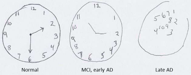
Delirium: an acute brain failure with a cause you can usually find
Delirium is an acute, fluctuating disturbance of attention and awareness caused by a physiologic insult. It is not a psychiatric diagnosis and it is not a personality problem; it is organ failure of the brain, and like any organ failure it has a cause. It occurs in roughly 20 to 30 percent of hospitalised older medical patients, in more than half of patients after hip fracture repair, and in the majority of ventilated intensive-care patients. It is associated with longer stays, higher mortality, new nursing-home placement and lasting cognitive decline — which means preventing and treating it is not a comfort measure, it is life-saving care.
The bedside tool is the Confusion Assessment Method. Delirium is present when feature 1 and feature 2 are present, plus either feature 3 or feature 4. Feature 1 is acute onset with a fluctuating course — ask the family or the previous shift whether this is different from two days ago. Feature 2 is inattention, tested by asking the patient to recite the months of the year backwards or to spell a five-letter word backwards. Feature 3 is disorganised thinking — rambling, illogical, unpredictable. Feature 4 is an altered level of consciousness — anything other than alert, whether hypervigilant, drowsy, stuporous or comatose. Inattention is the hallmark: a patient who cannot hold the thread of your sentence is delirious until proved otherwise.
Delirium has three motor subtypes. Hyperactive delirium — agitated, pulling at lines, climbing out of bed — is obvious and gets attention. Hypoactive delirium — quiet, withdrawn, sleepy, slow to answer, described in report as “pleasant, no problems” — is the most common form in older adults, is missed most of the time, and carries the worse prognosis. Mixed delirium alternates between the two, which is why a single reassuring assessment at 1400 does not rule delirium out. If you screen only the patients who bother you, you will find only the minority of cases.
Finding the cause, and treating without sedating
Delirium is a symptom, so the work is a search. The mnemonic DELIRIUM organises it: Drugs, especially anticholinergics, benzodiazepines, opioids and sedative-hypnotics; Electrolytes, particularly sodium, calcium and glucose; Lack of drugs — withdrawal from alcohol, benzodiazepines or nicotine, or untreated pain; Infection; Reduced sensory input — missing glasses and hearing aids, windowless rooms, darkness; Intracranial events including stroke, subdural haematoma and seizure; Urinary and faecal retention; and Myocardial and pulmonary causes — hypoxia, myocardial infarction, heart failure, pulmonary embolism.
Your first-pass workup is fast and mostly at the bedside: full vital signs including an accurate temperature, oxygen saturation, point-of-care glucose, a pain assessment, a medication review looking specifically at anything new or newly increased in the last 48 hours, a bladder scan for retention, and a check for impaction. Then send basic chemistry, calcium, magnesium, complete blood count, urinalysis with culture if there are urinary symptoms, and an electrocardiogram. Neuroimaging is not routine; obtain a head computed tomography scan when there are focal neurological signs, a fall or head strike, anticoagulation, or when the search comes up empty.
Treatment is cause-directed plus a non-pharmacological bundle that has been shown to prevent roughly a third of cases. Reorient at every contact, using a visible clock, calendar and whiteboard. Restore sensory input — glasses on the face, hearing aids in with working batteries, dentures in. Mobilise early and often; a patient who walks does not deteriorate the way a patient in bed does. Remove tethers: unnecessary urinary catheters, telemetry leads, continuous pulse oximetry, intravenous lines and restraints all anchor the patient to the bed and worsen delirium. Protect sleep with lights on by day, dark and quiet at night, and vital signs clustered so the patient is not woken at 0200 for a blood pressure that could wait. Treat pain adequately — undertreated pain causes far more delirium than scheduled analgesia does. Involve family; a familiar voice at the bedside is a therapeutic intervention.
Medication is a last resort, used only when agitation threatens the patient’s safety or an essential treatment, and never as a substitute for finding the cause. When it is unavoidable, low-dose haloperidol (0.25 to 0.5 mg) is the usual choice, with attention to the QT interval — hold and escalate if the corrected QT exceeds 500 ms — and to extrapyramidal effects. Haloperidol and other typical antipsychotics are avoided in Parkinson disease and Lewy body dementia, where patients can have severe, sometimes fatal, neuroleptic sensitivity; quetiapine is preferred there. All antipsychotics carry a boxed warning for increased mortality in older adults with dementia-related psychosis. Benzodiazepines cause delirium and should not be used to treat it, with one exception: alcohol or benzodiazepine withdrawal, where symptom-triggered lorazepam guided by a CIWA protocol is the correct therapy.
Dementia and depression, and the mistakes that follow confusing them
Chapter 5 covered the neurodegenerative diseases themselves. What matters here is discrimination and care. Dementia is a chronic, insidious, progressive decline in two or more cognitive domains that impairs independent function, with a normal level of consciousness and preserved attention until late in the disease. A patient with moderate Alzheimer disease can look at you, attend to your question and give you a wrong answer confidently; a delirious patient cannot hold the question in mind long enough to be wrong. Alzheimer disease begins with recent-memory loss; vascular dementia declines in steps with focal signs; Lewy body dementia brings fluctuating cognition, visual hallucinations, parkinsonism and dream enactment; frontotemporal dementia begins with personality, behaviour or language change, often before sixty-five.
Depression in the older adult is frequently missed because it presents as somatic complaint, apathy, poor concentration and memory difficulty — a picture once called pseudodementia. The distinguishing features are a subacute onset over weeks, intact attention, and a patient who is distressed by memory problems and answers “I don’t know” rather than confabulating. Screen with the fifteen-item Geriatric Depression Scale, where a score of 5 or more is a positive screen, or the PHQ-9. Then take suicide risk seriously: men aged 75 and older have the highest suicide rate of any group in the United States, most have seen a healthcare provider within the preceding month, and lethality is high because means are lethal. Ask directly and plainly. Asking does not plant the idea.
Treatment favours a selective serotonin reuptake inhibitor such as sertraline or escitalopram, started at half the usual adult dose and titrated slowly; response may take six to twelve weeks. Check serum sodium two to four weeks after starting, because SSRI-induced hyponatraemia from syndrome of inappropriate antidiuretic hormone is common in this population and presents as — predictably — confusion and falls. Avoid paroxetine (strongly anticholinergic) and tricyclic antidepressants. Remember that all three D’s coexist: delirium is superimposed on dementia in a large share of hospitalised patients, and depression is common in both. The clinical question is never which one, but what has changed acutely.
| Feature | Delirium | Dementia | Depression |
|---|---|---|---|
| Onset | Hours to days — acute | Months to years — insidious | Weeks to months — subacute |
| Course | Fluctuates, worse at night | Steadily progressive (or stepwise) | Persistent low mood, diurnal variation |
| Attention | Impaired — the hallmark | Normal until late | Poor concentration, but attention intact |
| Level of consciousness | Altered — hypervigilant or drowsy | Normal | Normal |
| Memory complaint | Unable to engage with testing | Minimises or confabulates | Distressed; answers “I don’t know” |
| Perception | Illusions and hallucinations common | Late, or early in Lewy body disease | Usually absent |
| Reversibility | Usually reversible if the cause is found | Not reversible | Highly treatable |
| First nursing action | Search for the physiologic cause | Support function, safety, routine | Screen, ask about suicide, refer |
Caring for the patient with established dementia in an acute setting is largely environmental. Keep routine, staff and room consistent. Give one instruction at a time, in short sentences, from the front, at eye level. Do not argue with or correct a false belief; redirect and validate the emotion behind it. Anticipate sundowning — late-afternoon agitation — by increasing light, reducing noise, toileting, offering a snack and planning activity before it starts rather than sedating after it does. Screen for the unmet need behind every behaviour: pain, hunger, thirst, a full bladder, constipation, cold, boredom, fear. Behaviour in dementia is communication, and the intervention is to answer what is being communicated.
Acute inattention with a fluctuating course is delirium until proved otherwise — a medical emergency requiring a search for hypoxia, hypoglycaemia, infection, retention, drugs and pain, treated first with reorientation, sensory aids, mobility and sleep rather than sedation.
Section 3Polypharmacy, Altered Pharmacology and Safer Prescribing
What the ageing body does to the drug
Absorption changes least. Gastric acid falls and transit slows, which mostly alters the rate rather than the extent of absorption — but achlorhydria genuinely impairs absorption of vitamin B12, calcium carbonate and iron, and reduced first-pass metabolism can substantially raise the bioavailability of drugs like propranolol, morphine and nitrates.
Distribution changes a great deal. Total body water falls from about 60 percent of body weight to roughly 50 percent in men and 45 percent in women, while fat mass rises. Water-soluble drugs — digoxin, gentamicin, lithium, ethanol — therefore have a smaller volume of distribution and reach higher peak concentrations from the same dose. Fat-soluble drugs — diazepam, amiodarone, most anaesthetic agents — have a larger volume of distribution and a much longer half-life, so they accumulate. Serum albumin (normal 3.5 to 5.0 g/dL) often falls with illness, leaving a larger free, active fraction of highly protein-bound drugs such as warfarin, phenytoin and naproxen — which is why a “therapeutic” phenytoin level in a patient with an albumin of 2.4 g/dL may in fact be toxic.
Metabolism slows selectively. Reduced hepatic blood flow and reduced phase I oxidation prolong the half-life of drugs cleared that way; phase II glucuronidation holds up. Excretion is the biggest single issue: falling glomerular filtration prolongs the half-life of every renally cleared drug — digoxin, most antibiotics, gabapentin, metformin, direct oral anticoagulants, morphine metabolites, allopurinol, methotrexate. Finally, pharmacodynamics change: the ageing brain is more sensitive to opioids, benzodiazepines and anticholinergics at any given concentration, while beta-receptor responsiveness to both agonists and blockers declines. The practical rule is start low and go slow — but go. Undertreated pain, untreated depression, unanticoagulated atrial fibrillation and untreated osteoporosis harm older adults just as reliably as overtreatment does.
Polypharmacy, the prescribing cascade and the Beers Criteria
Polypharmacy is usually defined as five or more regular medications, and hyperpolypharmacy as ten or more. Risk of an adverse drug event climbs steeply with each addition, and adverse drug events account for a substantial share of older-adult emergency admissions — most commonly from anticoagulants, antiplatelets, insulins, oral hypoglycaemics and opioids. The critical clinical habit is to treat every new symptom in an older adult as a possible drug effect until proved otherwise.
That habit prevents the prescribing cascade, in which an adverse effect is misread as a new disease and treated with another drug. The classic sequences are worth memorising: metoclopramide causes parkinsonism, which earns a levodopa prescription; amlodipine causes ankle oedema, which earns furosemide; a cholinesterase inhibitor causes urinary urgency, which earns oxybutynin, which causes confusion; an NSAID raises blood pressure, which earns an antihypertensive. Each cascade adds cost, interaction risk and a new set of adverse effects, and each one is undone by asking what the patient was started on shortly before the new symptom appeared.
The American Geriatrics Society Beers Criteria list medications that are potentially inappropriate in adults aged 65 and older. “Potentially inappropriate” means the risk usually exceeds the benefit and an alternative usually exists — not that the drug is forbidden. Complementary tools are the STOPP criteria (drugs to stop) and START criteria (drugs wrongly omitted, such as anticoagulation in atrial fibrillation or bisphosphonates after a fragility fracture).
| Drug or class | Why it is a problem in older adults | Safer direction |
|---|---|---|
| First-generation antihistamines (diphenhydramine, hydroxyzine) | Strongly anticholinergic; confusion, retention, falls; the hidden ingredient in “PM” sleep aids | Cetirizine or loratadine; sleep hygiene, not a pill |
| Benzodiazepines (all) | Falls, hip fracture, delirium, prolonged sedation; long-acting agents accumulate for days | Non-drug insomnia and anxiety management; taper existing use slowly |
| “Z-drugs” (zolpidem, eszopiclone, zaleplon) | Same fall and delirium profile as benzodiazepines, plus complex sleep behaviours | Sleep hygiene, daytime light and activity |
| Antipsychotics for dementia-related behaviour | Boxed warning: increased mortality and stroke; do not treat wandering or calling out | Identify and meet the unmet need driving the behaviour |
| Tricyclic antidepressants (amitriptyline, imipramine) | Anticholinergic, orthostatic, arrhythmogenic | SSRI or SNRI; duloxetine for neuropathic pain |
| Skeletal muscle relaxants (cyclobenzaprine, methocarbamol, carisoprodol) | Sedation, anticholinergic effects, falls; poorly effective at tolerated doses | Heat, physiotherapy, topical agents, scheduled acetaminophen |
| Chronic NSAIDs (ibuprofen, naproxen) | Gastrointestinal bleeding, acute kidney injury, fluid retention, heart failure and hypertension worsening | Acetaminophen up to 3 g/day as the analgesic base; topical NSAID |
| Ketorolac (oral or parenteral) | Avoid entirely, at any duration, in adults 65 and older — high rates of gastrointestinal bleeding, peptic ulceration and acute kidney injury even with a short course, and a “normal” creatinine can hide a clearance near 30 mL/min | Acetaminophen or a topical NSAID; a renally dose-adjusted opioid for severe short-term pain |
| Long-acting sulfonylureas (glyburide, chlorpropamide) | Prolonged, sometimes fatal hypoglycaemia that presents as confusion or a fall | Glipizide if a sulfonylurea is needed; relax the HbA1c target |
| Oxybutynin (immediate-release) and other bladder anticholinergics | Confusion, dry mouth, constipation, retention | Timed voiding, pelvic floor training; mirabegron if drug therapy is needed |
| Digoxin above 0.125 mg/day | Narrow therapeutic index, renally cleared, toxicity at “normal” creatinine | Lower dose, monitor level and potassium |
| Meperidine | Normeperidine accumulates in renal impairment causing tremor, myoclonus and seizures | Any other opioid, renally dose-adjusted |
| Proton pump inhibitors beyond 8 weeks without indication | C. difficile, fracture, hypomagnesaemia, B12 deficiency | Reassess indication; step down or stop |
| Nitrofurantoin with creatinine clearance under 30 mL/min | Inadequate urinary concentration plus systemic toxicity | Culture-directed alternative |
| Sliding-scale insulin as the sole regimen | Chases hyperglycaemia, produces swings and hypoglycaemia | Basal plus correction, individualised targets |
Monitoring, adherence and the nurse’s part in deprescribing
Some drugs demand active nursing surveillance in this population. Digoxin has a therapeutic range of 0.5 to 2.0 ng/mL, with a target of 0.5 to 0.9 ng/mL in heart failure. Toxicity presents as anorexia, nausea, visual halos or yellow-green colour disturbance, confusion, bradycardia and arrhythmias, and hypokalaemia potentiates it — so keep potassium between 3.5 and 5.0 mEq/L and be alert whenever a loop diuretic is added. Warfarin requires an INR of 2.0 to 3.0 for most indications and interacts with almost everything, including antibiotics and dietary change. Direct oral anticoagulants need renal dose adjustment, and the reduction rule is indication-specific: in nonvalvular atrial fibrillation apixaban is reduced to 2.5 mg twice daily when two of three criteria are met — age 80 or older, weight 60 kg or less, or serum creatinine 1.5 mg/dL or higher. Those criteria do not apply to venous thromboembolism treatment, which is 10 mg twice daily for 7 days and then 5 mg twice daily regardless of age, weight or creatinine. Metformin is contraindicated below an estimated glomerular filtration rate of 30 mL/min/1.73 m². Opioids require a scheduled bowel regimen from day one, not on request.
Adherence in this population is a mechanical problem as often as a motivational one. Ask to see the bottles — the “brown-bag review” — including over-the-counter products, herbals and anything belonging to a spouse. Check whether the patient can open the container, read the label, split the tablet, afford the refill, get to the pharmacy and remember the schedule. Simplify: once-daily dosing where possible, one pharmacy, a pill organiser filled weekly, alarms, and a single written schedule in large print. Ask about cost-related non-adherence directly, because patients rarely volunteer that they are splitting tablets to make a prescription last.
Deprescribing is a team activity that the nurse frequently initiates. For each drug, ask what the indication is, whether it is still present, whether the benefit will be realised within the patient’s remaining life expectancy and goals, whether a safer option exists, and whether a taper is needed. Benzodiazepines, opioids, beta-blockers, gabapentinoids, corticosteroids, proton pump inhibitors and antidepressants all require gradual tapering rather than abrupt discontinuation. The nurse’s specific contribution is the observation: you are the one who sees the patient sway on standing after the new alpha-blocker, sleep through breakfast after the new hypnotic, and stop eating after the new drug for something else.
In an older adult, treat any new symptom as an adverse drug effect until proved otherwise — this single habit prevents prescribing cascades and catches the falls, confusion, retention and hypoglycaemia that medications, not diseases, are causing.
Section 4Falls, Frailty and the Hazards of Hospitalisation
Falls: predicting them, preventing them, and what to do after one
About one in four adults aged 65 and older falls each year, and falls are the leading cause of injury-related death in that age group. The consequences run beyond the fracture: a fall generates fear of falling, which produces activity restriction, which produces deconditioning, which produces more falls. Hospitalisation is itself a fall risk factor because it combines an unfamiliar environment, new sedating drugs, tethering equipment, acute illness and a bed that is the wrong height.
The strongest single predictor of a fall is a previous fall, so ask everyone, on every admission, whether they have fallen in the past year and whether they feel unsteady or fear falling. Then assess the modifiable contributors: orthostatic hypotension, psychoactive and cardiovascular medications, vision, footwear, continence and urgency, cognitive impairment, peripheral neuropathy, pain, vitamin D status and environment. Take orthostatic vital signs properly — supine for five minutes, then repeat at one and three minutes of standing, recording symptoms as well as numbers. Use a validated risk tool such as the Morse Fall Scale or Hendrich II, and add a performance test: the Timed Up and Go asks the patient to rise from a chair, walk three metres, turn, return and sit, and a time of 12 seconds or more indicates increased fall risk.
Prevention on the unit is unglamorous and effective. Round hourly using the four P’s — pain, potty, position, possessions — because most falls happen on the way to the bathroom. Keep the bed low and locked with the call light, phone, water, glasses and assistive device in reach. Provide non-skid footwear, adequate lighting with a night light, a clear path and a raised toilet seat. Offer scheduled toileting rather than waiting for urgency. Review the medication list for anything sedating, hypotensive or anticholinergic. Ensure vitamin D repletion and refer to physiotherapy for strength and balance training, which has the best evidence of any single intervention. Bed and chair alarms have not been shown to reduce falls and are no substitute for a person.
After a fall, assess before you move the patient. Check responsiveness, airway, breathing and circulation, then take a full set of vital signs including orthostatics when it is safe, perform a neurological check, and examine for pain, deformity, and a shortened, externally rotated leg suggesting hip fracture. Establish whether the head was struck and whether the patient is anticoagulated — an anticoagulated patient who strikes the head needs imaging and observation even if they look completely well, because a subdural haematoma can declare itself hours to weeks later. Notify the provider and the family, document objectively what was found, and complete the incident report as a separate quality document that is not referenced in the medical record. Then ask why it happened, because the fall you investigate is the second one you prevent.
Immobility, sarcopenia and nutrition
Bed rest is a treatment with a dose-dependent toxicity. An older adult loses roughly one to three percent of muscle strength for every day of immobility, and the losses accelerate under acute illness. Within days you also see orthostatic intolerance, atelectasis, venous thromboembolism risk, constipation and impaction, insulin resistance, bone resorption with hypercalciuria, pressure injury and delirium. A patient admitted for pneumonia who walks twice a shift goes home; one who stays in bed for the same pneumonia goes to a skilled nursing facility. Mobility is not the physiotherapist’s job alone — it is nursing care, and it belongs on the plan every shift.
Sarcopenia — the age-related loss of muscle mass, strength and performance — is the substrate. It is worsened by immobility, protein deficiency, inflammation and untreated illness, and improved by resistance exercise plus adequate protein. Healthy older adults need about 1.0 to 1.2 g/kg/day of protein, and 1.2 to 1.5 g/kg/day during acute illness or wound healing — substantially more than the general adult recommendation, and something to advocate for when a patient is sitting in front of a clear-liquid tray for a third day.
Malnutrition is common and under-detected. Unintentional loss of 5 percent of body weight in one month or 10 percent in six months is significant. Screen with a validated tool, weigh the patient on the same scale at the same time, and record actual intake as a percentage rather than “ate well.” Be careful with laboratory shortcuts: albumin and prealbumin fall with inflammation and are markers of illness severity, not of nutritional status, in an acutely ill patient. Address the causes you can fix — ill-fitting dentures, dry mouth, dysphagia, depression, constipation, unappetising restricted diets, being fed by a rushed person, eating alone. Liberalising a diet is often the highest-yield nutrition intervention in a frail older adult.
Dehydration deserves separate attention because the older adult has both a reduced thirst drive and a reduced ability to concentrate urine. Weight is the most reliable monitor — a change of 1 kg equals roughly 1 L of fluid. Look for orthostatic hypotension, a dry axilla, longitudinal tongue furrows, urine output below 0.5 mL/kg/hr, and a BUN-to-creatinine ratio above 20:1. Skin turgor is unreliable in aged skin; if you use it, test over the sternum or forehead rather than the back of the hand. Offer fluids proactively on a schedule, because a patient who is not thirsty will not ask.
Skin, continence and the geriatric syndromes that end independence
Pressure injury risk rises with thinning subcutaneous tissue, reduced mobility, incontinence, poor nutrition and reduced sensation. Screen with the Braden Scale, which scores six subscales for a total of 6 to 23 — 18 or below indicates risk in an older adult, and 12 or below indicates high risk. Prevention is mechanical and relentless: reposition on a schedule, use a 30-degree lateral tilt rather than direct trochanteric pressure, keep the head of the bed at or below 30 degrees where clinically permitted to limit shear, float the heels off the bed entirely, use a pressure-redistributing surface, manage moisture with a barrier product, and feed the patient. Inspect the skin under every device — oxygen tubing, cervical collars, catheters, compression sleeves — because device-related injury is a large share of what occurs on med-surg floors. Handle aged skin gently: use silicone dressings rather than adhesive tape, lift rather than drag, and use a slide sheet to prevent skin tears.
Urinary incontinence is not normal ageing and should never be managed by simply applying a brief. Identify the type. Stress incontinence leaks with cough or laughter and responds to pelvic floor training. Urge incontinence is a sudden compelling need and responds to timed voiding and bladder retraining. Overflow incontinence is constant dribbling from retention — confirm with a bladder scan, where a post-void residual under 50 mL is normal and one over 200 mL is abnormal. Functional incontinence means the bladder works but the person cannot reach the toilet in time, which is a mobility, clothing, lighting or staffing problem. Avoid indwelling catheters, which cause infection, tethering, delirium and deconditioning; if one is in place, ask on every shift whether it can come out today.
Two more syndromes belong on your radar. Sensory impairment — presbycusis, the high-frequency sensorineural hearing loss of ageing, and presbyopia and cataract — masquerades as cognitive impairment and worsens delirium; the fix is glasses, working hearing aids, a pocket amplifier and good lighting. And constipation, which is so ordinary it is ignored, causes delirium, urinary retention, anorexia, abdominal pain and, in impaction, overflow diarrhoea that gets mistaken for infection. Track bowel movements deliberately and act at 48 hours rather than at five days.
| Screen or tool | What it measures | Threshold that changes your plan |
|---|---|---|
| Timed Up and Go | Functional mobility and fall risk | 12 seconds or more indicates increased fall risk |
| Orthostatic vital signs | Blood pressure response to standing | Drop of 20 mmHg systolic or 10 mmHg diastolic within 3 minutes |
| Braden Scale | Pressure injury risk (total 6–23) | 18 or below at risk; 12 or below high risk |
| Mini-Cog | Cognitive screen: three-word recall plus clock draw | Score of 0–2 out of 5 is a positive screen for impairment |
| Geriatric Depression Scale (15-item) | Depressive symptoms | 5 or more is a positive screen |
| Confusion Assessment Method | Delirium | Features 1 and 2, plus 3 or 4 |
| Post-void residual (bladder scan) | Urinary retention | Under 50 mL normal; over 200 mL abnormal |
| Katz ADL / Lawton IADL | Baseline and current function | Any new dependency is an acute finding |
Hospitalisation itself injures older adults through bed rest, tethers, sedatives and missed meals, so mobility, continence, skin, nutrition, hydration and sleep are not comfort tasks — they are the interventions that decide whether the patient goes home or to a facility.
Section 5Elder Mistreatment, Caregivers and the Safe Transition Home
Recognising elder mistreatment
Elder mistreatment affects roughly one in ten community-dwelling older adults each year, and only a small fraction of cases is ever reported. It takes six forms. Physical abuse is the infliction of injury or inappropriate restraint or drugging. Sexual abuse is non-consensual contact of any kind, including with a person who cannot consent. Psychological or emotional abuse includes threats, humiliation, isolation and controlling contact with others. Neglect is the failure of a responsible caregiver to provide food, hydration, hygiene, shelter, medication or medical care, and is the most commonly substantiated form. Abandonment is desertion by a caregiver. Financial exploitation — misuse of funds, coerced signatures, new joint accounts, unpaid bills despite adequate income — is the most common form overall and among the least reported. Self-neglect, in which a person cannot or does not maintain their own safety, health or hygiene, is the single largest category referred to Adult Protective Services in many states.
Risk rises with cognitive impairment, functional dependence, social isolation and shared living arrangements, and with a caregiver who has substance use, mental illness, or financial dependence on the older adult. The physical red flags are injuries that do not match the stated mechanism, delayed presentation, bruising in atypical locations such as the inner arms, neck, ears, or genital area, patterned or bilateral injuries, repeated emergency visits to different facilities, and malnutrition, dehydration, poor hygiene, untreated pressure injuries or medication non-adherence that has no medical explanation. The behavioural red flags are just as important: a caregiver who answers for the patient, refuses to leave the room, minimises or contradicts the patient, or is hostile toward staff; a patient who is fearful, withdrawn, watches the caregiver before answering, or apologises constantly.
Your action is defined and non-negotiable. Interview the patient alone. Create a legitimate reason to separate them — a scan, a bath, a specimen — and ask directly, calmly and without judgement: has anyone hurt you, threatened you, touched you in a way you did not want, taken your money, or failed to help you when you needed it? Do you feel safe where you live? Document objectively: the patient’s own words in quotation marks, a body map, measurements, photographs per facility policy, and the caregiver’s behaviour as observed rather than interpreted. Ensure immediate safety first — treat injuries, and do not discharge into a suspected unsafe situation. Then report. In most United States jurisdictions nurses are mandated reporters; the threshold is reasonable suspicion, not proof, and reports go to Adult Protective Services, with law enforcement for immediate danger. You do not investigate, and you do not confront the suspected abuser. A cognitively intact adult with capacity may refuse services, and you may still be required to report; the two things are separate.
Caregivers, ageism and communication that works
Most long-term care in the community is delivered unpaid by family, most often by a spouse or daughter who is herself over sixty. Caregiver strain is a clinical problem with clinical consequences: depression, worsened physical health, and a strong association with both nursing-home placement and mistreatment. Screen for it — the Modified Caregiver Strain Index and the Zarit Burden Interview take minutes — and ask the caregiver directly how they are sleeping, whether they get any time away and whether they feel they can keep doing this. Then connect them to something real: respite care, adult day programmes, home health aides, meal delivery, area agency on ageing services, support groups and social work. Teaching the caregiver a safe transfer technique is as much a nursing intervention as teaching the patient a medication.
Communication has to be adapted deliberately, and adaptation is not the same as condescension. Avoid elderspeak — the sing-song, high-pitched, diminutive-laden speech (“sweetie,” “honey,” “are we ready for our bath?”) that measurably increases resistance to care and communicates that the person is not an adult. Address patients by their title and surname unless invited otherwise, speak to the patient rather than the family member, and do not assume confusion until you have tested for it. For presbycusis, lower your pitch instead of raising your volume, face the patient in good light so lips are visible, reduce background noise, and confirm hearing aids are in and switched on. For vision, use printed material in at least 14-point sans-serif type with high contrast, avoid glare, and make sure glasses are clean and on.
Teach in small pieces. Cover one topic at a time, put the most important information first and repeat it last, use plain language rather than clinical vocabulary, and verify with teach-back: “I want to be sure I explained this clearly — tell me how you will take this new water pill when you get home.” A patient who nods and says “yes” has told you nothing. Include the caregiver with the patient’s permission, and give everything in writing to take home.
Transitions, discharge and knowing when to escalate
The transition out of hospital is where older adults are most often harmed. Medication discrepancies occur in a large proportion of transitions, follow-up frequently fails to happen, and readmission within thirty days is common. A safe discharge is built, not announced on the morning of departure. Perform a full medication reconciliation against the pre-admission list, name every drug that was started, stopped or changed and say why, and make sure the patient does not go home holding both the old bottle and a new one containing the same drug under a different name. Confirm that a follow-up appointment is scheduled before the patient leaves, and that transport, equipment, home health and prescriptions are actually arranged rather than merely recommended.
Discharge teaching should name specific warning signs and the number to call: the weight gain, the shortness of breath, the confusion, the fever, the bleeding, the new leg pain. Assess the home — stairs, bathroom, lighting, rugs, whether there is anyone there at night — and address driving, firearm access and alcohol use when the assessment raises them. Advance care planning is covered in depth in Chapter 14, but at minimum confirm whether an advance directive and a healthcare proxy exist, whether the documents are in the chart, and whether the family knows what they say.
Escalation follows the same priority logic as the rest of med-surg: airway, breathing and circulation first, then unstable before stable, then acute before chronic. Report with a complete SBAR. Situation: “This is the nurse on 4 West calling about Mrs. Alvarez in 412, an 84-year-old admitted yesterday with a hip fracture. She has become acutely confused in the last three hours.” Background: “She was alert and oriented on admission. She received oxycodone at 1400 and diphenhydramine for itching at 1600. She has not voided since 1100.” Assessment: “Temperature 37.9, heart rate 108, blood pressure 98/56, respirations 24, oxygen saturation 91 percent on room air, glucose 84. Bladder scan shows 620 mL. CAM is positive for acute onset, fluctuation and inattention.” Recommendation: “I think she has hypoxia and urinary retention with possible sepsis. I have put her on oxygen and I would like an order for in-and-out catheterisation, a chest film, blood cultures, a lactate and a basic metabolic panel, and I would like the diphenhydramine discontinued. Can you come and see her?”
Delegation in geriatric care follows the same boundary as everywhere else. Assistive personnel may take vital signs on stable patients, assist with hygiene, feeding, ambulation and toileting, apply non-skid footwear, reposition on a schedule, record intake and output, weigh the patient, and report observations to you. What may never be delegated is assessment, teaching, evaluation and any unstable patient. The aide can tell you that Mr. Beckett would not eat breakfast and seemed sleepy; only the registered nurse can decide whether that is depression, delirium, hypoglycaemia, a new opioid or an evolving sepsis — and only the registered nurse can act on it.
Interview the older adult alone whenever mistreatment is suspected and report on reasonable suspicion rather than proof, and treat discharge as a clinical procedure — reconciled medications, arranged follow-up, named red flags and teach-back — because the transition is where this population is most often harmed.
The chapter in one breath
Ageing does not remove function; it removes reserve. That is why acute illness in an older adult announces itself as confusion, a fall, incontinence or a refusal to eat rather than as chest pain, fever or classic dysuria, and why a normal serum creatinine can hide a creatinine clearance of 30 mL/min. Learn the systems that change — stiff arteries and blunted baroreceptors, weak cough and low reserve lungs, falling glomerular filtration and impaired concentrating ability, slowed phase I hepatic metabolism, blunted fever, fragile skin, sarcopenic muscle — and the presentations follow logically. Acute inattention with a fluctuating course is delirium, a medical emergency whose commonest causes are drugs, hypoxia, infection, retention, electrolytes and pain, and whose first-line treatment is reorientation, glasses and hearing aids, mobility, sleep and removal of tethers, not sedation. Treat every new symptom as a possible adverse drug effect, know the Beers list and the anticholinergic burden, and refuse to let a prescribing cascade start. Prevent falls, immobility, malnutrition, dehydration, pressure injury and catheter dependence, because hospitalisation is itself the hazard. Watch for mistreatment, interview alone, report on suspicion. Then discharge deliberately: reconciled medicines, arranged follow-up, red flags in writing and teach-back to prove it landed. Escalate by ABC, unstable over stable, acute over chronic — and keep assessment, teaching, evaluation and every unstable patient for yourself.
Words worth knowing
| Term | What it means |
|---|---|
| Homeostenosis | Age-related narrowing of physiologic reserve; baseline function is preserved but the capacity to respond to stress is lost |
| Frailty | A syndrome of reduced reserve across systems; the Fried phenotype requires 3 or more of weight loss, exhaustion, weak grip, slow gait and low activity |
| Sarcopenia | Age-related loss of muscle mass, strength and performance; worsened by bed rest and protein deficiency, improved by resistance exercise |
| Atypical presentation | Acute illness announcing itself as confusion, a fall, incontinence or anorexia rather than the classic symptom — the rule, not the exception, in this population |
| Orthostatic hypotension | Fall of 20 mmHg or more systolic, or 10 mmHg or more diastolic, within 3 minutes of standing |
| Delirium | Acute, fluctuating disturbance of attention and awareness from a physiologic cause; reversible when the cause is found |
| Hypoactive delirium | The quiet, withdrawn, drowsy subtype — the most common form in older adults, the most often missed, and the one with the worse prognosis |
| Confusion Assessment Method | Bedside delirium tool requiring acute onset with fluctuation plus inattention, and either disorganised thinking or altered consciousness |
| Sundowning | Late-afternoon and evening agitation in dementia; prevented with light, routine, toileting, food and activity rather than treated with sedation |
| Apathetic thyrotoxicosis | Hyperthyroidism in the older adult presenting as weight loss, atrial fibrillation, heart failure and withdrawal instead of agitation and heat intolerance |
| Polypharmacy | Five or more regular medications; each addition raises the risk of interaction, adverse event, falls and non-adherence |
| Prescribing cascade | An adverse drug effect misdiagnosed as a new disease and treated with another drug — for example amlodipine oedema treated with furosemide |
| Beers Criteria | American Geriatrics Society list of potentially inappropriate medications in adults aged 65 and older; risk usually exceeds benefit and an alternative exists |
| Anticholinergic burden | The additive effect of multiple anticholinergic drugs, producing dry mouth, blurred vision, constipation, urinary retention, tachycardia, confusion and falls |
| Cockcroft–Gault equation | Estimates creatinine clearance from age, weight, sex and serum creatinine; required because normal creatinine hides low clearance when muscle mass is low |
| Timed Up and Go | Rise, walk 3 metres, turn, return and sit; 12 seconds or more indicates increased fall risk |
| Braden Scale | Pressure injury risk score of 6 to 23; 18 or below indicates risk in an older adult, 12 or below high risk |
| Presbycusis | Age-related high-frequency sensorineural hearing loss; lower your pitch rather than raising your volume |
| Elder mistreatment | Physical, sexual or psychological abuse, neglect, abandonment or financial exploitation of an older adult; reportable on reasonable suspicion |
| Capacity versus competence | Capacity is a clinical, decision-specific judgement of understanding, appreciation, reasoning and communication; competence is a global legal status set by a court |
Check yourself
Rehabilitation, Mobility and Chronic Illness
Rehabilitation begins the hour the crisis is survived, not the day the patient is discharged to a rehab unit. The stroke is treated in minutes, the femur is nailed in hours — and then somebody has to teach a frightened adult to stand, to empty a bladder that no longer signals, to eat with the hand that still works, to be a parent from a wheelchair. That work is nursing work, and it is graded on function rather than on cure. This chapter gives you the practical spine of it: how the rehabilitation team divides labour, how function is measured with tools you will actually chart in, how to transfer a patient without injuring either of you, which gait pattern belongs to which device, what a person with a C6 injury can and cannot do, and how to recognise autonomic dysreflexia fast enough to prevent a stroke. It ends where chronic illness actually lives — in the patient’s house, in their marriage, in their bank account.
By the end of this chapter you can
- Describe the roles of the interdisciplinary rehabilitation team and the criteria distinguishing an inpatient rehabilitation facility from a skilled nursing facility, home health and outpatient therapy.
- Perform and document a functional assessment using ADLs, IADLs, the Barthel Index, the FIM/Section GG scale and the Timed Up and Go, and translate the score into a nursing goal.
- Execute a safe transfer using a gait belt, correct body mechanics and safe patient handling equipment, and state the manual lifting limit and the axilla rule.
- Fit a cane, walker and crutches correctly and teach the four-point, two-point, three-point and swing-through gaits plus the stair rule — up with the good, down with the bad.
- Predict retained function by spinal cord injury level, distinguish spinal shock from neurogenic shock, and classify injuries using the ASIA Impairment Scale and the incomplete cord syndromes.
- Recognise autonomic dysreflexia and carry out the intervention sequence in order — sit the patient up first, then remove the trigger — with correct SBAR escalation.
- Design neurogenic bladder and neurogenic bowel programmes for upper and lower motor neuron injury, including intermittent catheterisation volumes and digital stimulation technique.
- Prevent contractures, venous thromboembolism, pressure injury, orthostatic hypotension and disuse osteoporosis, and manage spasticity drugs with their monitoring parameters.
- Support psychosocial adjustment to chronic illness and disability, apply self-management and teach-back teaching, and state which rehabilitation tasks may and may not be delegated.
Section 1What Rehabilitation Is, and How Function Is Measured
The team, the goal and the setting
Acute care asks “what is wrong and how do we fix it?” Rehabilitation asks a harder question: “given what cannot be fixed, what can this person still do, and what has to change so they can do it?” The World Health Organization’s International Classification of Functioning frames disability not as a property of a body but as the gap between what a body can do and what an environment demands. A T10 paraplegic on a level campus with curb cuts is far less disabled than the same person in a third-floor walk-up. That is not a philosophical aside — it is why a rehabilitation nurse spends as much time on the discharge environment as on the patient’s quadriceps.
The rehabilitation team is genuinely interdisciplinary, meaning the disciplines set one shared goal rather than five parallel ones. The physiatrist (physical medicine and rehabilitation physician) leads medically. Physical therapy owns gross mobility, gait, transfers, strength and endurance. Occupational therapy owns the upper extremity, activities of daily living, adaptive equipment, cognition applied to task, and home modification. Speech-language pathology owns swallowing, speech, language and cognitive-communication. The rehabilitation nurse — the certified credential is CRRN — is the one discipline present for all twenty-four hours, and the job is to carry therapy into life: the transfer PT taught at ten in the morning must happen the same way at two in the morning, or it is not learned. Add the social worker or case manager (funding, placement, benefits), the dietitian, the psychologist, the chaplain, the vocational counsellor and the recreational therapist, and you have the weekly team conference.
Where the patient goes after acute care is a matter of tolerance and skilled need, not of enthusiasm. An inpatient rehabilitation facility requires that the patient can participate in and benefit from about three hours of therapy a day, five days a week, from at least two therapy disciplines, with physician oversight several days a week and a formal weekly team conference. If the patient cannot sit up for ninety minutes at a stretch, they cannot yet do IRF. A skilled nursing facility delivers a lower dose — often one to two hours a day — and is the right answer for the deconditioned eighty-eight-year-old after a hip fracture who needs time more than intensity. Long-term acute care takes the ventilator weans and complex wounds. Home health and outpatient therapy carry the tail end. Getting this recommendation right at the team conference is one of the most consequential things a nurse contributes, because the level of care determines the ceiling of recovery.
| Setting | Therapy dose | Typical candidate | Nursing focus |
|---|---|---|---|
| Inpatient rehabilitation facility (IRF) | About 3 h/day, 5 days/week; two or more disciplines | Stroke, spinal cord injury, brain injury, amputation, multiple trauma, hip fracture with good potential | Carry-over of therapy techniques 24/7; bowel and bladder programmes; skin; family training |
| Skilled nursing facility (SNF) | Roughly 1–2 h/day as tolerated | Deconditioned older adult, slower progress, medically fragile, lower endurance | Restorative nursing, fall prevention, nutrition, medication reconciliation |
| Long-term acute care (LTACH) | Variable; medically driven | Ventilator wean, complex wounds, prolonged IV therapy | Airway, secretion clearance, wound care, gradual mobility |
| Home health | Two to three visits/week | Homebound, has a caregiver, safe home environment | Environmental assessment, caregiver teaching, medication safety |
| Outpatient / day rehab | Two to five visits/week | Independent transport, community re-entry, return to work | Reinforcement, self-management, vocational goals |
ADLs, IADLs and the scored tools
Activities of daily living are the six survival tasks of the body: bathing, dressing, toileting, transferring, continence and feeding — the Katz index. Instrumental activities of daily living are the eight tasks of running a household and a life: using a telephone, shopping, preparing food, housekeeping, laundry, arranging transportation, managing medications and managing finances — the Lawton scale. The distinction matters clinically because IADLs fail first. The earliest reliable sign of cognitive decline in an older adult is usually not forgetting a name; it is unopened mail, a bounced cheque, a pill organiser that no longer matches the bottles. Ask about IADLs explicitly and ask the family separately, because patients systematically over-report independence.
Three scored tools appear on the chart and on the exam. The Barthel Index scores ten ADL items in increments of five to a total of 100; higher is more independent, and a perfect 100 means independent in those ten tasks, not that the person can safely live alone. The Functional Independence Measure (FIM) scores eighteen items — thirteen motor and five cognitive — from 1 to 7 for a total of 18 to 126. Learn the anchors: 7 is complete independence, 6 modified independence (device, extra time or safety consideration but no helper), 5 supervision or set-up only, 4 minimal assistance (the patient does 75 percent or more of the effort), 3 moderate assistance (patient does 50 to 74 percent), 2 maximal assistance (patient does 25 to 49 percent), 1 total assistance (patient does under 25 percent). The line between 5 and 4 is the line between hands-off and hands-on, and it is where reimbursement, staffing and discharge safety all pivot. United States rehabilitation facilities now report the six-level Section GG self-care and mobility items on the assessment instrument, which uses the same logic with different numbers; whichever tool your unit uses, score what you saw, not what the patient claims and not what the last shift wrote.
Mobility and balance get their own quick screens. The Timed Up and Go — rise from a standard armchair, walk three metres, turn, return, sit — is normal under about ten seconds, and twelve seconds or more flags increased fall risk. The 30-second chair stand and the four-stage balance test round out the fall screen. The Berg Balance Scale runs 0 to 56, with scores under about 45 associated with falls. The 6-minute walk test measures endurance and is the one that changes most visibly over a rehab stay. Do not administer any of these to a patient who has not yet been cleared for weight bearing, and never do a first TUG alone.
Goals, carry-over and what you may delegate
Rehabilitation goals are written the way objectives are written: measurable, time-bound, and owned by the patient. “Patient will improve mobility” is not a goal. “By 14 March, patient will transfer bed to wheelchair with stand-by assist of one and a gait belt, four out of five attempts, verbalising the sequence unprompted” is. Then you make the environment enforce it: the wheelchair parked on the strong side, the call bell within reach of the working hand, the urinal where a C7 patient can actually get it. Learned helplessness is iatrogenic. Every time a rushed nurse does for a patient what the patient could do in four extra minutes, a little function is taken back.
Delegation in rehabilitation is a frequent test point because the tasks look routine and are not. Assistive personnel may ambulate and transfer a stable patient with a device the patient has already been taught, perform passive and active range of motion after the RN has instructed them, position and turn, apply compression stockings, feed a patient with no swallowing precautions, weigh, measure intake and output, and report what they observe. The RN retains assessment, teaching, evaluation and any unstable patient — that includes the first ambulation after any change in status, medication, surgery or neurological event; the initial gait training and device fitting; evaluating whether the patient tolerated the activity; and every part of the response to autonomic dysreflexia. A useful bedside test: if the task requires nursing judgement about whether it is safe to continue, it is not delegable.
Older adults deserve a specific note here. Muscle strength falls roughly one to one-and-a-half percent per day of bed rest, and faster during critical illness. A previously independent eighty-year-old admitted for pneumonia can lose the ability to rise from a toilet in five days — not from the pneumonia but from the bed. The countermeasure is unglamorous and enormously effective: get them up. Sit them in a chair for meals, remove the tethers that anchor them (unnecessary Foley catheters, telemetry, continuous pulse oximetry, IV pumps that could be saline-locked), protect sleep, and mobilise at least three times a day unless there is a written reason not to.
Rehabilitation nursing is graded in function, so assess with a scored tool, chart the exact level of assist, and remember that the round-the-clock carry-over of what therapy taught is the nurse’s unique contribution.
Section 2Safe Transfers, Gait Belts and Assistive Devices
Before the first step
Most mobility disasters are failures of the thirty seconds before the patient stood up. Run a fixed pre-mobility check every time. First, the order: what is the weight-bearing status, and is there a brace, an immobiliser or a spinal precaution? Non-weight-bearing means the limb does not touch the floor at all. Touch-down or toe-touch weight bearing means the toe may rest for balance only, roughly the weight of the leg. Partial weight bearing is a specified percentage, commonly thirty to fifty percent, which patients cannot estimate without practice on a scale. Weight bearing as tolerated lets the patient titrate by pain. Full is full. If the order is ambiguous, do not interpret it — call.
Second, the physiology. Check vital signs and check orthostatics if the patient has been down for more than a day or two, is on antihypertensives or diuretics, or has any spinal cord or autonomic involvement: measure supine, then sitting, then standing at one and three minutes. A drop of 20 mmHg systolic or 10 mmHg diastolic within three minutes of standing, or any symptomatic drop, means dangle longer and use an abdominal binder and compression stockings. Look at haemoglobin — a haemoglobin of 7 g/dL will not tolerate what 12 will. Check oxygenation, glucose (a capillary glucose of 60 mg/dL is not a walking glucose), pain control timed so the patient is comfortable but not sedated, and recent sedating or anticoagulant medications.
Third, the environment. Bed at the height where the patient’s feet are flat on the floor with hips slightly higher than knees. Brakes locked on bed and chair. Non-skid footwear on, not socks. Lines, drains and tubing gathered and long enough. Chair positioned on the patient’s strong side at roughly a forty-five degree angle so the pivot is short. Floor dry. Enough people — and enough is decided before, not during. Then tell the patient the plan in the exact words you will use: “On three, you push down on the bed with your hands and stand. One, two, three, stand.”
The transfer itself
Apply the gait belt around the waist over clothing, snug enough that only two flat fingers fit underneath, buckle to the side or front and away from any incision, ostomy, feeding tube or fresh graft. Grasp it underhand at the sides or back. The belt is a handle for guiding and for lowering — it is not a hoist, and it will not stop a full-weight fall. The single most important prohibition in this chapter after autonomic dysreflexia is this: never lift a patient by the axillae. Pulling under the arms stretches the brachial plexus, dislocates hemiplegic shoulders whose rotator cuff no longer holds the joint, and has produced permanent nerve injury. Never let the patient grab around your neck either.
Mechanics: stand facing the patient with your feet apart, one foot slightly forward, knees and hips bent, back straight, your centre of gravity low and close to theirs. Block the patient’s weak knee with your knee, or brace their feet with yours. Have the patient scoot to the edge, place both feet flat and slightly back under the knees, lean forward — nose over toes — and push up from the mattress or armrests rather than pulling on you or on the walker. Rise, pause and let blood pressure catch up, then step and turn. Do not twist your spine; move your feet. If the patient begins to fall, do not fight it. Widen your stance, pull them toward you, protect the head, and ease them down your leg to the floor.
Modern practice is not to depend on human strength at all. Safe patient handling guidance sets the maximum recommended manual lift at roughly 35 pounds under ideal conditions; almost no adult patient qualifies. Use the equipment: friction-reducing slide sheets and air-assist mattresses for lateral transfers, sit-to-stand powered lifts for the patient who has some weight-bearing and can follow directions, full-body ceiling or floor lifts for the dependent patient, and a transfer (slide) board for the seated patient with strong arms — the classic technique for spinal cord injury at C7 and below. Bariatric patients require bariatric-rated equipment and more helpers, never more effort. The nurse who injures their back does not get to help anyone tomorrow.
Canes, walkers and crutches
Fit first. For a cane, with the patient standing upright in their shoes and arms relaxed, the handgrip should sit at the level of the greater trochanter, which is roughly the wrist crease, producing about 20 to 30 degrees of elbow flexion. The cane is held in the hand opposite the affected leg, because that reproduces normal reciprocal gait and widens the base of support toward the weak side. A quad cane gives more support than a single-point cane; position the base so the two short legs sit closest to the patient and the two longer legs face away from the foot, or the patient trips over the wide side of the base. For a walker, the same wrist-crease rule applies. A standard pick-up walker gives the most stability and the slowest gait; a two-wheeled rolling walker suits the patient who cannot lift; a four-wheeled rollator with hand brakes and a seat suits the endurance-limited patient with good cognition but is not appropriate for someone who leans hard on the frame, because it will roll away.
For axillary crutches, the top pad sits two to three finger-widths — about 1.5 to 2 inches — below the axilla, with the handgrips again at wrist-crease height and 20 to 30 degrees of elbow flexion, and the crutch tips resting about 6 inches lateral and 6 inches anterior to the foot. The weight goes through the hands, never the armpit. Weight borne on the axillary pads compresses the brachial plexus and produces “crutch palsy” — wrist drop and numbness. Teach patients to check tips for wear and to keep both hands free by using a backpack.
| Device / gait | Who it is for | Sequence |
|---|---|---|
| Cane | Mild unilateral weakness, balance | Cane in hand opposite the weak leg. Cane and weak leg advance together, then strong leg |
| Walker | Bilateral weakness, poor balance, partial weight bearing | All four points down, advance walker one step length, then weak leg, then strong leg. Never on stairs; never pull on it to stand |
| Four-point gait | Weight bearing on both legs; weak or unsteady; slowest and most stable | Right crutch, left foot, left crutch, right foot — three points on the ground at all times |
| Two-point gait | Partial weight bearing both legs; faster, mimics normal walking | Right crutch with left foot together, then left crutch with right foot together |
| Three-point gait | One leg non-weight-bearing or partial (fracture, new amputation); needs strong arms | Both crutches and the affected leg advance together, then the strong leg swings through |
| Swing-to / swing-through | Bilateral lower-limb paralysis with braces | Both crutches forward, then both legs swing to the crutches (swing-to) or past them (swing-through) |
| Stairs — UP | All devices except walker | Strong leg steps up FIRST, then crutches or cane with the weak leg |
| Stairs — DOWN | All devices except walker | Crutches or cane with the weak leg go down FIRST, then the strong leg |
The mnemonic students remember at three in the morning is “up with the good, down with the bad” — the good leg goes up to heaven, the bad leg goes down to the other place. The logic is mechanical: climbing requires the leg that can generate force, so the strong leg lifts the body up; descending is controlled lowering, so the weak leg and the device absorb the descent while the strong leg controls it from above. When a handrail is available, use it — both crutches go into the opposite hand held horizontally, one hand on the rail. Teach patients to back a wheelchair down a ramp or curb and to approach the seat until the backs of both legs touch it before sitting.
When a patient falls
Falls are the most common adverse event in rehabilitation because the whole enterprise is deliberately pushing the edge of safety. Screen everyone with a validated tool (Morse, Hendrich II or STEADI), and treat the modifiable causes: orthostatic hypotension, polypharmacy — especially benzodiazepines, sedative-hypnotics, opioids, anticholinergics, antihypertensives and hypoglycaemics — vision, footwear, urinary urgency, delirium and environmental hazards. Bed alarms and non-skid socks are the least effective interventions on the list; scheduled toileting, hourly rounding and a bedside commode are among the best.
If a fall happens, the sequence is assessment before movement. Do not lift the patient up first. Assess responsiveness, airway, breathing and circulation, then check for head strike, neck or back pain, and obvious deformity or shortening and external rotation of a leg, which suggests hip fracture. Take vital signs and a neurological check on the floor. Only when you have ruled out spinal or fracture injury do you move the patient, with enough people and usually with a lift. Then notify the provider, notify the family, perform neurological checks per protocol — more urgently if the patient is anticoagulated, because a delayed intracranial bleed is the classic killer — document objectively what you found, and file the incident report separately from the chart.
Fit the device, use the gait belt at the waist and never under the axillae, remember cane opposite the weak leg and up with the good, down with the bad — and reach for a mechanical lift rather than your own back.
Section 3Spinal Cord Injury: Levels, Syndromes and Retained Function
The first hours: spinal shock and neurogenic shock
Two different phenomena share a confusing name. Spinal shock is a neurological event: immediately after cord injury, everything below the lesion goes flaccid and areflexic — no deep tendon reflexes, no bulbocavernosus reflex, atonic bladder and bowel, loss of temperature regulation. It is not a circulatory state and it tells you nothing about final prognosis, because a completely transected cord and a badly bruised one look identical during it. Spinal shock resolves over days to weeks, and the return of reflexes — classically the bulbocavernosus — marks the end. Only then can the injury be accurately classified as complete or incomplete, which is why families should be counselled that the early picture is not the final one.
Neurogenic shock is a circulatory event, and it occurs with injuries at roughly T6 and above, where sympathetic outflow is interrupted. The result is unopposed vagal tone: vasodilation with pooled blood, and — critically — the heart cannot mount a compensatory tachycardia. The presentation is hypotension with bradycardia and warm, dry, flushed skin below the lesion. Contrast that with the trauma patient’s haemorrhagic shock: hypotension with tachycardia and cool, clammy, pale skin. A trauma patient can have both at once, so never assume the low blood pressure is “just neurogenic” until bleeding is excluded. Management is measured fluid resuscitation (over-resuscitation causes pulmonary oedema in a patient with a compromised cough), a vasopressor that also supports heart rate such as norepinephrine or dopamine — avoid pure alpha-agonists such as phenylephrine, which trigger reflex bradycardia in a heart that already cannot speed up — and atropine for symptomatic bradycardia, with a common target of maintaining mean arterial pressure around 85 to 90 mmHg for the first week to perfuse the injured cord.
The other first-hours priorities follow airway-breathing-circulation exactly. Injuries at C3 to C5 threaten the diaphragm — the old teaching “C3, 4 and 5 keep the diaphragm alive” is worth the rote memorisation. Watch for a rising respiratory rate with falling tidal volume, a weakening cough, paradoxical abdominal movement, and a fall in vital capacity; ascending oedema in the first 24 to 72 hours can convert a C5 patient who was breathing into a C4 patient who is not. Keep the neck immobilised until cleared, log-roll with enough staff, watch for ileus and gastric distension (an NG tube early is common), and expect poikilothermia — these patients take on the room’s temperature and will become hypothermic on a cold trauma bay table. One thing you should not expect to see is routine high-dose methylprednisolone: prophylactic steroids are no longer recommended in acute spinal cord injury, because the infection, pneumonia, gastrointestinal bleeding and wound complications outweigh any functional gain.
Level by level: what is retained
The neurological level of injury is the most caudal segment with normal sensation and with motor strength of at least 3 out of 5 (antigravity), provided the segment above is normal. Function is predicted by what is preserved at and above that level. Tetraplegia (formerly quadriplegia) means all four limbs and the trunk are involved, from a cervical lesion; paraplegia means the trunk and legs, from a thoracic, lumbar or sacral lesion. Learn the table below the way you learn cranial nerves — it is the backbone of every SCI question you will be asked.
| Level | Key muscle gained | Practical function |
|---|---|---|
| C1–C3 | Neck accessory muscles only | Ventilator dependent (phrenic outflow lost). Sip-and-puff or chin-controlled power chair. Total assist for ADLs |
| C4 | Diaphragm, trapezius | May wean from ventilator; shoulder shrug; head and neck control; power chair with chin or head control. Total assist for ADLs |
| C5 | Deltoid, biceps (elbow flexion, shoulder abduction) | Can feed and groom with a mobile arm support and universal cuff; no wrist or hand function; power chair; dependent for transfers |
| C6 | Wrist extensors | Tenodesis grasp — wrist extension passively closes the fingers. Dressing upper body, self-catheterisation with adaptations, slide-board transfers with assist, drives with hand controls |
| C7–C8 | Triceps (elbow extension), then finger flexors | The independence level. Can push up, do independent transfers, propel a manual wheelchair, perform most ADLs and self-care, live independently with adaptations |
| T1–T6 | Full arms and hands; intercostals begin | Fully independent in self-care and manual wheelchair. At risk for autonomic dysreflexia. Limited trunk stability; weak cough |
| T7–T12 | Abdominals, trunk extensors | Good trunk control and cough; independent transfers including floor-to-chair; standing frame or braces for exercise |
| L1–L2 | Hip flexors | Household ambulation with long leg braces (KAFOs) and crutches; wheelchair for community distances |
| L3–L4 | Quadriceps (knee extension) | Functional ambulation with ankle-foot orthoses and canes or crutches |
| L5–S1 | Dorsiflexion, then plantar flexion | Ambulates with an AFO or minimal device; footdrop pattern if L4–L5 involved |
| S2–S4 | Bowel, bladder, sexual reflex arcs | Ambulation preserved; lesions here cause a flaccid bowel and bladder and saddle anaesthesia |
Complete, incomplete and the cord syndromes
The ASIA Impairment Scale grades severity. A is complete: no motor or sensory function in the lowest sacral segments S4–S5. B is sensory incomplete: sensation but no motor function below the level, including at S4–S5. C is motor incomplete with more than half the key muscles below the level weaker than grade 3. D is motor incomplete with at least half the key muscles at grade 3 or better — most of these patients walk. E is normal. Everything turns on sacral sparing, which is why the examination always ends with light touch and pinprick at the anal mucocutaneous junction, deep anal pressure and voluntary anal contraction. A patient with any sacral sparing is incomplete and has a materially better prognosis, so never document “complete” without that part of the exam.
Four incomplete patterns recur. Central cord syndrome is the most common: a hyperextension injury in an older adult with cervical spondylosis, producing weakness that is worse in the arms than the legs — the patient walks in with useless hands. Anterior cord syndrome follows flexion injury or anterior spinal artery infarct: motor function and pain and temperature are lost below the lesion while proprioception and vibration are preserved through the intact posterior columns; prognosis is the worst of the group. Brown-Séquard syndrome follows hemisection, classically penetrating trauma: ipsilateral loss of motor function, proprioception and vibration with contralateral loss of pain and temperature beginning a segment or two below. Cauda equina and conus medullaris syndromes involve the lumbosacral roots and produce flaccid weakness, saddle anaesthesia, and areflexic bowel and bladder with urinary retention and overflow — a new saddle numbness with retention in a back-pain patient is a surgical emergency that needs imaging within hours, not a rehabilitation referral.
Predict function from the neurological level — C4 diaphragm, C6 tenodesis, C7 independence, T6 and above autonomic dysreflexia risk — and never call an injury complete until sacral sparing has been checked after spinal shock resolves.
Section 4Autonomic Dysreflexia and Neurogenic Bladder and Bowel
Autonomic dysreflexia: the emergency you must never miss
Autonomic dysreflexia occurs in patients with spinal cord injury at T6 or above — occasionally as low as T8 — once spinal shock has resolved. A noxious stimulus below the level of injury (a distended bladder, an impacted rectum, a pressure injury, an ingrown toenail, a tight leg bag strap, labour contractions, even a fracture the patient cannot feel) fires afferent signals into the cord. Below the lesion, a massive sympathetic discharge produces intense vasoconstriction and blood pressure rises steeply. Baroreceptors in the carotid and aortic arch detect the surge and send a parasympathetic correction — but that correction can only reach the body above the lesion, because the descending inhibitory signal cannot cross the injured segment. The result is a split body, and it is exactly what you see on examination.
Above the lesion: pounding headache, flushed and sweating face and neck, nasal congestion, blurred vision or spots, anxiety and a sense of doom, and a bradycardia that is often the giveaway. Below the lesion: pale, cold, dry skin with piloerection — goosebumps. The blood pressure is the diagnosis. Baseline systolic pressure in a person with a high cord injury is often only 90 to 110 mmHg, so a rise of 20 to 40 mmHg above their own baseline is significant even when the number would look unremarkable in another patient. Untreated, this is a hypertensive emergency that causes intracerebral haemorrhage, seizure, retinal haemorrhage, myocardial ischaemia and death. Some patients have no headache at all — silent dysreflexia — so any unexplained bradycardia, sweating or blood pressure jump in a T6-or-above patient is dysreflexia until you have excluded it.
The order of actions is a favourite examination item because it is counter-intuitive: this is the one hypertensive patient you sit up. Gravity is the fastest antihypertensive you own.
| Step | Action | Why |
|---|---|---|
| 1 | Sit the patient upright, head of bed high, legs dependent — do not lay them flat | Orthostatic pooling drops blood pressure within seconds |
| 2 | Loosen or remove anything constrictive: abdominal binder, compression stockings, leg-bag straps, tight clothing, shoes | Removes the stimulus and reduces venous return |
| 3 | Check blood pressure every 2–5 minutes and stay with the patient; call for help | Blood pressure is the whole monitor here |
| 4 | Bladder first. Check the catheter for kinks, obstruction and a full bag. If blocked, irrigate gently with 10–15 mL warm saline or replace the catheter. If no catheter, instil 2 percent lidocaine jelly and wait 2 minutes before catheterising; drain slowly | Bladder distension or blockage causes the large majority of episodes |
| 5 | Bowel second. If bladder is clear, instil lidocaine jelly into the rectum, wait 2–5 minutes, then gently check for and remove impaction. Stop if the blood pressure climbs further | Disimpaction is itself a noxious stimulus; anaesthetise first |
| 6 | Skin third. Head-to-toe survey for pressure injury, burn, ingrown toenail, fracture, tight dressing, sunburn | Any nociceptive input below the lesion will do it |
| 7 | If systolic pressure remains at or above about 150 mmHg, give a rapid-onset short-acting antihypertensive per order — immediate-release nifedipine (bite and swallow), nitroglycerin paste applied above the level of injury, or sublingual captopril | Short-acting and, for paste, removable — wipe it off when pressure falls |
| 8 | Monitor for at least 2 hours after resolution | Rebound hypotension and recurrence are both common |
Two drug warnings. Never give nitrates — paste, sublingual or otherwise — if the patient has taken a phosphodiesterase-5 inhibitor (sildenafil, vardenafil within 24 hours; tadalafil within 48 hours). Many men with spinal cord injury use these; ask before you reach for the nitroglycerin, because the combination causes refractory, potentially fatal hypotension. And note that the once-taught “bite and squirt under the tongue” nifedipine technique is no longer recommended; current guidance is bite the capsule and swallow. Prevention is where nursing actually wins: a bladder programme that never lets volumes exceed roughly 500 mL, a bowel programme that prevents impaction, meticulous skin surveillance, and a wallet card so that the emergency department, the dentist and the labour and delivery unit all know what this patient is at risk for.
Neurogenic bladder
Where the lesion sits relative to the sacral micturition centre at S2–S4 decides the bladder’s personality. An upper motor neuron (reflexic, spastic) bladder follows a lesion above the conus: the reflex arc is intact but disconnected from the brain, so the detrusor contracts involuntarily at small volumes and often fights a sphincter that does not relax — detrusor-sphincter dyssynergia — generating dangerously high storage pressures that back up into the kidneys. A lower motor neuron (areflexic, flaccid) bladder follows conus or cauda equina injury: the arc itself is destroyed, so the bladder fills passively, distends enormously and leaks by overflow. The clinical distinction is not academic. The first bladder is treated by relaxing it; the second is treated by emptying it.
Clean intermittent catheterisation is the standard of care for both, and it is superior to an indwelling catheter for infection, stones, urethral erosion and quality of life. Catheterise every four to six hours, adjusting frequency and fluid so that drained volumes stay under about 400 to 500 mL — the number to remember is that consistently exceeding 500 mL means catheterise more often or drink less, not use a bigger catheter. Total fluid around 2 litres a day, spread evenly and tapered in the evening. Home technique is clean, not sterile; in hospital, follow your facility’s policy. Pharmacology supports it: antimuscarinics such as oxybutynin or tolterodine quiet the overactive detrusor (watch anticholinergic burden in older adults — dry mouth, constipation, blurred vision, urinary retention, confusion); the beta-3 agonist mirabegron does the same without antimuscarinic activity, which is why it is often preferred in older adults, but it has its own parameter — monitor blood pressure and avoid it in severe uncontrolled hypertension; alpha-blockers such as tamsulosin lower outlet resistance (watch orthostatic hypotension and floppy iris at cataract surgery); onabotulinumtoxinA injected into the detrusor is used for refractory overactivity and requires the patient be willing and able to catheterise afterwards.
Teach the surveillance points. Asymptomatic bacteriuria is nearly universal in people who catheterise and should not be treated — treating it breeds resistance without helping the patient. Treat when there are symptoms, and remember that in spinal cord injury the symptoms are atypical: fever, cloudy or foul urine, new or worsening spasticity, new incontinence between catheterisations, malaise, flank or suprapubic discomfort, or an episode of autonomic dysreflexia. Long-term risks needing annual follow-up are hydronephrosis and reflux, stones, and — with decades of indwelling catheterisation — bladder cancer.
Neurogenic bowel
The same upper-versus-lower logic applies, and the bowel programme is arguably the single greatest determinant of whether a person with spinal cord injury leaves the house. A reflexic (UMN) bowel has an intact anal reflex and a tight sphincter, so it responds to digital stimulation: a gloved, well-lubricated finger inserted and rotated gently against the rectal wall for 15 to 20 seconds, repeated every 5 to 10 minutes, triggers reflex peristalsis and evacuation. Combine it with a bisacodyl or glycerin suppository, and schedule the whole programme 20 to 30 minutes after a meal to exploit the gastrocolic reflex, ideally with the patient sitting upright on a padded commode so gravity helps. Daily or every-other-day, at the same time, every time. An areflexic (LMN) bowel has a patulous, flaccid sphincter and does not respond to digital stimulation at all; it needs gentle manual evacuation, usually daily, with the stool kept firm rather than soft so it does not simply leak out.
Support the programme with the boring things that make it work: 15 to 30 g of fibre daily, adequate fluid, activity as tolerated, a stool softener such as docusate, a stimulant such as senna, and polyethylene glycol titrated to a formed but soft stool. Avoid chronic enemas and avoid escalating laxatives to fix a timing problem. The measurable goals are a complete evacuation in under about 60 minutes, no more than one unplanned stool a month, and no impaction. In older adults, review the medication list first — opioids, anticholinergics, calcium-channel blockers, iron and calcium supplements sabotage more bowel programmes than any dietary failure. And note the safety limit: digital stimulation and manual evacuation in a T6-or-above patient can themselves precipitate autonomic dysreflexia, which is why lidocaine jelly and a blood-pressure check belong in the routine, not just in the emergency.
In a patient injured at T6 or above, sudden hypertension with bradycardia and a pounding headache means autonomic dysreflexia — sit them upright first, loosen everything constrictive, then hunt the trigger starting with the bladder, then the bowel, then the skin.
Section 5Preventing Immobility Complications and Living with Chronic Illness
The immobility syndrome and how to interrupt it
Immobility is a disease with predictable organ-by-organ complications, and almost all of them are preventable by nursing action rather than by prescription. Contractures begin within days: soft tissue shortens in whatever position the limb is left. Perform passive range of motion through the full arc at least twice daily unless contraindicated, position deliberately — a trochanter roll to stop hip external rotation, no pillow under the knee, a resting hand splint to preserve the functional arch, an ankle-foot orthosis or a footboard to prevent plantar flexion contracture and footdrop, prone lying to stretch hip flexors in the patient who can tolerate it. Beware the exception: in a patient with a C6 injury who relies on tenodesis, the finger flexors are deliberately allowed to tighten a little, so follow the therapist’s hand-positioning plan rather than stretching them out.
Venous thromboembolism risk is highest in the first three months after spinal cord injury and is high after any major orthopaedic or neurological event. Pharmacological prophylaxis with low-molecular-weight heparin is generally started within the first 72 hours once bleeding risk allows, combined with sequential compression devices, and continued for weeks to months by risk. Remember that the patient cannot feel a calf. Measure thigh and calf circumference daily, look for unilateral swelling, warmth and a low-grade fever, and treat unexplained shortness of breath or a drop in oxygen saturation as pulmonary embolism until proven otherwise. Orthostatic hypotension is managed with an abdominal binder and compression stockings applied before sitting up, gradual tilt-table or reclining-chair progression, adequate salt and fluid, and, when needed, midodrine or fludrocortisone.
| System | Complication | Prevention and nursing action |
|---|---|---|
| Musculoskeletal | Contracture, footdrop | PROM twice daily, splints, AFO, trochanter roll, no knee pillow, prone positioning |
| Musculoskeletal | Disuse osteoporosis, fragility fracture; hypercalcaemia in young men early after SCI | Weight-bearing/standing frame, vitamin D, hydration; watch for lethargy, nausea, confusion |
| Musculoskeletal | Heterotopic ossification (warm, swollen joint with lost ROM, raised alkaline phosphatase) | Gentle ROM, report early; NSAIDs or bisphosphonates, surgery only after maturation |
| Vascular | DVT and pulmonary embolism | LMWH plus SCDs, daily limb measurement, mobilise early |
| Cardiovascular | Orthostatic hypotension, deconditioning | Binder and stockings before sitting, gradual upright progression, midodrine |
| Integumentary | Pressure injury | Braden on admission and per policy (18 or below at risk); turn every 2 h in bed; weight shift every 15–30 min in the chair; pressure-redistributing cushion, never a donut; heels floated; head of bed 30 degrees or less; protein 1.25–1.5 g/kg/day when healing |
| Respiratory | Atelectasis, pneumonia, retained secretions | Incentive spirometry, assisted (quad) cough, upright positioning, vital capacity trending |
| Genitourinary | Stones, UTI, reflux | Catheterisation schedule, fluid 2 L/day, annual upper-tract surveillance |
| Gastrointestinal | Constipation, impaction, ileus | Scheduled bowel programme, fibre, fluid, medication review |
| Metabolic | Insulin resistance, weight gain, negative nitrogen balance | Dietitian referral, adjusted calorie needs, resistance exercise |
| Neuropsychiatric | Depression, delirium, sleep disruption, isolation | PHQ-9 screening, sleep protection, peer support, remove tethers, mobilise |
Spasticity, pain and the messages they carry
Spasticity emerges after spinal shock resolves and is not purely an enemy — a degree of tone helps with transfers, standing and venous return, and preserves muscle bulk. It becomes a target when it interferes with function, positioning, sleep, hygiene or skin. First-line management is non-pharmacological: daily stretching, positioning, weight-bearing, cold, and above all removal of noxious triggers. Drugs are added when that is not enough. Baclofen, a GABA-B agonist, is the workhorse; oral doses are titrated slowly and cause sedation and weakness. An intrathecal baclofen pump gives far greater effect at a fraction of the dose, and it carries one lesson you must not forget: abrupt intrathecal baclofen withdrawal — from a pump failure, empty reservoir, catheter kink or missed refill — produces high fever, rebound spasticity far worse than baseline, altered mental status, rhabdomyolysis, and death. It mimics sepsis and malignant hyperthermia. Treat it as an emergency: escalate immediately, get the pump interrogated urgently, restart baclofen by mouth and give a benzodiazepine as ordered as a bridging GABA agent while intrathecal delivery is restored, and never let a refill appointment be treated as optional.
Tizanidine, an alpha-2 agonist, sedates and drops blood pressure, so warn about orthostasis and check liver enzymes. Dantrolene works at the muscle rather than the cord and is the one with dose-related hepatotoxicity — baseline and serial liver function tests are mandatory. Diazepam helps nocturnal spasms but adds falls, sedation and dependence, particularly in older adults. Botulinum toxin targets a focal problem muscle without systemic sedation.
Here is the clinical reflex that matters more than any of those drugs: a sudden increase in spasticity is a symptom, not a diagnosis. In a patient whose sensation is absent, escalating tone may be the only way the body can report a urinary tract infection, a full bladder, an impacted rectum, a developing pressure injury, an ingrown toenail, a fracture or an acute abdomen. Late in the course, ascending sensory loss with new pain and rising spasticity months to years after injury suggests post-traumatic syringomyelia and needs MRI. Investigate the change before you increase the dose. The same applies to pain: neuropathic, burning, below-level pain responds to gabapentin or pregabalin (renally dosed, sedating, watch for oedema and dizziness) or duloxetine, while nociceptive shoulder pain in a manual wheelchair user is an overuse injury that needs a seating and propulsion review, not an opioid.
The psychosocial work of chronic illness
Every patient in this chapter is being asked to do something harder than any exercise on the therapy schedule: to become a different person than the one who woke up on the day of the injury or the diagnosis. Grief here is real but is not tidy. The old stage models are useful only as vocabulary; adjustment is non-linear, and chronic sorrow — recurring waves of grief triggered by milestones, anniversaries, a friend’s wedding, a child’s first steps — is a normal response to a permanent loss, not a failure of coping. Depression is genuinely common after stroke, spinal cord injury, amputation and progressive neurological disease, at roughly a fifth to a third of patients, and it is undertreated because staff mistake it for realism. Screen formally with the PHQ-9, ask directly about suicidal thinking, and treat it, because untreated depression is the strongest predictor of failed rehabilitation participation.
The clinical model to work from is Corbin and Strauss’s idea that chronic illness has a trajectory with phases — onset, crisis, stable, unstable, downward, comeback — and that the patient and family are doing three simultaneous jobs: managing the illness, managing their everyday life, and managing the identity change. Self-management education, in the Lorig tradition, targets all three: problem-solving skills, decision-making, resource use, partnership with clinicians, and above all self-efficacy. Practically, that means teaching in short chunks, at a fifth-to-eighth-grade reading level, using teach-back (“show me how you would do your catheterisation tonight”), building an action plan the patient writes in their own words, and using motivational interviewing rather than lecturing when behaviour is the target. Address sexuality explicitly rather than waiting to be asked: reflexogenic erections are often preserved in upper motor neuron injury, PDE-5 inhibitors are commonly used, women usually resume menses within six months and remain fertile, and pregnancy in a woman with a T6-or-above injury carries a real risk of autonomic dysreflexia in labour that the obstetric team must know about in advance.
Do not treat the family as visitors. Caregiver strain is measurable, predicts placement and predicts the caregiver’s own morbidity; ask the caregiver how they are sleeping, whether they get any time away, and what they are afraid of. Train them in the actual tasks — transfers, bowel programme, catheterisation, skin checks, medication schedules — with return demonstration before discharge, not with a handout on the day. Connect patients with peer mentors, because a person who has lived ten years with the same injury teaches things no professional can. Anticipate the financial and vocational dimensions: refer to vocational rehabilitation early, know that reasonable workplace accommodation is a legal right, and get the social worker involved with the durable medical equipment and benefits paperwork long before the discharge date.
Immobility complications are prevented by nursing routines — range of motion, pressure redistribution, VTE prophylaxis, bowel and bladder programmes — while the harder half of rehabilitation is helping a person rebuild an identity, a household and a role around a condition that is not going away.
The chapter in one breath
Rehabilitation is the branch of med-surg nursing that measures success in function. Start by scoring it: ADLs and IADLs, Barthel, FIM or Section GG, Timed Up and Go, and chart the exact level of assist rather than an adjective, because the difference between supervision and hands-on assist decides where the patient lives next. Move patients safely — check weight-bearing status and orthostatics first, use a gait belt at the waist, never lift by the axillae, use mechanical equipment rather than your back, put the chair on the strong side. Fit devices correctly: cane opposite the weak leg, walker never on stairs, crutch pads two to three finger-widths below the axilla, four-point for stability, three-point for a non-weight-bearing leg, and up with the good, down with the bad. In spinal cord injury, predict function from level — C4 diaphragm, C6 tenodesis, C7 independence — distinguish neurogenic shock (hypotension, bradycardia, warm dry skin) from haemorrhage, and treat autonomic dysreflexia by sitting the patient up first, then hunting the trigger: bladder, bowel, skin. Build bowel and bladder programmes around the upper-versus-lower motor neuron distinction. Prevent contractures, VTE and pressure injury by routine. Then do the real work: identity, family, money and role.
Words worth knowing
| Term | What it means |
|---|---|
| Activities of daily living (ADLs) | The six basic self-care tasks — bathing, dressing, toileting, transferring, continence, feeding. |
| Instrumental ADLs (IADLs) | Household and community tasks — phone, shopping, cooking, housekeeping, laundry, transport, medications, finances. These fail first. |
| Functional Independence Measure (FIM) | Eighteen items scored 1 to 7 (total 18–126); 7 is complete independence, 1 is total assistance. The 5-to-4 boundary separates hands-off from hands-on help. |
| Barthel Index | Ten ADL items scored to 100; higher is more independent, but 100 does not equal safe to live alone. |
| Timed Up and Go (TUG) | Rise, walk 3 metres, turn, return, sit. Twelve seconds or more flags increased fall risk. |
| Gait belt | A webbed belt around the waist over clothing, two fingers of slack, used to guide and lower — never a hoist, and never applied under the axillae. |
| Three-point gait | Crutch pattern for a non-weight-bearing leg: both crutches and the affected leg advance together, then the strong leg swings through. |
| Tenodesis grasp | Passive finger closure produced by active wrist extension; the functional hand pattern of a C6 injury. |
| Neurological level of injury | The most caudal cord segment with normal sensation and at least antigravity (3 out of 5) motor strength, with normal function above it. |
| Spinal shock | Transient flaccid paralysis and areflexia below the lesion after acute cord injury; ends with the return of reflexes and says nothing about prognosis. |
| Neurogenic shock | Distributive shock from lost sympathetic outflow at T6 or above: hypotension with bradycardia and warm, dry skin below the lesion. |
| Autonomic dysreflexia | Uninhibited sympathetic surge from a noxious stimulus below a T6-or-above lesion causing dangerous hypertension, bradycardia, headache, flushing above and pallor below. Sit the patient up first. |
| ASIA Impairment Scale | A (complete) through E (normal), determined by motor and sensory testing and by sacral sparing at S4–S5. |
| Central cord syndrome | Hyperextension injury in cervical spondylosis producing weakness worse in the arms than the legs. |
| Brown-Séquard syndrome | Cord hemisection: ipsilateral loss of motor function, proprioception and vibration; contralateral loss of pain and temperature. |
| Cauda equina syndrome | Lumbosacral root compression with saddle anaesthesia, flaccid bowel and bladder and urinary retention — a surgical emergency. |
| Neurogenic bladder (UMN vs LMN) | Reflexic and spastic with small capacity above the conus; areflexic and flaccid with overflow at or below S2–S4. |
| Digital stimulation | Gentle rotation of a lubricated gloved finger in the rectum for 15 to 20 seconds to trigger reflex evacuation in an upper motor neuron bowel. |
| Heterotopic ossification | Bone formation in soft tissue around a joint after injury; warm swollen joint, lost range, elevated alkaline phosphatase. |
| Chronic sorrow | Recurring, situation-triggered waves of grief over a permanent loss — a normal response, not failed coping. |
Check yourself
End-of-Life Care, Ethics and Complex Decisions
Every other chapter in this book asks how to keep a patient alive. This one asks what to do when that is no longer the goal, or no longer possible, and it asks it on a floor where you have three other patients and a discharge pending. End-of-life nursing is not a soft speciality bolted onto med-surg; it is a technical discipline with its own pharmacology, its own assessment findings, its own legal instruments and its own error modes. You will titrate opioids for dyspnoea and be accused, silently, of hastening death. You will find a living will in the chart that nobody has read and a family insisting on full code. You will be the one who notices mottling at the knees and knows the family has hours, not days. This chapter gives you the frameworks — capacity, surrogacy, advance directives, double effect, hospice, death by neurologic criteria — and the bedside moves that go with them, including how to survive doing this work repeatedly.
By the end of this chapter you can
- Distinguish decision-making capacity from competence and apply the four-ability assessment to a patient refusing treatment.
- Rank the surrogate decision-maker hierarchy and apply expressed wishes, substituted judgement and best interest standards in the correct order.
- Compare a living will, a durable power of attorney for health care and a POLST, and state which one an EMS crew can act on.
- Defend why withholding and withdrawing life-sustaining treatment are ethically and legally equivalent, and structure a time-limited trial.
- Titrate opioids and adjuvants for pain, dyspnoea, terminal secretions, terminal delirium and nausea, naming the monitoring parameter for each drug class.
- Apply the doctrine of double effect and distinguish palliative sedation from euthanasia and from medical aid in dying.
- State hospice eligibility, the four levels of care, and teach a family the signs of imminent death.
- Differentiate brain death, coma, vegetative state and minimally conscious state, and describe the nurse’s role in organ donation referral and decoupling.
- Recognise moral distress and disenfranchised grief in yourself and colleagues and name two structural remedies.
Section 1Ethical ground, capacity, and who speaks when the patient cannot
The principles, and where they actually collide
Six principles carry almost all of the ethical weight in med-surg practice. Autonomy is the patient’s right to direct their own care, including the right to refuse treatment that you believe would help. Beneficence is the duty to act for the patient’s good. Nonmaleficence is the duty to avoid harm, and it is the principle most often invoked when we stop doing something. Justice is fair allocation — the ICU bed, your time, the last unit of platelets. Veracity is truth-telling, and fidelity is keeping promises, including the promise that you will not abandon someone once treatment stops.
These do not conflict in the abstract; they conflict in room 412. A patient with metastatic disease refuses a transfusion for a haemoglobin of 6.2 g/dL because she wants to go home today. Autonomy says honour it. Beneficence says argue. The resolution is almost never to pick a principle and steamroll the other — it is to establish whether the refusal is informed and capacitated, and then to honour it while managing symptoms aggressively. Autonomy does not oblige you to provide treatment that cannot achieve its physiologic aim; a family cannot demand CPR for a patient in refractory shock on three pressors and expect it to work, though how that conversation is handled matters enormously and is a clinician-team decision, not a unilateral bedside one.
Note also what autonomy is not. It is not a licence to withhold information because a family asks you to. If a patient with capacity asks a direct question, veracity governs; you may explore how much they want to know and who they want present, but you do not lie. Culturally, some patients delegate decision-making to a family member, and that delegation is itself an exercise of autonomy — document it.
Capacity versus competence, and the four abilities
Competence is a legal status determined by a court. Capacity is a clinical judgement, made at the bedside, about a specific decision at a specific moment. A patient can lack capacity to consent to a Whipple procedure at 0300 while delirious and have full capacity for the same decision at 1000 after their urinary retention is relieved. Capacity is decision-specific and time-specific, and it slides along a scale — the threshold rises with the risk of the decision. Consenting to a flu shot needs less than refusing dialysis.
The working test has four abilities: the patient can understand the relevant information, appreciate that it applies to them personally, reason with it by comparing options against their own values, and express a consistent choice. Ask directly. “Tell me in your own words what the doctor said is going on.” “What do you think will happen if you don’t have the surgery?” “What matters to you as you weigh this?” “So what would you like to do?” A patient who says “I have cancer but it isn’t really in me” fails appreciation. A patient who cannot explain any consequence fails understanding. A patient who changes their answer every time you ask fails expression of a stable choice.
An unwise decision is not incapacity. That single sentence prevents most of the errors students make. Refusing amputation for a gangrenous foot is not evidence of incapacity if the patient can articulate that they would rather die intact than live with the loss. Conversely, agreeing with everything you say is not evidence of capacity — a pleasantly demented patient will nod at anything. Screen the reversible contributors first: pain, hypoxia, hypoglycaemia, urinary retention, faecal impaction, benzodiazepines and anticholinergics, hyponatraemia, infection, hearing loss. In older adults, uncorrected presbycusis is the single most common thing mistaken for incapacity. Put in the hearing amplifier, turn on the light, and ask again.
| Ability | Question that tests it | Failure looks like |
|---|---|---|
| Understanding | “In your own words, what is the problem and what is being offered?” | Cannot restate the diagnosis, option or main risk |
| Appreciation | “What do you think this means for you?” | Denies the condition applies to them; delusional framing |
| Reasoning | “How did you weigh the choices? What matters most to you?” | No comparison offered; answer untethered from stated values |
| Expressing a choice | “So what would you like us to do?” | No choice, or a choice that reverses each time asked |
The surrogate hierarchy and the three standards
When a patient lacks capacity, someone else decides. An agent named in a durable power of attorney for health care outranks everyone, because the patient personally chose them; a court-appointed guardian steps in only where no agent was named, or where a court has expressly limited or revoked the agent’s authority. Absent a named agent, most state statutes default to a hierarchy resembling: spouse or registered domestic partner, then adult children (usually by majority), then parents, then adult siblings, then other relatives, then a close friend familiar with the patient’s values. Learn your own state’s order, because they differ, and never assume the person doing the most talking is the legal decision-maker.
The surrogate’s job is not to say what they want. It is, in strict order: follow the patient’s expressed wishes if known (written or clearly stated); if not, apply substituted judgement — what would this patient have chosen, given what they valued; and only if the patient’s values are unknowable, apply the best interest standard, weighing benefits and burdens as a reasonable person would. Reframing the question is the single most useful thing a nurse does in a family meeting. “What would your mother say if she could sit up and listen to all of this?” lifts the decision off the daughter’s shoulders and puts it back where it belongs.
Conflicts get escalated, not adjudicated at the bedside. Two adult children disagreeing, a surrogate demanding treatment the team judges non-beneficial, a surrogate who appears to be deciding against the patient’s known wishes — all of these go to the provider and the ethics committee. Ethics consults can be requested by anyone, including the nurse, including the family, and requesting one is not an accusation.
Capacity is a decision-specific clinical judgement with four testable abilities; when it is absent, the named agent decides using the patient’s expressed wishes first, substituted judgement second, and best interest only as a last resort.
Section 2Advance directives, POLST, and withholding versus withdrawing
Three documents that do three different jobs
The living will is an instructional directive written by the patient. It says what they would want under specified conditions — typically a terminal condition or permanent unconsciousness — and it only takes effect once the patient lacks capacity and those trigger conditions are certified. It is a statement of wishes, not an order. No paramedic can act on it in a driveway at 0200.
The durable power of attorney for health care (health care proxy, medical power of attorney) names a person. It is more useful than a living will in practice, because no document can anticipate every clinical scenario but a well-briefed agent can reason through novel ones. Encourage patients to do both: name a person and tell that person what they value.
The POLST (also MOLST, POST, MOST depending on state) is a portable medical order, signed by a physician, APRN or PA, printed on distinctive paper, intended for patients who are seriously ill or frail — the practical screen is that you would not be surprised if this person died within a year. It converts wishes into actionable orders across settings: CPR or DNR, the level of medical intervention (comfort-focused, selective, full treatment), and often antibiotics and artificially administered nutrition. EMS honours it. It goes home with the patient, and it is reviewed whenever the clinical picture changes or the patient transfers between settings. A POLST is for the seriously ill now; an advance directive is for every adult, healthy or not.
The Patient Self-Determination Act of 1990 requires facilities receiving Medicare or Medicaid funds to ask on admission whether the patient has an advance directive, to give information about them, to document the answer in the record, and to not condition care on whether one exists. That admission question is a nursing responsibility and it is not a formality — it is the moment the document actually gets into the chart, which is where it is useless if it stays in a safe deposit box at home.
| Living will | DPOA for health care | POLST / MOLST | |
|---|---|---|---|
| What it is | Written statement of wishes | Legal appointment of an agent | Signed portable medical order set |
| Who completes it | Any adult with capacity | Any adult with capacity | Clinician, after conversation with patient or surrogate |
| Who it is for | Everyone | Everyone | Serious illness or frailty; prognosis roughly a year or less |
| When it takes effect | Only when capacity is lost and trigger conditions met | Only when capacity is lost | Immediately, in every setting |
| EMS can act on it | No | No | Yes |
| Travels with patient | Rarely in practice | Rarely in practice | Yes — that is the point |
Code status language, and what DNR does not mean
A DNR (or DNAR, or AND — allow natural death) order says one thing: do not attempt chest compressions and defibrillation if the heart stops. DNI separately addresses intubation. Full code means everything. These are separable, and the sloppiest error on a med-surg floor is treating DNR as a global withdrawal order. A DNR patient still gets antibiotics if that is the plan, still gets their heart failure treated, still goes to the OR for a hip fracture to control pain and permit repositioning, still gets a rapid response called for a rising respiratory rate. Perioperative DNR orders in particular must be explicitly reconsidered, not automatically suspended, before anaesthesia.
Code status must match the medical orders and the POLST. When they disagree — a POLST from home says comfort measures, the admission order set says full code — that is a red flag requiring escalation before anything else happens. SBAR: “Situation: Mr Alvarez in 412 is a full code in our orders. Background: he arrived with a signed POLST dated last month specifying DNR and comfort-focused treatment, and his daughter is his named agent. Assessment: our orders contradict a valid portable order and he is now hypotensive at 84/50 with a respiratory rate of 30. Recommendation: I need you at the bedside now to reconcile code status and clarify goals before he arrests.”
Withholding, withdrawing, and the time-limited trial
Withholding a treatment (never starting the ventilator) and withdrawing it (stopping the ventilator) are ethically and legally equivalent. This is settled in ethics, in law, and in every major professional position statement, and it is nonetheless the belief students most often carry in wrong. The reasoning is simple: in both cases the patient dies of the underlying disease, not of the act; the intent in both cases is to stop imposing a burden that no longer serves the patient’s goals; and consent to start a treatment was never consent to continue it forever. A patient with capacity, or a surrogate acting on their wishes, may stop any treatment, including one keeping them alive.
The practical consequence is enormous. If withdrawing felt like killing and withholding did not, clinicians would refuse to start treatments that might work — and patients would be denied trials that could have saved them. That is why the time-limited trial exists: start the pressors, the ventilator, the dialysis, but agree in advance on the specific markers and the specific date at which the team and family will reconvene and decide whether it is working. “We will give five days of full support. If he is not off pressors and following commands by Friday, that tells us the disease is not reversible, and we will change to comfort.” Write it in the chart. Say it out loud in the family meeting. Trials without endpoints become indefinite treatment by default.
Specific withdrawals have specific mechanics. Ventilator withdrawal: pre-medicate with opioid and often a benzodiazepine before touching the tube, ensure any neuromuscular blocking agent has fully worn off or been reversed — paralysing a dying patient masks distress and is absolutely prohibited — silence alarms, remove the monitor from the family’s line of sight, and choose terminal extubation or a terminal wean with the family prepared for noisy breathing. Implantable cardioverter-defibrillators should be deactivated when goals turn to comfort so the patient is not shocked while dying; a magnet suspends shocks temporarily and formal reprogramming is definitive, while the pacing function is usually left alone because it is not prolonging dying. Dialysis withdrawal typically leads to death in about seven to ten days, with uraemic somnolence, and needs a symptom plan for dyspnoea from volume and for pruritus. Feeding tubes may be withdrawn on the same reasoning as any other treatment.
A living will states wishes and a POLST states orders; withholding and withdrawing life-sustaining treatment are ethically identical, which is exactly why time-limited trials are safe to start.
Section 3Symptom control at the end of life and the doctrine of double effect
Pain and dyspnoea: the two that define the death
Volume I covered pain assessment and the analgesic ladder. What changes at the end of life is route, titration speed and tolerance for side effects. Oral remains first choice while swallowing lasts; when it fails, concentrated morphine solution 20 mg/mL given sublingually or buccally is absorbed adequately and needs no pump, and subcutaneous is preferred over intravenous once access is a burden. There is no analgesic ceiling for pure mu agonists — the correct dose is the one that relieves the symptom without unacceptable toxicity, and it may be many times a textbook starting dose in a tolerant patient.
Know the arithmetic. Oral morphine 30 mg is roughly equivalent to intravenous morphine 10 mg, oral oxycodone 20 mg, oral hydromorphone 7.5 mg and intravenous hydromorphone 1.5 mg. When rotating between opioids, reduce the calculated equianalgesic dose by 25–50 percent for incomplete cross-tolerance, then titrate up. A reasonable breakthrough dose is 10–15 percent of the total 24-hour dose, offered every one to two hours as needed. In renal failure, morphine is a trap: the active metabolites morphine-6- and morphine-3-glucuronide accumulate and produce sedation, myoclonus and hyperalgesia — rotate to hydromorphone or fentanyl. Fentanyl patches are for stable, established pain only, never for opioid-naive patients or rapid titration, and they need 12–24 hours to reach effect and the same to wear off.
Dyspnoea is treated with low-dose opioids, and this is the intervention students most reliably under-use. In an opioid-naive patient, morphine 2.5–5 mg orally or 1–2 mg intravenously, repeated and titrated, reduces the sensation of breathlessness at doses well below those causing respiratory depression. A fan directed at the cheek stimulates the trigeminal nerve and measurably reduces breathlessness. Supplemental oxygen helps only if the patient is actually hypoxaemic; in a normoxaemic dyspnoeic patient it performs no better than air and adds a mask nobody wants on their face. Benzodiazepines are an adjunct for the anxiety that rides along with air hunger, not a first-line treatment for the dyspnoea itself. Positioning upright, treating a pleural effusion or bronchospasm if reversible, and reducing fluids all belong in the plan.
| Symptom | First-line drug class and example | Nursing monitoring |
|---|---|---|
| Pain | Mu opioid agonist — morphine, hydromorphone, fentanyl | Symptom score, sedation score, respiratory pattern, bowel movements, myoclonus |
| Dyspnoea | Low-dose opioid; fan; oxygen only if hypoxaemic | Patient report of air hunger, accessory muscle use, respiratory rate trend, SpO2 only if it changes the plan |
| Terminal secretions | Antimuscarinic — glycopyrrolate 0.2 mg SC, scopolamine patch, atropine 1% drops SL | Audible rattle, family distress, delirium (scopolamine and atropine cross the blood–brain barrier; glycopyrrolate does not) |
| Terminal delirium | Antipsychotic — haloperidol 0.5–2 mg PO/SC/IV | Agitation, reversible causes, QT if monitored, extrapyramidal signs |
| Nausea, gastric stasis | Prokinetic dopamine antagonist — metoclopramide | Emesis frequency, bowel sounds, dystonic reaction — avoid in complete obstruction |
| Malignant bowel obstruction | Octreotide plus dexamethasone plus antiemetic | Output volume, distension, glucose on steroids |
| Constipation on opioids | Stimulant laxative — senna, with or without docusate; methylnaltrexone for refractory — contraindicated in known or suspected mechanical bowel obstruction because of the risk of gastrointestinal perforation | Last bowel movement, distension, overflow diarrhoea from impaction; rule out obstruction and impaction before giving methylnaltrexone |
| Anxiety | Benzodiazepine — lorazepam 0.5–1 mg | Paradoxical agitation, especially in older adults and delirium |
Secretions, delirium, mouth and skin
Terminal secretions — the “death rattle” — come from saliva and airway secretions pooling in the hypopharynx once the patient can no longer swallow or cough. The evidence strongly suggests it distresses families far more than patients. Reposition to a lateral or semi-prone position so secretions drain, stop or markedly reduce artificial hydration, and give an antimuscarinic early, because these drugs dry new secretions but do nothing for the pool already sitting there. Deep suctioning is usually the wrong answer: it stimulates more secretion, can cause gagging and bleeding, and it is done for the room, not for the patient. Gentle oral suction of visible pooled secretions is reasonable. Say to the family, clearly: “That sound is from a little fluid in the back of the throat. It is loud and it is hard to hear, but there is no evidence it is uncomfortable for him. His face is relaxed, and that is what we watch.”
Terminal delirium occurs in most dying patients and must first be screened for reversible causes: uncontrolled pain, urinary retention, faecal impaction, hypoxia, hypercalcaemia, hypoglycaemia, and drugs — anticholinergics, corticosteroids, benzodiazepines and opioid neurotoxicity. Palpate the bladder and check the last bowel movement before you reach for a drug; a full bladder is a routinely missed cause of terminal agitation. Nonpharmacologic measures come first: daylight, quiet, one familiar voice, removing tethers and unnecessary lines, glasses and hearing aids in place. Haloperidol is the first-line drug. Benzodiazepines given alone for hyperactive delirium frequently worsen it and should be added only for a specific indication such as seizure risk, alcohol withdrawal, or as part of a deliberate sedation plan.
Mouth care is not a comfort measure to be delegated and forgotten — in a mouth-breathing dying patient it is the single highest-yield hourly intervention. Moist swabs, lip balm, artificial saliva, ice chips if safe. Eyes need lubricating drops if the lids stay open. Skin: pressure redistribution and repositioning are still due, but as circulation fails the goal shifts from prevention to comfort, and turning schedules should be relaxed if turning causes pain. The Kennedy terminal ulcer — a rapidly appearing, pear- or butterfly-shaped, purple or maroon sacral lesion in the last days of life — represents skin failure from hypoperfusion, not neglect, and families should be told so before they see it.
Double effect, palliative sedation, and what is not euthanasia
The doctrine of double effect is the ethical framework that permits an action with a foreseen but unintended harmful consequence. Four conditions: the act itself must be good or morally neutral (giving an analgesic); the intention must be the good effect (relieving suffering), not the bad one; the bad effect must not be the means by which the good is achieved (you do not relieve dyspnoea by causing apnoea); and there must be proportionate reason — the good sought must be grave enough to accept the risk. Refractory suffering in a dying patient is proportionate reason.
Apply it honestly, then apply the clinical evidence, which is reassuring: appropriately titrated opioids given for pain or dyspnoea, with the dose escalated in response to the symptom rather than in jumps, do not measurably shorten survival. Respiratory depression is a risk of large jumps in an opioid-naive patient, not a risk of proportionate titration in a symptomatic one. Document the symptom, the dose, and the response — “RR 34 with visible air hunger and accessory muscle use; morphine 2 mg IV; 20 minutes later RR 20, relaxed, family reports she looks comfortable” — and your defence is written before anyone asks for it. Withholding an indicated opioid from a dyspnoeic dying patient because of a number on a monitor is the actual ethical failure in that room.
Palliative sedation is the proportionate use of sedatives, most often midazolam, to reduce consciousness to the degree required to relieve a symptom that has proved refractory — meaning every reasonable alternative has failed or is intolerable. It requires an established refractory symptom, team consensus often including palliative care, informed consent from patient or surrogate, imminent dying (usually hours to days), and continuation of all comfort medications and basic care. It is titrated to comfort, not to a depth of coma, and it is documented meticulously. Its intent is relief of suffering; death remains caused by the disease.
None of this is euthanasia. Euthanasia is a clinician administering a substance with the intent of causing death; it is illegal throughout the United States. Medical aid in dying, legal in a minority of states under strict statutory conditions, involves a clinician prescribing a lethal dose that the patient with capacity self-administers; nurses do not administer it, and the American Nurses Association position preserves the right of conscientious objection while insisting that the nurse must not abandon the patient. Know your state law and your employer’s policy before the conversation arrives, not during it.
Artificial nutrition and hydration
Anorexia and reduced intake are normal, expected features of dying, not causes of it. In advanced dementia, tube feeding does not prolong survival, does not prevent aspiration — it may increase it — does not heal pressure injuries, and requires restraints or sedation more often than not. In the last days of any terminal illness, artificial hydration tends to increase peripheral oedema, pulmonary congestion, ascites and terminal secretions, and dying patients do not report hunger or thirst when mouth care is meticulous. Careful hand feeding for pleasure, at whatever pace the patient wants, is the alternative — and it preserves the human act of being fed.
The teaching conversation is the hard part, because feeding is love in almost every culture and family. Do not open with data. Open with “Tell me what you are worried about,” and let them say the sentence they are carrying: “I don’t want her to starve.” Then reframe: “She isn’t hungry — her body is winding down and it can no longer use food. What we can do is keep her mouth moist and let her taste anything she wants. You can still feed her. Would you like me to show you how to swab her mouth?” Giving a family a job at the bedside is a nursing intervention.
Titrate opioids to the symptom and document symptom-to-response, because proportionate treatment of pain and dyspnoea — and even sedation for genuinely refractory suffering — is protected by the doctrine of double effect and is not euthanasia.
Section 4Hospice, the last days, and what families need to hear
Palliative care is not hospice
Palliative care is specialised symptom and communication support for anyone with serious illness, at any stage, of any prognosis, delivered alongside disease-directed treatment. A 34-year-old starting curative chemotherapy should have palliative care. It requires no prognosis, no giving anything up, and early referral in advanced cancer improves quality of life and in some trials survival. Say this to patients in exactly those words, because most of them hear “palliative” and think “dying next week.”
Hospice in the United States is a specific insurance benefit with specific rules. Two physicians — usually the attending and the hospice medical director — certify that the prognosis is six months or less if the disease follows its expected course. The patient elects the benefit and, in the traditional model, forgoes treatment intended to cure the terminal diagnosis; unrelated conditions are still treated normally. Certification runs in benefit periods of 90 days, 90 days, then unlimited 60-day periods with recertification and a required face-to-face encounter. Patients who live longer are not thrown out and patients may revoke the benefit at any time and return later. Roughly a quarter of hospice enrolments happen in the last three days of life, which is a system failure, not a patient preference.
The benefit covers the interdisciplinary team — RN case manager, hospice aide, social worker, chaplain, volunteers, medical director — plus medications related to the terminal diagnosis, durable medical equipment, supplies, and bereavement support for the family for 13 months after the death. Four levels of care exist: routine home care, continuous home care for crisis symptom management, general inpatient care for symptoms uncontrollable at home, and inpatient respite of up to five days to relieve the caregiver. Knowing that respite exists, and offering it before the caregiver collapses, is a concrete nursing contribution.
Common non-cancer eligibility anchors are worth carrying: heart failure with NYHA class IV symptoms at rest despite optimal therapy; end-stage lung disease with dyspnoea at rest, recurrent infections and hypoxaemia on oxygen; dementia at FAST stage 7 with loss of meaningful speech, non-ambulation and a serious comorbid event such as aspiration pneumonia or a stage 3–4 pressure injury; and end-stage renal disease off dialysis. A Palliative Performance Scale of 40 percent or less, unintentional weight loss above 10 percent, and repeated hospitalisations all support the picture.
The last days: what you will see and what you will say
Weeks out, the trajectory shows increasing sleep, withdrawal from the world, reduced intake and profound fatigue. Days out, the patient is mostly bed-bound and sleeping most of the day, taking sips only, and may have periods of confusion. In the last hours to days the physical findings converge: mottling beginning at the knees and feet and spreading proximally, cool and dusky extremities, a narrowed pulse pressure, tachycardia giving way to bradycardia, decreased urine output with dark concentrated urine, and eventually anuria. Breathing becomes irregular — Cheyne–Stokes cycles of crescendo–decrescendo with apnoeic pauses, then slow agonal breaths with jaw movement. Terminal secretions appear. Consciousness declines, the swallow disappears, and the eyes may remain partly open with a fixed gaze.
Teach families before they witness it, because unexplained findings become traumatic memories. Tell them the mottling is the circulation drawing in to the core and it is not painful. Tell them the long pauses in breathing are expected and that the gasping breaths at the very end are a reflex, not a struggle for air. Tell them hearing is thought to persist late, so they should keep talking, keep using their name, and say what they need to say. Tell them that a brief return of alertness and appetite — the rally — happens and is not recovery. Tell them, honestly, that we predict timing badly: “Everything I see tells me hours to a couple of days. I could be wrong in either direction. If there is someone who needs to come, they should come now.” That last sentence is one of the most important things a nurse ever says.
What to do first at a dying bedside follows the same logic as anywhere else, reframed: airway and breathing first, but comfort is now the endpoint rather than oxygenation. Grimacing, moaning, tachypnoea with accessory muscles, restlessness and a furrowed brow with turning are the assessment data. In a non-verbal or demented patient use a behavioural scale such as PAINAD — breathing, negative vocalisation, facial expression, body language, consolability. Then continence and skin, then position, then the environment, then the family. Stop the vital signs, the glucose checks, the daily weights and the routine labs that no longer change a decision, and get the orders discontinued so night shift does not wake the patient to document numbers nobody will act on.
At the moment of death and after
Pronouncement rules vary. In many states an RN may pronounce death in hospice, long-term care or under facility protocol, while a physician or APRN certifies the cause on the death certificate. The assessment is deliberate and unhurried: no response to verbal or tactile stimulus, no spontaneous respirations observed over a full minute, no heart sounds auscultated over a full minute, no palpable pulse, pupils fixed and dilated. Note the time. Then stop and let the room breathe before you start doing tasks — many units now use a formal pause, a brief moment of silence at the bedside acknowledging the person who died and the people who cared for them.
Ask before you act. Bathing, positioning, who may touch the body, whether the window is opened, whether specific prayers or rituals are required and by whom, whether the family wishes to participate in care — all of this varies by culture and faith and is asked, never assumed. Postmortem care generally involves aligning the body supine with the head elevated to reduce discolouration, closing eyes and mouth, removing lines and tubes, replacing soiled dressings, and applying identification per policy. The critical exception: if the case is a medical examiner or coroner case — unexpected or unattended death, trauma, suspected homicide or suicide, death within 24 hours of admission, occupational deaths, deaths in custody, and other categories set by state law — leave every line, tube, drain and endotracheal tube in place, do not wash the body, and preserve any restraints or devices. Know that list.
Bereavement follow-up belongs to the plan of care. Hospice provides it for 13 months; hospitals should at minimum give written information on what happens next, whom to call, and how to obtain the death certificate. Anticipatory grief in the weeks before, and the disorganised early weeks after, are normal; a grief that becomes rigidly stuck with persistent yearning, functional impairment and inability to accept the death beyond a year meets criteria for prolonged grief disorder and warrants referral.
Hospice requires a certified prognosis of six months or less and provides an interdisciplinary team plus 13 months of bereavement care; the family’s experience of the death is shaped almost entirely by whether someone explained mottling, Cheyne–Stokes breathing and the death rattle before they happened.
Section 5Death by neurologic criteria, donation, and the nurse who carries it
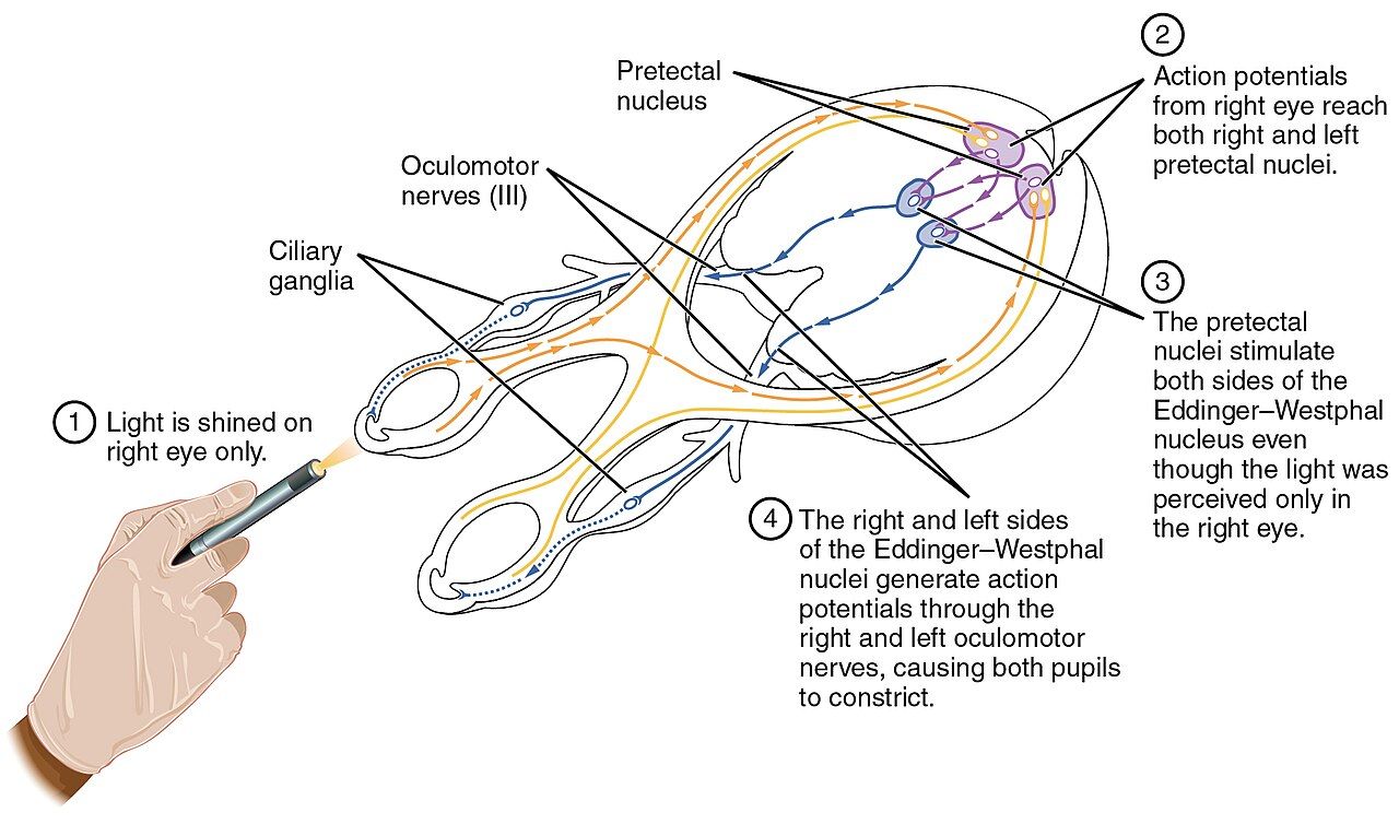
Four states that look similar and are not
Brain death — death determined by neurologic criteria — is the irreversible cessation of all functions of the entire brain, including the brainstem. Under the Uniform Determination of Death Act this is legal death. The patient is dead. The heart may still beat because the ventilator maintains oxygenation and the myocardium has intrinsic automaticity, and this is precisely why the language you use matters: say “he died” and “the ventilator,” never “life support” or “we are keeping him alive.” The time of death is the time of the determination, not the time the ventilator is later disconnected.
Coma is a state of unarousable unresponsiveness with eyes closed and no sleep–wake cycles; brainstem function is variably preserved and the state usually evolves within two to four weeks into recovery, vegetative state or death. Vegetative state (unresponsive wakefulness syndrome) is wakefulness without awareness: eyes open, sleep–wake cycles present, brainstem reflexes and spontaneous breathing intact, no reproducible purposeful response. Reflexive movements, grimacing and even sounds occur, which is why families so often believe there is more there than there is. Minimally conscious state shows inconsistent but reproducible evidence of awareness — following a simple command sometimes, tracking, localising to pain — and carries a meaningfully better prognosis than vegetative state.
| Brain death | Coma | Vegetative state | Minimally conscious | |
|---|---|---|---|---|
| Eyes / sleep–wake | Closed, none | Closed, none | Open, cycles present | Open, cycles present |
| Awareness | None | None | None | Inconsistent but reproducible |
| Brainstem reflexes | All absent | Variably present | Present | Present |
| Spontaneous breathing | Absent (apnoea test positive) | Usually present | Present | Present |
| Legal status | Dead | Alive | Alive | Alive |
How the determination is made, and what families will see
Prerequisites come before any examination. There must be a known, irreversible, catastrophic cause on imaging or history sufficient to explain the findings. Confounders must be excluded: core temperature at or above 36 °C, systolic blood pressure at or above 100 mmHg (or mean arterial pressure at or above 75 mmHg), no drug or alcohol intoxication and no residual sedatives — allow at least five half-lives, longer with hypothermia or organ failure — no neuromuscular blockade, confirmed with train-of-four, and correction of severe metabolic, electrolyte, acid–base and endocrine derangements. Skipping the prerequisites invalidates everything that follows.
The examination demonstrates coma with no response to noxious central stimulus and absence of every brainstem reflex: pupils fixed and non-reactive, no corneal reflex, no oculocephalic response, no oculovestibular response to cold caloric irrigation, no gag, no cough with deep tracheal suctioning, no facial movement to supraorbital pressure. The apnoea test follows: preoxygenate to a PaO2 above roughly 200 mmHg, establish a normal baseline PaCO2, disconnect from the ventilator with oxygen delivered by cannula into the airway, and observe for eight to ten minutes for any respiratory effort. The test is positive for apnoea when the PaCO2 reaches 60 mmHg or rises 20 mmHg above baseline with no respiratory effort. It is aborted for hypotension, desaturation or arrhythmia, and an ancillary test is used instead — cerebral angiography, nuclear perfusion scanning, or in some protocols EEG or transcranial Doppler. Ancillary tests are for when the clinical exam cannot be completed, not a substitute for it.
Your bedside job during all of this is family. Explain each step before it happens and stay with them if they choose to watch. Prepare them for two things especially. First, spinal reflexes survive brain death — triple flexion, deep tendon reflexes, and occasionally the dramatic Lazarus sign of arm elevation and adduction. Say in advance: “You may see his arm or leg move. Those movements come from the spinal cord, not from the brain, and they do not mean he can feel or respond.” Second, diabetes insipidus is common as the pituitary fails: expect urine output above 300 mL/hour with a rising serum sodium above 145 mmol/L and dilute urine of low specific gravity, requiring desmopressin and careful replacement.
Organ donation: refer early, decouple always
Federal Conditions of Participation require hospitals to notify their organ procurement organisation of every death and every imminent death, in a timely manner, using defined clinical triggers — typically a ventilated patient with a severe neurologic injury, a Glasgow Coma Scale at or below a set threshold, or any plan to discuss withdrawal of life-sustaining treatment. Notification is a call, not a conversation with the family. Make it early, make it per policy, and make it even if you are certain the family will decline — eligibility is not yours to determine.
Then decouple. The news that the patient has died, or that further treatment is futile, must be separated in time from any discussion of donation; families given both at once consent less and grieve worse. The donation request itself should be made by a trained designated requestor — an OPO coordinator or a hospital-designated, specifically trained staff member — not by the bedside nurse acting alone, and ideally in collaboration with the team the family already trusts. If a family asks you directly about donation before the OPO arrives, answer factually and briefly, then bring in the coordinator. Do not raise it yourself, do not offer opinions about the recipient, and do not tell a family it is what their loved one would have wanted.
First-person authorisation matters: if the patient registered as a donor on a state registry or driver’s licence, that is a legally binding gift under the Uniform Anatomical Gift Act and the family cannot revoke it, though skilled OPO staff work to bring families along rather than override them. Donation after circulatory death applies to patients who do not meet brain death criteria but whose family has independently decided to withdraw life-sustaining treatment; the withdrawal decision must be made first and entirely independently of donation, after which withdrawal occurs in a controlled setting and organs are recovered after a mandated hands-off period following asystole. Between referral and recovery, the nurse maintains donor management goals — normothermia, mean arterial pressure typically above 60–70 mmHg, oxygenation, glucose control, electrolyte and diabetes insipidus management — and does so with the same meticulous care as for any patient, because that care becomes someone else’s life.
Moral distress, grief, and staying in this work
Moral distress is the specific pain of knowing the ethically right action and being constrained from taking it — running a code on a patient you know had a POLST, feeding a contracted patient through a tube against everything you would want, watching a family demand escalation that only prolongs dying. It is not burnout and it is not the same as a hard shift. It accumulates: repeated episodes produce a rising baseline called the crescendo effect, and what remains after each episode is moral residue. Unaddressed, it is a leading reason nurses leave the bedside.
Naming it is the first intervention. The classic Four A’s framework — Ask (am I distressed, and about what), Affirm (this matters and I will act), Assess (what specifically is the constraint), Act (what change do I request, and of whom) — converts a vague sick feeling into a specific request. Individual coping is necessary but insufficient; the durable remedies are structural: accessible ethics consultation that any staff member can trigger, routine post-death debriefs, unit-based ethics rounds, schedules that do not assign the same nurse to three consecutive deaths, and leaders who treat a nurse’s ethical objection as data rather than insubordination.
Grief is separate. Nurses experience real grief for patients and rarely have permission to show it — disenfranchised grief, unacknowledged by the culture around them. Compassion fatigue and secondary traumatic stress come from absorbing others’ suffering; burnout comes from workload and system dysfunction; they compound each other. The protective factors that show up repeatedly are peer connection, ritual (the pause, a condolence card the team signs, attending a funeral when you can), supervision or debriefing that is scheduled rather than heroic, sleep, and the deliberate practice of remembering the deaths that went well. You will do this work for decades. Build the habits now, in your first year, and not in the year you are thinking about leaving.
Brain death is legal death and must be determined with prerequisites, full brainstem examination and a formal apnoea test; refer every imminent death to the OPO early, keep the death notification decoupled from a request made by a trained requestor, and treat your own moral distress as a problem with structural solutions.
The chapter in one breath
End-of-life care is technical work with a legal skeleton. Capacity is a decision-specific clinical judgement resting on four abilities — understanding, appreciation, reasoning and expressing a choice — and an unwise decision is not incapacity; competence, by contrast, is decided by a court. When capacity is gone, the named health care agent decides, then the statutory hierarchy, applying expressed wishes, then substituted judgement, then best interest. A living will records wishes and needs capacity to be lost before it applies; a POLST is a portable signed order that EMS will honour. DNR means no compressions and no defibrillation, nothing else. Withholding and withdrawing are ethically identical, which makes time-limited trials safe to start and mandatory to bound with an endpoint. Symptoms are treated proportionately: opioids for pain and dyspnoea with a fan and oxygen only if hypoxaemic, glycopyrrolate for secretions, haloperidol after reversible causes for terminal delirium, meticulous mouth care always. The doctrine of double effect protects proportionate titration; palliative sedation for refractory suffering is not euthanasia. Hospice requires a certified six-month prognosis and returns an interdisciplinary team plus 13 months of bereavement care. Recognise imminent death by mottling, narrowed pulse pressure, Cheyne–Stokes breathing and anuria, and say so to families before they see it. Brain death is legal death; refer early, decouple, let trained requestors ask. Then tend to your own moral residue.
Words worth knowing
| Term | What it means |
|---|---|
| Decision-making capacity | Clinical, decision-specific ability to understand, appreciate, reason and express a choice; can fluctuate and can be restored |
| Competence | Global legal status determined by a court, not by a clinician |
| Substituted judgement | Surrogate standard asking what this particular patient would have chosen, used when wishes were never expressed directly |
| Best interest standard | Last-resort surrogate standard weighing benefits against burdens when the patient’s values are unknowable |
| Living will | Instructional advance directive stating treatment wishes; takes effect only after capacity is lost and trigger conditions are met |
| Durable power of attorney for health care | Advance directive naming an agent to decide when the patient cannot; outranks the default surrogate hierarchy |
| POLST / MOLST | Portable, clinician-signed medical order set for the seriously ill that is actionable in every setting, including by EMS |
| Patient Self-Determination Act | 1990 federal law requiring facilities to ask about, document and inform patients regarding advance directives on admission |
| Allow natural death (AND) | Alternative phrasing for DNR that frames the order as permitting dying rather than withholding rescue |
| Time-limited trial | Agreed period of full treatment with pre-specified markers and a set date to reassess and change course |
| Doctrine of double effect | Ethical principle permitting an act with a foreseen but unintended harmful effect when intent, means and proportionality are satisfied |
| Palliative sedation | Proportionate sedation for a refractory symptom in an imminently dying patient; titrated to comfort, not to death |
| Medical aid in dying | Statutorily permitted prescription of a lethal dose that a patient with capacity self-administers; nurses do not administer it |
| Terminal secretions | Pooled hypopharyngeal secretions producing audible rattle; treated with repositioning, reduced fluids and antimuscarinics, not deep suctioning |
| Cheyne–Stokes respiration | Crescendo–decrescendo breathing with apnoeic pauses, a common and expected pattern in the last hours |
| Kennedy terminal ulcer | Rapidly appearing purple sacral lesion of the last days reflecting skin failure from hypoperfusion, not poor care |
| Brain death | Irreversible cessation of all brain and brainstem function; legal death under the Uniform Determination of Death Act |
| Vegetative state | Wakefulness without awareness — eyes open with sleep–wake cycles, brainstem intact, no reproducible purposeful response |
| Decoupling | Deliberate separation in time of the death or futility notification from the organ donation request |
| Moral distress | Knowing the ethically right action while being constrained from taking it; accumulates as moral residue over repeated episodes |