Innate Immunity
Long before the body knows what hit it, it is already fighting back. Innate immunity is the defense you are born with. It is a set of barriers, cells, and chemical alarms. They answer in seconds to minutes. They answer the same way every time, against almost anything that does not belong. Innate immunity has never met the microbe before, and it will not remember it after. It runs from the skin that keeps microbes out, to the neutrophils that swarm a cut, to the fever that spikes at three in the morning. It is the fast, blunt, always-on first responder of the immune system. This chapter walks through its layers in the order the body meets an invader.
By the end of this chapter you can
- Contrast innate and adaptive immunity by speed, specificity, and memory.
- Describe the first-line defenses, which are skin, mucous membranes, and normal flora, and say how each one keeps microbes out.
- Identify the major phagocytic cells and walk through the steps of phagocytosis.
- Explain the inflammatory response. Tie the four cardinal signs to what is happening in the tissue.
- Describe how pyrogens reset the hypothalamic thermostat to make a fever. Weigh what it buys and what it costs.
- Summarize the outcomes of the complement cascade, which are opsonization, inflammation, and cytolysis.
- Explain how interferons protect cells from viral infection.
- Link each of these defenses to something that can be checked in a real case.
Section 1First line: barriers
Two immune systems, one goal
The body defends itself with two systems that overlap. Innate immunity is there from birth. It answers in seconds to minutes. It uses the same non-specific weapons on every invader. It has no memory, so the tenth exposure gets the same answer as the first. Adaptive immunity is the subject of the next chapter. It is slower to start. It is exactly tuned to one particular microbe. And it remembers, which is why a second exposure is met faster and harder. Innate immunity buys the days that adaptive immunity needs to gear up. In the body the two work as one team. Learning them apart makes each one far easier to hold onto.
The immune system is often taught as three lines of defense. Innate immunity supplies the first two. The first line is the set of physical, chemical, and biological barriers that stop microbes from ever getting in. That is this section. The second line is the response inside the body, once something breaks through a barrier. It is phagocytes, inflammation, fever, and antimicrobial proteins, and it fills Sections 2 to 5. Adaptive immunity is the third line.
Skin and mucous membranes
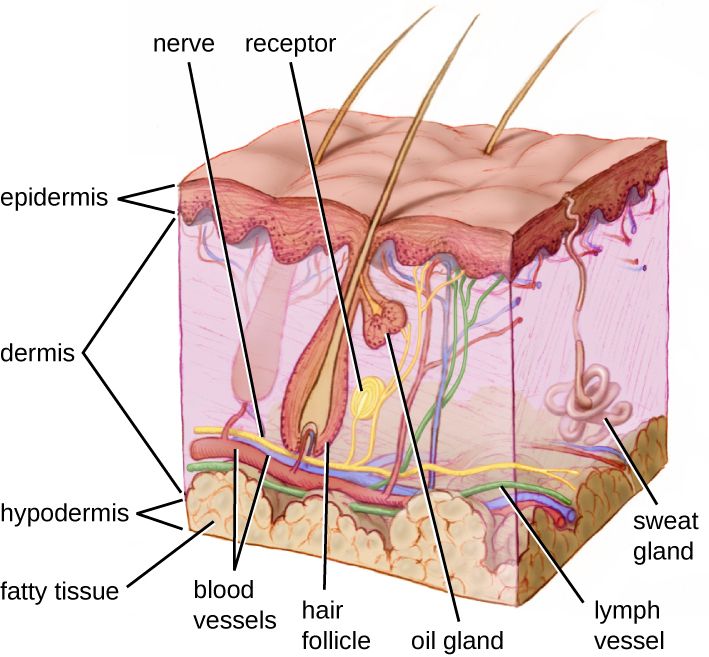
The skin is the largest organ in the body. It is also the most important physical barrier there is. Its outer layer is the epidermis. That layer is built of tightly packed cells stuffed with keratin. They stack up into dead, shingled sheets, and those sheets shed all day long. Any microbe stuck to a shed sheet goes with it. Most bacteria simply cannot get through intact skin. So every break in the skin matters. An IV site, a surgical cut, a pressure injury, even a scratch, is a door in the wall. Keeping that door clean and covered is frontline infection control.
Some surfaces open to the outside world. The respiratory, digestive, urinary, and reproductive tracts all do. There the barrier is not dry skin. It is a mucous membrane. These surfaces stay moist and carry a coat of mucus, a sticky gel. Mucus traps microbes before they reach the cells underneath. In the airway, cells carry tiny hair-like cilia. The cilia beat in waves and sweep the trapped debris up and out of the lungs. That moving belt is the mucociliary escalator. Tears, saliva, and the flushing action of urine wash microbes away as well. This is why a breathing tube raises the risk of pneumonia so sharply. The tube runs straight past the mucociliary escalator. It is also why coughing, deep breathing, and enough fluid are worth the trouble.
Chemical and microbial reinforcements
Barriers are more than walls. They are hostile chemical places. The skin puts out sebum, which is oily and acidic, along with sweat. Together they hold the surface at a low, unfriendly pH. Lysozyme is an enzyme found in tears, saliva, and mucus. It chews apart the peptidoglycan of bacterial cell walls. It hits Gram-positive cells hardest, because their wall sits bare on the surface. Stomach acid is brutally low in pH. The vagina is held acidic by resident lactobacilli. On top of all of this sits the normal flora, the harmless microbes that already live on skin and mucosal surfaces. They take up the space and eat the food, so they crowd out would-be pathogens. That protection is called competitive exclusion. Wipe the flora out, with a broad-spectrum antibiotic for instance, and opportunists move into the empty space. Candida and Clostridioides difficile are the two classic examples.
| First-line defense | Type | How it stops microbes |
|---|---|---|
| Skin (epidermis) | Physical | Keratinized, dry, shedding cell layers block entry |
| Mucous membranes | Physical | Trap microbes in mucus; cilia and flushing remove them |
| Sebum & sweat | Chemical | Low pH and fatty acids hold growth down |
| Lysozyme | Chemical | Enzyme that digests bacterial cell walls |
| Stomach acid / vaginal acidity | Chemical | Low pH kills or blocks many microbes |
| Normal flora | Biological | Competitive exclusion — crowd out pathogens |
- Innate immunity is fast, non-specific, and has no memory. Adaptive is slow, specific, and remembers.
- The first line is physical, chemical, and microbial barriers that keep invaders out.
- Skin and mucous membranes are the physical walls. Lysozyme, acid, and sebum are the chemical defenses.
- Normal flora protect by competitive exclusion. Any break in the skin is a breach that care has to cover.
Section 2Second line: cells
The white blood cell responders
Once a microbe slips past the barriers, it meets the cellular second line. These are the white blood cells, also called leukocytes. They are made in the bone marrow, and they patrol the blood and the tissues. Two of them do most of the eating, and those two are the ones to know. Neutrophils are the most numerous white cell and the first to arrive at an infection. They are fast, aggressive, short-lived front-line soldiers. Macrophages are the larger, longer-lived cleanup crew. They grow out of circulating monocytes that mature after leaving the blood. Macrophages patrol the tissues, engulf microbes and debris, and help kick off other immune responses. A microbe-eating cell of either kind is a phagocyte. The act of eating is phagocytosis, Greek for cell eating.
These cells do not reach a wound by luck. Chemical signals are released at the site of infection. They come from damaged cells, from the microbes themselves, and from complement in Section 5. Those signals draw phagocytes toward the trouble. That directed march is chemotaxis. To get out of the bloodstream, a circulating white cell first sticks to the vessel wall, which is margination. Then it squeezes between the cells of the vessel lining and into the tissue. That move is diapedesis, also called extravasation.
How a phagocyte recognizes an enemy
How does a phagocyte tell self from invader? It carries pattern-recognition receptors (PRRs). These are sensors that latch onto molecular signatures shared by whole classes of microbes. Human cells do not carry those signatures. The patterns themselves are called pathogen-associated molecular patterns (PAMPs). The classic PAMP is the lipopolysaccharide (LPS) in the outer membrane of Gram-negative bacteria. Because the patterns are broad, one receptor can spot many microbes. That breadth is the hallmark of innate, non-specific immunity. Recognition gets easier still when a microbe is coated with tags called opsonins. Antibodies and the complement protein C3b are the main ones. That coating is opsonization. It is like painting "eat me" on the microbe, and it speeds engulfment up enormously.
Phagocytosis, step by step
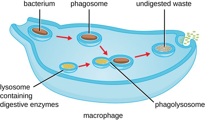
The killing sequence is worth memorizing, because it comes back all through immunology. (1) Chemotaxis. The phagocyte migrates toward the microbe. (2) Adherence. It binds the microbe, helped by opsonins. (3) Ingestion. The cell membrane wraps around the microbe and pinches off an inside bubble called a phagosome. (4) Fusion. The phagosome merges with a lysosome, which is full of digestive enzymes, to make a phagolysosome. (5) Killing and digestion. Enzymes, reactive oxygen from the respiratory burst, and low pH destroy the microbe. (6) Exocytosis. The leftovers that cannot be digested are dumped back out. What stays behind is spent neutrophils, dead microbes, and tissue fluid. That mixture is what we call pus.
| Cell | Comes from | Role |
|---|---|---|
| Neutrophil | Bone marrow (granulocyte) | First responder; most abundant WBC; fast, short-lived phagocyte |
| Monocyte → macrophage | Bone marrow; matures in tissue | Large, long-lived phagocyte; cleanup and immune signaling |
| Dendritic cell | Bone marrow | Phagocyte that alerts adaptive immunity |
| Natural killer (NK) cell | Lymphoid line | Kills virus-infected and tumor cells (not classic phagocytosis) |
| Eosinophil | Bone marrow (granulocyte) | Attacks parasites; a role in allergy |
- Neutrophils are the first and most numerous phagocytes. Macrophages, from monocytes, are the long-lived cleanup phagocytes.
- Phagocytes reach an infection by chemotaxis, margination, and diapedesis.
- They spot microbes when PRRs bind PAMPs, and they engulf opsonized microbes fastest.
- Phagocytosis runs chemotaxis → adherence → ingestion (phagosome) → fusion (phagolysosome) → digestion → exocytosis. The leftover debris is pus.
Section 3Inflammation
The four cardinal signs
Inflammation is the tissue's coordinated answer to injury or infection. It makes four classic signs, and they have been recognized since ancient times. They are redness, heat, swelling, and pain. In Latin, rubor, calor, tumor, dolor. A fifth is often added, loss of function, or functio laesa. Far from being the enemy, these signs are visible proof that the second line has arrived and is working. Look at a wound, an IV site, or a swollen joint, and you are reading the four cardinal signs straight off the body. They are the everyday language of checking for infection.
What is actually happening in the tissue
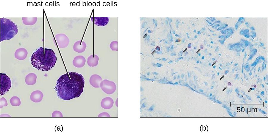
Each sign has a mechanism. Cells get injured, or microbes get detected. Resident mast cells and damaged tissue then release chemical mediators. The biggest one is histamine. Prostaglandins, bradykinin, and cytokines come with it. These mediators do two things. First they cause vasodilation. Blood vessels widen, so more blood floods the area. That extra warm blood is the redness and the heat. Second they raise vascular permeability. The vessel walls turn leaky, so fluid, proteins, and cells pour out into the tissue. That buildup of fluid is edema, and it is the swelling. The swelling then presses on nerve endings, and prostaglandins and bradykinin make those nerves more sensitive. Together, that is the pain. The whole point of all this is to deliver phagocytes and clotting proteins to the site, and to wall the infection off in a mesh of fibrin so it cannot spread.
| Cardinal sign | Latin | What causes it |
|---|---|---|
| Redness | rubor | Vasodilation — more blood flowing in |
| Heat | calor | Warmth of that increased blood flow |
| Swelling | tumor | Fluid leaking from leaky vessels (edema) |
| Pain | dolor | Pressure on nerves plus pain-sensitizing mediators |
| Loss of function | functio laesa | Swelling and pain limit use of the part |
Acute, chronic, and when inflammation turns dangerous
Acute inflammation is short. It settles as soon as the threat is cleared. Some threats cannot be cleared. Then inflammation turns chronic. Macrophages stay on the scene, and they may wall the invader off inside a ball of immune cells called a granuloma. Tuberculosis is the classic case. Inflammation in one spot protects. The same mediators spilled into the bloodstream body-wide can be a disaster. That is sepsis, a body-wide inflammatory response to infection. Vessels widen and leak everywhere at once. Blood pressure drops away and organs are starved of oxygen. The very tools that heal one small wound can kill when they are unleashed everywhere.
- The four cardinal signs are redness, heat, swelling, and pain, plus loss of function.
- Mast cells and injured tissue release histamine and other mediators that cause vasodilation and more vascular permeability.
- Inflammation delivers phagocytes and walls the infection off with fibrin.
- Inflammation in one spot heals. Body-wide inflammation, which is sepsis, is life-threatening.
Section 4Fever
Resetting the thermostat
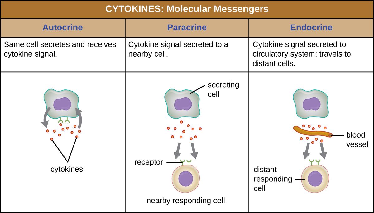
Fever, also called pyrexia, is a core body temperature that is abnormally high. It is not the infection getting the upper hand. It is a deliberate, regulated defense. Body temperature is run by a thermostat in the hypothalamus, which normally holds things near 37°C (98.6°F). Fever happens when substances called pyrogens raise the hypothalamic set-point. There are two kinds. Exogenous pyrogens come from outside the body. The most famous is the LPS of Gram-negative bacteria, also called endotoxin. Endogenous pyrogens are made by the body's own immune cells. They are the cytokines interleukin-1 (IL-1), IL-6, and tumor necrosis factor (TNF), released by macrophages when they meet a microbe. These pyrogens drive the making of prostaglandin E₂ in the hypothalamus, and that is what actually pushes the set-point up.
Here is the key idea. Once the set-point is raised, the body is not overheating. It now believes 39°C is the correct temperature, and it works to get there. That is why the onset of a fever brings shivering and a feeling of cold. Muscles contract to make heat. Blood vessels narrow to hold it in. All of it is a climb toward the new, higher target.
The three phases of a fever
A fever runs a predictable course, and knowing the phase changes what should be done. In the onset, or chill phase, the set-point jumps. The person feels cold, shivers, and has cool, pale skin, while the body drives its temperature up. In the plateau, also called the stadium, the body has reached the new set-point. The skin is warm, flushed, and dry, and the temperature holds steady up high. In defervescence, the flush or crisis, pyrogens fall and the set-point drops back to normal. Now the body is too hot, so it sweats and flushes to shed the extra heat. Reading these phases is practical. Someone who is shivering is still climbing toward a set-point the hypothalamus has already chosen. Warmth from outside will not push the temperature past it. So add blankets and cut the drafts to stop the shivering. Shivering burns a lot of oxygen. It drives up the metabolic rate and the work of the heart, in exactly the people who can least afford it, the ones with heart disease, lung disease, or sepsis. What must not happen in the chill phase is cooling. A tepid sponge bath or a cooling blanket during shivering fights the set-point and makes the shivering worse. Cooling measures belong to the plateau and defervescence phases. Antipyretics work a different way. They lower the set-point itself.
| Phase | Set-point | What you see | What to do |
|---|---|---|---|
| Onset (chill) | Rising | Shivering, cold, pale, “goosebumps” | Temp is climbing; warm the room and add blankets to stop the shivering — do not cool now |
| Plateau (stadium) | Reached (high) | Warm, flushed, dry skin | Watch fluids; temp is holding high |
| Defervescence (flush) | Falling | Sweating, flushed, feels hot | Replace the fluid lost in sweat |
Friend or foe?
A moderate fever helps. Higher temperatures slow the growth of many pathogens. They speed up the enzymes of the immune response. They sharpen phagocytes and other defenders. So wiping out every low fever with a drug is not always wise, because the fever is doing work. But fever has costs and limits too. It raises metabolic demand, heart rate, and fluid loss. In young children a fast rise can set off a febrile seizure. And a very high temperature, roughly above 40 to 41°C (104 to 106°F), begins to denature the body's own proteins. That is a medical emergency. Antipyretics such as acetaminophen and NSAIDs work by blocking prostaglandin production, which lowers the set-point back down.
- Fever is a regulated rise in the hypothalamic set-point, not a loss of control.
- Exogenous pyrogens such as LPS and endogenous pyrogens such as the cytokines IL-1, IL-6, and TNF both act through prostaglandin E₂.
- The phases run onset (chills, climbing) → plateau (steady, high) → defervescence (sweating, falling).
- A moderate fever aids defense. A very high fever above 40°C is dangerous, and some people cannot mount one at all.
Section 5Complement & interferons
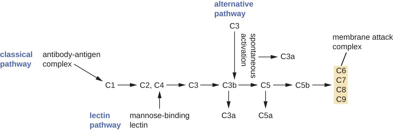
Complement: a cascade of ready-made proteins
Complement is a set of about 30 blood proteins. The liver makes most of them. They circulate in an inactive form and wait. Once triggered, they activate one another in order. That is a cascade, where each activated protein cuts and switches on the next, so the signal grows at every step. Three pathways can set it off. The classical pathway is triggered by antibodies already bound to a microbe. The alternative pathway is triggered straight off microbial surfaces. The lectin pathway is triggered by carbohydrates on microbes. All three converge on the central protein, C3, which is split into C3a and C3b. That split is the hinge of the whole system.
From there, complement delivers three life-saving outcomes. Opsonization. C3b coats the microbe as an opsonin, tagging it so phagocytes grab it far more easily. That ties this section straight back to phagocytosis. Inflammation. The small fragments C3a and C5a are anaphylatoxins. They make mast cells release histamine. C5a is also a powerful chemotactic signal that calls phagocytes to the scene. Cytolysis. The terminal proteins assemble into a ring called the membrane attack complex (MAC), which punches a hole in the microbe's membrane. Its contents leak out and it bursts. Three jobs, easy to hold onto: tag, recruit, and destroy.
| Complement outcome | Key players | What it does |
|---|---|---|
| Opsonization | C3b | Coats the microbe so phagocytes engulf it faster |
| Inflammation | C3a, C5a | Anaphylatoxins — trigger histamine release and attract phagocytes |
| Cytolysis | MAC (C5b–C9) | Forms pores that lyse the microbe |
Interferons: the antiviral alarm
Barriers, phagocytes, and complement are formidable against bacteria. A virus is a different problem. It hides inside a host cell, where those defenses cannot reach. For viruses, innate immunity has another tool. Interferons (IFNs) are a family of cytokines, named for the way they interfere with viral replication. When a cell is infected by a virus, it releases type I interferons, which are IFN-α and IFN-β. These do not save the dying cell that made them. Instead they act as a warning to the neighboring cells. A neighbor that picks up the interferon signal switches on an antiviral state. It makes enzymes that chew up viral genetic material and shut protein building down if a virus gets in. The virus then finds no factory to hijack. A separate interferon, IFN-γ, mainly activates macrophages and bridges over to adaptive immunity.
This matters in two directions. The dragging tiredness, the muscle aches, and the fever of a viral illness like influenza are largely the work of your own interferons, not of the virus directly. And because interferons are natural antivirals and immune modulators, purified versions are used as medicines. They treat certain viral hepatitis infections, some cancers, and multiple sclerosis.
- Complement is a cascade of about 30 proteins. Three pathways, classical, alternative, and lectin, converge on C3.
- Its three outcomes are opsonization (C3b), inflammation (C3a and C5a), and cytolysis (the MAC).
- Interferons are antiviral cytokines. A virus-infected cell releases type I IFN to put its neighbors into an antiviral state.
- Interferons cause much of the sick feeling of a viral illness, and they are used as medicines.
The chapter in one breath
- Innate immunity is the fast, non-specific, memory-free defense you are born with. It buys time for adaptive immunity.
- The first line keeps microbes out with skin and mucous membranes, chemical defenses such as lysozyme, acid, and sebum, and the normal flora.
- When barriers fail, phagocytes, neutrophils first and then macrophages, find microbes by chemotaxis and destroy them by phagocytosis.
- Inflammation, which is redness, heat, swelling, and pain, comes from histamine-driven vasodilation and leaky vessels. Body-wide, it becomes sepsis.
- Fever is a raised hypothalamic set-point driven by pyrogens, meaning LPS and the cytokines IL-1, IL-6, and TNF, acting through prostaglandin E₂. It helps in moderation and turns dangerous when very high.
- Complement tags (opsonization), recruits (inflammation), and destroys (MAC). Interferons warn neighboring cells to resist viruses.
- Nearly every innate defense shows up in something that can be checked: skin integrity, the white cell differential, wound signs, the fever phase, and the early signs of sepsis.
ReferenceWords worth knowing
A quick reference for the terms in this chapter. Skim it now, or circle back whenever a word trips you up.
- Antipyretic
- A drug such as acetaminophen or an NSAID that lowers fever by blocking prostaglandin production and resetting the hypothalamic set-point down.
- Chemotaxis
- The directed movement of a cell, such as a phagocyte, toward a chemical signal released at a site of infection.
- Complement
- A cascade of about 30 blood proteins that, once activated, cause opsonization, inflammation, and cell lysis.
- Cytokine
- A signaling protein released by cells to talk to one another; includes the interleukins, the interferons, and TNF.
- Diapedesis
- The squeezing of white blood cells between the cells of a vessel wall to enter infected tissue; also called extravasation.
- Fever (pyrexia)
- A regulated rise in core body temperature, caused by pyrogens raising the hypothalamic set-point.
- Inflammation
- The tissue response to injury or infection, marked by redness, heat, swelling, and pain.
- Innate immunity
- The non-specific defenses present from birth that answer fast and without memory.
- Interferon (IFN)
- An antiviral cytokine; a virus-infected cell releases type I IFN to put neighboring cells into an antiviral state.
- Lysozyme
- An enzyme in tears, saliva, and mucus that digests the peptidoglycan of bacterial cell walls.
- Macrophage
- A large, long-lived phagocyte grown from a monocyte; cleans up microbes and debris and signals other immune cells.
- Membrane attack complex (MAC)
- The ring of terminal complement proteins, C5b to C9, that forms pores and lyses a target cell.
- Neutrophil
- The most abundant white blood cell and the first phagocyte to arrive at an infection.
- Opsonization
- The coating of a microbe with tags, antibody or C3b, that make phagocytosis faster and easier.
- PAMP
- Pathogen-associated molecular pattern — a molecular signature such as LPS, shared by many microbes and read by innate receptors.
- Pattern-recognition receptor (PRR)
- A receptor on innate immune cells that binds PAMPs to detect microbes.
- Phagocytosis
- The engulfment and destruction of a microbe by a phagocyte inside a phagolysosome.
- Pyrogen
- A fever-inducing substance; exogenous, such as LPS, or endogenous, such as the cytokines IL-1, IL-6, and TNF.
- Sepsis
- A body-wide, dysregulated inflammatory response to infection that can cause dangerously low blood pressure and organ failure.
- Vasodilation
- Widening of blood vessels; in inflammation it raises blood flow, which causes redness and heat.